. 2014 Jan 23;16(1):R21.
doi: 10.1186/ar4449.

High mechanical strain of primary intervertebral disc cells promotes secretion of inflammatory factors associated with disc degeneration and pain

High mechanical strain of primary intervertebral disc cells promotes secretion of inflammatory factors associated with disc degeneration and pain

Rahul Gawri et al. Arthritis Res Ther. .

Abstract

Introduction: Excessive mechanical loading of intervertebral discs (IVDs) is thought to alter matrix properties and influence disc cell metabolism, contributing to degenerative disc disease and development of discogenic pain. However, little is known about how mechanical strain induces these changes. This study investigated the cellular and molecular changes as well as which inflammatory receptors and cytokines were upregulated in human intervertebral disc cells exposed to high mechanical strain (HMS) at low frequency. The impact of these metabolic changes on neuronal differentiation was also explored to determine a role in the development of disc degeneration and discogenic pain.

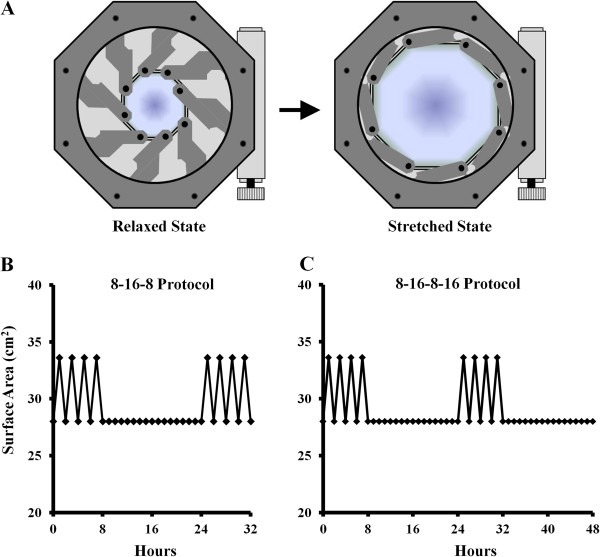
Methods: Isolated human annulus fibrosus (AF) and nucleus pulposus (NP) cells were exposed to HMS (20% cyclical stretch at 0.001 Hz) on high-extension silicone rubber dishes coupled to a mechanical stretching apparatus and compared to static control cultures. Gene expression of Toll-like receptors (TLRs), neuronal growth factor (NGF) and tumour necrosis factor α (TNFα) was assessed. Collected conditioned media were analysed for cytokine content and applied to rat pheocromocytoma PC12 cells for neuronal differentiation assessment.

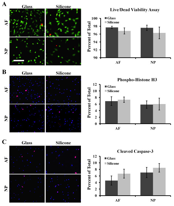
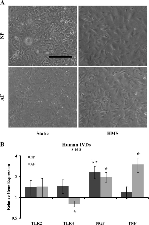
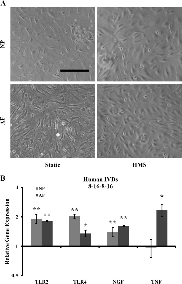
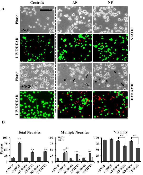
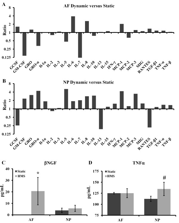
Results: HMS caused upregulation of TLR2, TLR4, NGF and TNFα gene expression in IVD cells. Medium from HMS cultures contained elevated levels of growth-related oncogene, interleukin 6 (IL-6), IL-8, IL-15, monocyte chemoattractant protein 1 (MCP-1), MCP-3, monokine induced by γ interferon, transforming growth factor β1, TNFα and NGF. Exposure of PC12 cells to HMS-conditioned media resulted in both increased neurite sprouting and cell death.

Conclusions: HMS culture of IVD cells in vitro drives cytokine and inflammatory responses associated with degenerative disc disease and low-back pain. This study provides evidence for a direct link between cellular strain, secretory factors, neoinnervation and potential degeneration and discogenic pain in vivo.


Figures

Figure 1
Figure 1
Viability and proliferation of intervertebral disc cells cultured on modified silicone surfaces. (A) Representative images show living (green) and dead (red) cells, with quantification shown at right. (B) Representative immunofluorescence images of phosphohistone H3 stained cells (red) and 4′,6-diamidino-2-phenylindole (DAPI) nuclear stain (blue), with quantification shown at right. (C) Representative immunofluorescence images of cleaved caspase 3–stained cells (red) and DAPI nuclear stain (blue), with quantification shown at right. AF, Annulus fibrosus; NP, Nucleus pulposus. Scale bar: 200 μm. Error bars represent SEM. Three independent experiments were performed. Significance was calculated by Student’s t-test.
Figure 2
Figure 2
Cell-stretching device and strain protocols. (A) Schematic representation of mechanical stretching device used to apply low-frequency, high-magnitude strains. Graphical representation of the 8 hours/16 hours/8 hours stretch protocol (B) and the 8 hours/16 hours/8 hours/16 hours (C) stretch protocol applied to cells.
Figure 3
Figure 3
Application of the 8 hours/16 hours/8 hours stretch protocol to human intervertebral disc cells. (A) Representative morphological images of static and high mechanical strain (HMS) cultured human annulus fibrosus (AF) and nucleus pulposus (NP) cells. Scale bar: 200 μm. (B) Gene expression analysis of both AF and NP cells immediately after the stretch protocol ended. IVD, Intervertebral disc; NGF, Neuronal growth factor; TLR, Toll-like receptor; TNF, Tumour necrosis factor. Error bars represent SEM. Three independent experiments were performed. *P < 0.05 and **P < 0.01 by Student’s t-test.
Figure 4
Figure 4
Application of 8 hours/16 hours/8 hours/16 hours stretch protocol. (A) Representative morphological images of human static and high mechanical strain (HMS) cultured annulus fibrosus (AF) and nucleus pulposus (NP) cells. Scale bar: 200 μm. (B) Gene expression analysis of both NP and AF cells after the additional 16-hour resting period. IVD, Intervertebral disc; NGF, Neuronal growth factor; TLR, Toll-like receptor; TNF, Tumour necrosis factor. Error bars represent SEM. Three independent experiments were performed. *P < 0.05 and **P < 0.01 by Student’s t-test.
Figure 5
Figure 5
Media from nucleus pulposus and annulus fibrosus cells cultured under high mechanical strain promote neurite outgrowth of PC12 cells. (A) Conditioned media from static and high mechanical strain (HMS) nucleus pulposus (NP) and annulus fibrosus (AF) cells were incubated with PC12 cells, and neurite outgrowth and viability were observed and compared to vehicle (−NGF) and neural growth factor (NGF)-treated (+NGF) controls. Black arrows in phase images indicate neurites. Scale bar: 200 μm. (B) Quantification of total and multiple neurite outgrowth and viability. Error bars represent SEM three independent experiments were performed. *P < 0.05, **P < 0.01, and #P < 0.05 (all by Student’s t-test) for samples with three or more neurites per cell body. All samples were compared to –NGF controls.
Figure 6
Figure 6
Cytokine analysis of conditioned media. Cytokine array blots were used to compare high mechanical strain (HMS) conditioned media of nucleus pulposus (NP) cells (A) and annulus fibrosus (AF) cells (B) with their respective static controls. Quantitation was carried out by performing enzyme-linked immunosorbent assays with conditioned media for neural growth factor (NGF) (C) and tumour necrosis factor α (TNFα) (D). GCSF, Granulocyte colony-stimulating factor; GM-CSF, Granulocyte-macrophage colony-stimulating factor; GRO, growth-related oncogene; IFN, Interferon; MCP, monocyte chemoattractant protein; MIG, monokine induced by γ interferon; IL, Interleukin; TGF, Transforming growth factor. Error bars represent SEM. Three independent experiments were performed. *P < 0.05 and #P = 0.0504 by Student’s t-test (the latter statistic approaching significance).

Similar articles

Cited by

References

    1. Frymoyer JW, Cats-Baril WL. An overview of the incidences and costs of low back pain. Orthop Clin North Am. 1991;16:263–271. - PubMed
    1. Adams MA, Roughley PJ. What is intervertebral disc degeneration, and what causes it? Spine (Phila Pa 1976) 2006;16:2151–2161. doi: 10.1097/01.brs.0000231761.73859.2c. - DOI - PubMed
    1. Gardner-Morse MG, Stokes IA. Structural behavior of human lumbar spinal motion segments. J Biomech. 2004;16:205–212. doi: 10.1016/j.jbiomech.2003.10.003. - DOI - PubMed
    1. Räty HP, Kujala UM, Videman T, Impivaara O, Crites Battié M, Sarna S. Lifetime musculoskeletal symptoms and injuries among former elite male athletes. Int J Sports Med. 1997;16:625–632. doi: 10.1055/s-2007-972693. - DOI - PubMed
    1. Takahashi H, Suguro T, Okazima Y, Motegi M, Okada Y, Kakiuchi T. Inflammatory cytokines in the herniated disc of the lumbar spine. Spine (Phila Pa 1976) 1996;16:218–224. doi: 10.1097/00007632-199601150-00011. - DOI - PubMed

Publication types

Substances