. 2014 Jan 23;16(1):R11.
doi: 10.1186/bcr3603.

Inducible and coupled expression of the polyomavirus middle T antigen and Cre recombinase in transgenic mice: an in vivo model for synthetic viability in mammary tumour progression

Inducible and coupled expression of the polyomavirus middle T antigen and Cre recombinase in transgenic mice: an in vivo model for synthetic viability in mammary tumour progression

Trisha Rao et al. Breast Cancer Res. .

Abstract

Introduction: Effective in vivo models of breast cancer are crucial for studying the development and progression of the disease in humans. We sought to engineer a novel mouse model of polyomavirus middle T antigen (PyV mT)-mediated mammary tumourigenesis in which inducible expression of this well-characterized viral oncoprotein is coupled to Cre recombinase (TetO-PyV mT-IRES-Cre recombinase or MIC).

Methods: MIC mice were crossed to the mouse mammary tumour virus (MMTV)-reverse tetracycline transactivator (rtTA) strain to generate cohorts of virgin females carrying one or both transgenes. Experimental (rtTA/MIC) and control (rtTA or MIC) animals were administered 2 mg/mL doxycycline beginning as early as eight weeks of age and monitored for mammary tumour formation, in parallel with un-induced controls of the same genotypes.

Results: Of the rtTA/MIC virgin females studied, 90% developed mammary tumour with complete penetrance to all glands in response to doxycycline and a T50 of seven days post-induction, while induced or un-induced controls remained tumour-free after one year of induction. Histological analyses of rtTA/MIC mammary glands and tumour revealed that lesions followed the canonical stepwise progression of PyV mT tumourigenesis, from hyperplasia to mammary intraepithelial neoplasia/adenoma, carcinoma, and invasive carcinoma that metastasizes to the lung; at each of these stages expression of PyV mT and Cre recombinase transgenes was confirmed. Withdrawal of doxycycline from rtTA/MIC mice with end-stage mammary tumours led to rapid regression, yet animals eventually developed PyV mT-expressing and -non-expressing recurrent masses with varied tumour histopathologies.

Conclusions: We have successfully created a temporally regulated mouse model of PyV mT-mediated mammary tumourigenesis that can be used to study Cre recombinase-mediated genetic changes simultaneously. While maintaining all of the hallmark features of the well-established constitutive MMTV-PyV mT model, the utility of this strain derives from the linking of PyV mT and Cre recombinase transgenes; mammary epithelial cells are thereby forced to couple PyV mT expression with conditional ablation of a given gene. This transgenic mouse model will be an important research tool for identifying synthetic viable genetic events that enable PyV mT tumours to evolve in the absence of a key signaling pathway.


Figures

Figure 1
Figure 1
Induced rtTA/MIC animals develop mammary tumours with characteristic histopathological features of PyV mT-driven mammary tumourigenesis. (A) Kaplan-Meier mammary tumour onset curve as measured by physical palpation. A total of 87.1% (34/39) of mice developed multifocal mammary tumours with a T50 of 7 days and an average of 22.0 ± 7.1 days post-induction with doxycycline. Control animals (induced rtTA or MIC; un-induced rtTA/MIC) were monitored for at least one year. (B) Representative H&E-stained sections of normal ductal structures in a mammary gland from a control animal (an un-induced rtTA/MIC) followed by typical stages of PyV mT mammary tumour progression (hyperplasia, mammary intraepithelial neoplasia (MIN), and adenocarcinoma) in mammary glands and tumours from rtTA/MIC mice following doxycycline induction. (Scale bar: 100 μm).
Figure 2
Figure 2
PyV mT and Cre recombinase are expressed at all stages of tumourigenesis. (A) Immunohistochemical detection of PyV mT (middle row) and Cre recombinase (bottom row) in ductal structures at the indicated stages of mammary tumour progression in rtTA/MIC mice, contrasted by normal mammary gland histology from control (an un-induced rtTA/MIC) and wildtype mice. The corresponding H&E-stained sections are shown for comparison (top row). (Scale bar: 100 μm). (B) Immunoblot analysis of protein lysates from rtTA/MIC mammary glands (mg), mammary tumours (tum) and adjacent mammary glands (amg) from animals sacrificed at palpation or at end-stage (“late onset” refers to palpation after 16 days of induction) as indicated using antibodies directed to E-cadherin (epithelial content control), PyV mT, Cre recombinase and α-tubulin (loading control). Controls include (from left to right) mammary glands from two un-induced rtTA/MIC mice, one induced MIC mouse and one induced rtTA mouse. Positive controls for PyV mT and Cre recombinase expression were mammary tumours from MMTV-PyV mT (PyV mT) and MMTV-NIC (NIC) animals, respectively; arrowheads indicate specific bands for these proteins.
Figure 3
Figure 3
Cre recombinase expression and activity are uniform in the epithelium of rtTA/MIC mammary tumours. X-gal staining (blue) of mammary tumour sections from rtTA/MIC animals carrying the Cre recombinase-activated β-galactosidase reporter transgene (GTRosa). Negative controls include MMTV-PyV mT mammary tumours with either GTRosa (PyV mT/GTRosa) or MMTV-Cre recombinase transgenes (PyV mT/Cre recombinase). An MMTV-PyV mT mammary tumour with both transgenes (PyV mT/Cre recombinase/GTRosa) was used as a positive control. Samples were counterstained with nuclear fast red (pink). (Scale bar: 500 μm).
Figure 4
Figure 4
rtTA/MIC end-stage mammary tumours exhibit a high capacity for metastatic dissemination to the lungs. (A) Quantification of the number of metastatic lung lesions (left axis) and the percentage of lung tissue occupied by metastases (right axis) in rtTA/MIC animals at end-stage tumour burden. Bars represent the average value for each parameter. (B) H&E-stained whole lung sections representative of low, medium and high levels of metastasis. (C) Representative lung lesion from a tumour-bearing rtTA/MIC animal stained with H&E (top row) and PyV mT (bottom row). An MMTV-NIC (NIC) lung lesion is shown as a negative control for PyV mT staining (Scale bar: 200 μm).
Figure 5
Figure 5
Doxycycline withdrawal in rtTA/MIC tumours leads to rapid regression and eventual spontaneous recurrence of masses. (A) Total mammary tumour burden measured over time in rtTA/MIC mice prior to and following doxycycline withdrawal (indicated by the dotted line at time 0). Tumour-bearing mice were de-induced upon reaching burden endpoint. Each line represents an individual animal labeled by its ID number. (B) Quantification of the number of measurable masses detected pre-regression (pre), at the point of maximum regression (mid) and at sacrifice (recurrent). Bars represent the average value at each time-point. **P <0.01.
Figure 6
Figure 6
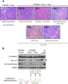
Recurrent masses from de-induced rtTA/MIC mice have variable histopathologies and some exhibit re-expression of PyV mT. (A) H&E-stained recurrent mammary tumours arising in rtTA/MIC mice post-doxycycline withdrawal. The mouse ID number (followed by the tumour location in the case of multiple recurrences, for example, “R1”) and histopathology of the tumour are indicated for each image. A pre-regression rtTA/MIC doxycycline-dependent mammary tumour exhibiting typical end-stage adenocarcinoma is shown for comparison. (Scale bar: 100 μm). (B) Immunoblot analysis of protein lysates from rtTA/MIC mammary tumours prior to and following doxycycline withdrawal using antibodies directed to E-cadherin (epithelial content control), PyV mT, Cre recombinase and Hsp90 (loading control); the arrowhead indicates the specific band for PyV mT protein. Resected mammary tumours were used for pre- and mid-regression time-points (pre- and mid-, respectively), while recurrent masses were harvested from mice sacrificed at clinical endpoint. The mouse ID numbers of the recurrent tumour lysates are indicated and correspond to the images labeled in (A). The incidence of adenocarcinoma in the corresponding histological section for each sample is indicated by a “+” symbol.

Similar articles

Cited by

References

    1. Fluck MM, Schaffhausen BS. Lessons in signaling and tumorigenesis from polyomavirus middle T antigen. Microbiol Mol Biol Rev. 2009;73:542–563. doi: 10.1128/MMBR.00009-09. - DOI - PMC - PubMed
    1. Cecena G, Wen F, Cardiff RD, Oshima RG. Differential sensitivity of mouse epithelial tissues to the polyomavirus middle T oncogene. Am J Path. 2006;168:310–320. doi: 10.2353/ajpath.2006.050443. - DOI - PMC - PubMed
    1. Guy CT, Cardiff RD, Muller WJ. Induction of mammary tumors by expression of polyomavirus middle T oncogene: a transgenic mouse model for metastatic disease. Mol Cell Biol. 1992;12:954–961. - PMC - PubMed
    1. Lin EY, Jones JG, Li P, Zhu L, Whitney KD, Muller WJ, Pollard JW. Progression to malignancy in the polyoma middle T oncoprotein mouse breast cancer model provides a reliable model for human diseases. Am J Path. 2003;163:2113–2126. doi: 10.1016/S0002-9440(10)63568-7. - DOI - PMC - PubMed
    1. White DE, Kurpios NA, Zuo D, Hassell JA, Blaess S, Mueller U, Muller WJ. Targeted disruption of β1-integrin in a transgenic mouse model of human breast cancer reveals an essential role in mammary tumor induction. Cancer Cell. 2004;6:159–170. doi: 10.1016/j.ccr.2004.06.025. - DOI - PubMed

Publication types

MeSH terms

LinkOut - more resources