. 2014 Mar;22(3):490-8.
doi: 10.1016/j.joca.2013.12.016. Epub 2014 Jan 21.

Primary cilia disassembly down-regulates mechanosensitive hedgehog signalling: a feedback mechanism controlling ADAMTS-5 expression in chondrocytes

Affiliations

Primary cilia disassembly down-regulates mechanosensitive hedgehog signalling: a feedback mechanism controlling ADAMTS-5 expression in chondrocytes

C L Thompson et al. Osteoarthritis Cartilage. 2014 Mar.

Abstract

Objective: Hedgehog signalling is mediated by the primary cilium and promotes cartilage degeneration in osteoarthritis. Primary cilia are influenced by pathological stimuli and cilia length and prevalence are increased in osteoarthritic cartilage. This study aims to investigate the relationship between mechanical loading, hedgehog signalling and cilia disassembly in articular chondrocytes.

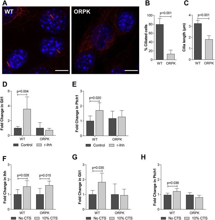
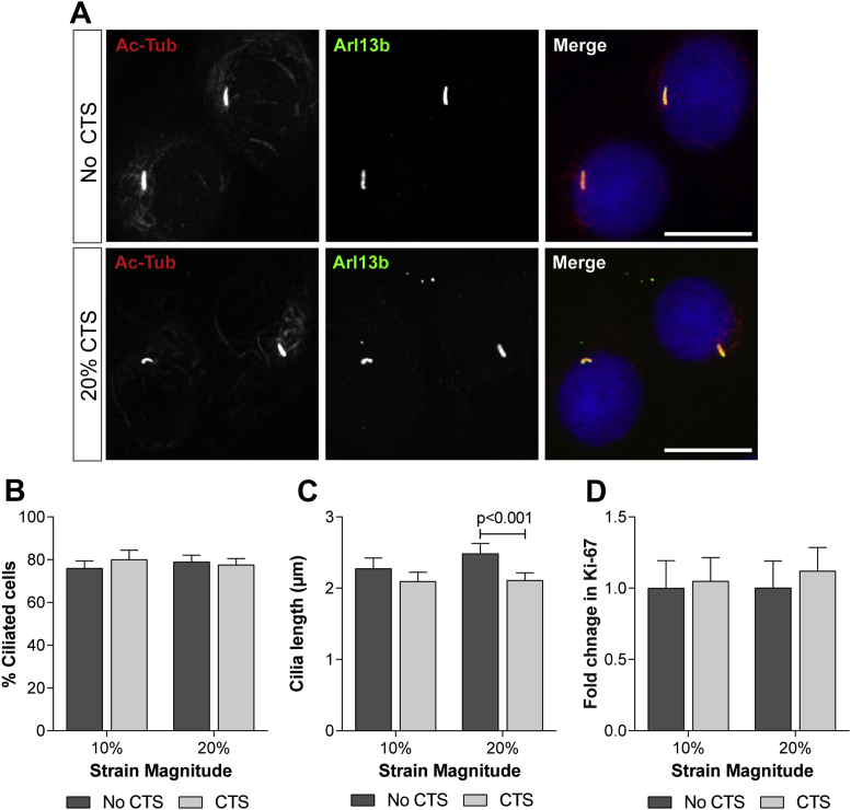
Methods: Primary bovine articular chondrocytes were subjected to cyclic tensile strain (CTS; 0.33 Hz, 10% or 20% strain). Hedgehog pathway activation (Ptch1, Gli1) and A Disintegrin And Metalloproteinase with Thrombospondin Motifs 5 (ADAMTS-5) expression were assessed by real-time PCR. A chondrocyte cell line generated from the Tg737(ORPK) mouse was used to investigate the role of the cilium in this response. Cilia length and prevalence were quantified by immunocytochemistry and confocal microscopy.

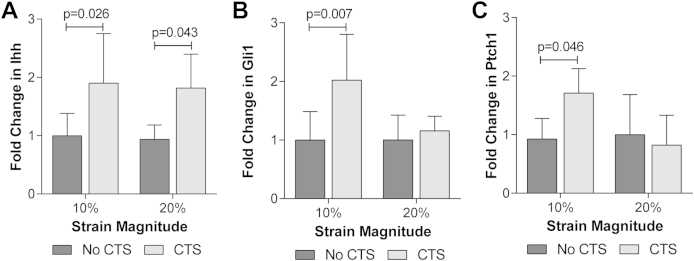
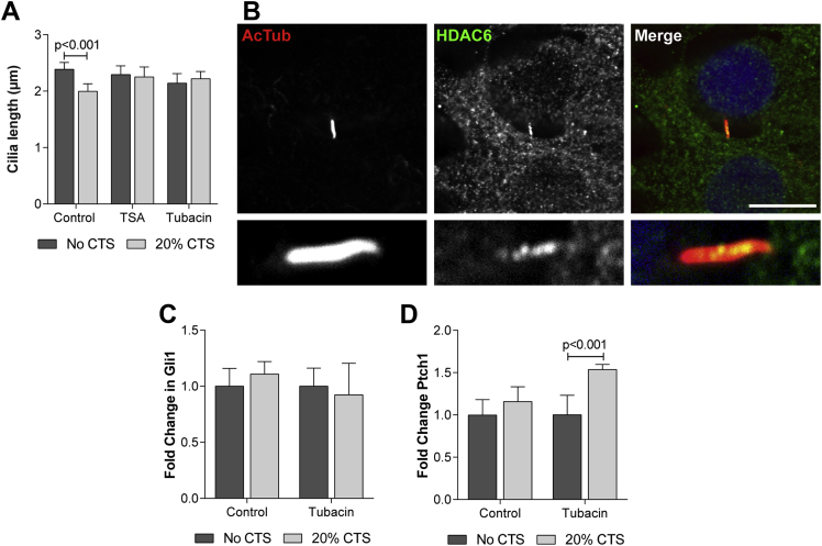
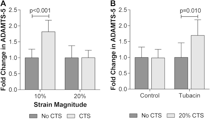
Results: Mechanical strain upregulates Indian hedgehog expression and activates hedgehog signalling. Ptch1, Gli1 and ADAMTS-5 expression were increased following 10% CTS, but not 20% CTS. Pathway activation requires a functioning primary cilium and is not observed in Tg737(ORPK) cells lacking cilia. Mechanical loading significantly reduced cilium length such that cilia became progressively shorter with increasing strain magnitude. Inhibition of histone deacetylase 6 (HDAC6), a tubulin deacetylase, prevented cilia disassembly and restored mechanosensitive hedgehog signalling and ADAMTS-5 expression at 20% CTS.

Conclusions: This study demonstrates for the first time that mechanical loading activates primary cilia-mediated hedgehog signalling and ADAMTS-5 expression in adult articular chondrocytes, but that this response is lost at high strains due to HDAC6-mediated cilia disassembly. The study provides new mechanistic insight into the role of primary cilia and mechanical loading in articular cartilage.

Keywords: ADAMTS-5; Chondrocyte; Cilia length; Hedgehog; Primary cilium.


Figures

Supplementary Fig. S1
GAPDH expression is not significantly altered by mechanical strain. Changes in GAPDH expression for (A) primary bovine articular chondrocytes and (B) WT and ORPK chondrocytes subjected to 10% or 20% CTS or unstrained control period for 1 h. Data represents mean ± CI (n = 8–10). Data is expressed as a fold change relative to the mean of the unstrained control group. Statistical significance was assessed by two-way ANOVA with post hoc Bonferroni corrected tests.
Fig. 1
Fig. 1
Mechanical strain upregulates Ihh expression and activates hedgehog signalling in articular chondrocytes. Changes in (A) Ihh, (B) Gli1 and (C) Ptch1 gene expression for primary bovine articular chondrocytes subjected to CTS for 1 h at 10% or 20% strain. Data is expressed as a fold change relative to the mean of the unstrained (noCTS) control group. Data represents mean ± CI (n = 8). Data was analysed by two-way ANOVA with post hoc Bonferroni corrected t-test.
Fig. 2
Fig. 2
The primary cilium is required for chondrocyte hedgehog signalling. (A) Representative confocal maximum intensity Z projections showing primary cilia immunofluorescently labelled in WT and ORPK chondrocytes cultured in the absence of strain. The cilium was labelled with antibodies directed to acetylated α-tubulin (red) while nuclei were counterstained with DAPI (blue). Scale bar represents 20 μm. (B) Primary cilia prevalence (n = 10) and (C) primary cilia length for WT and ORPK chondrocytes (n = 10). Statistical significance was assessed by Student's t test. Changes in (D) Gli1 and (E) Ptch1 gene expression for WT and ORPK chondrocytes treated with 1 μg/ml r-Ihh for 24 h. Changes in (F) Ihh, (G) Gli1 and (H) Ptch1 gene expression for WT and ORPK chondrocytes subjected to 10% CTS for 1 h. Gene expression data is expressed as a fold change relative to the mean of the untreated or unstrained control group. Data is presented as mean ± CI (n = 6–10). Statistical significance was assessed by two-way ANOVA with post hoc Bonferroni corrected t-test.
Fig. 3
Fig. 3
Primary cilia disassemble in response to mechanical strain. (A) Representative confocal maximum intensity Z projections of primary bovine articular chondrocytes showing primary cilia following 1 h 20% CTS and the noCTS control. Acetylated α-tubulin (red); Arl13b (green) and nuclei (blue). Scale bar represents 20 μm. Primary cilia (B) prevalence and (C) length for primary articular chondrocytes subjected to CTS for 1 h at 10% and 20% strain. (D) The % of Ki-67 positive cells in unstrained cells and those subjected to 10% and 20% CTS. Data is expressed as the fold change relative to the mean of the unstrained samples and represents mean ± CI (n = 15–20). Statistical significance was assessed by two-way ANOVA with post hoc Bonferroni corrected t-tests.
Fig. 4
Fig. 4
HDAC6 modulates strain-induced cilia disassembly and hedgehog signalling. (A) Primary cilia length for bovine articular chondrocytes subjected to 20% CTS for 1 h in the presence of TSA (7 nM), Tubacin (500 nM) or the carrier DMSO (control). Data represents mean ± CI (n = 15). Statistical significance was assessed by two-way ANOVA with post hoc Bonferroni corrected t-tests. (B) Representative confocal maximum intensity Z projection showing HDAC6 (green) localised to the primary cilium immunofluorescently labelled for acetylated α-tubulin (red). Nucleus counterstained with DAPI (blue). Scale bar represents 20 μm, lower panel shows magnified view of the cilium. Ptch1 (C) and (D) Gli1 gene expression for primary articular chondrocytes subjected to 20% CTS for 1 h in the presence of Tubacin (500 nM) or the carrier DMSO (control). Data represents mean ± CI (n = 6). Data is expressed as a fold change relative to the mean of the untreated or unstrained control group. Statistical significance was assessed by two-way ANOVA with post hoc Bonferroni corrected t-tests.
Fig. 5
Fig. 5
Primary cilia disassembly regulates mechanosensitive ADAMTS-5 expression. Changes in ADAMTS-5 expression for (A) bovine articular chondrocytes subjected to 10% or 20% CTS or unstrained control period for 1 h and (B) bovine articular chondrocytes subjected to 20% CTS for 1 h in the presence of Tubacin (500 nM) or the carrier DMSO (control). Data is expressed as a fold change relative to the mean of the untreated or unstrained control group. Data represents mean ± CI (n = 6–8). Statistical significance was assessed by two-way ANOVA with post hoc Bonferroni corrected t-tests.

Similar articles

Cited by

References

    1. Wheatley D.N., Wang A.M., Strugnell G.E. Expression of primary cilia in mammalian cells. Cell Biol Int. 1996;20:73–81. - PubMed
    1. Ross A.J., May-Simera H., Eichers E.R., Kai M., Hill J., Jagger D.J. Disruption of Bardet–Biedl syndrome ciliary proteins perturbs planar cell polarity in vertebrates. Nat Genet. 2005;37:1135–1140. - PubMed
    1. Schneider L., Clement C.A., Teilmann S.C., Pazour G.J., Hoffmann E.K., Satir P. PDGFR alpha alpha signaling is regulated through the primary cilium in fibroblasts. Curr Biol. 2005;15:1861–1866. - PubMed
    1. Corbit K.C., Aanstad P., Singla V., Norman A.R., Stainier D.Y., Reiter J.F. Vertebrate Smoothened functions at the primary cilium. Nature. 2005;437:1018–1021. - PubMed
    1. Huangfu D., Liu A., Rakeman A.S., Murcia N.S., Niswander L., Anderson K.V. Hedgehog signalling in the mouse requires intraflagellar transport proteins. Nature. 2003;426:83–87. - PubMed

Publication types