. 2014 Jan 15;5(1):211-23.
doi: 10.18632/oncotarget.1319.

AID downregulation is a novel function of the DNMT inhibitor 5-aza-deoxycytidine

Affiliations

AID downregulation is a novel function of the DNMT inhibitor 5-aza-deoxycytidine

Chiou-Tsun Tsai et al. Oncotarget. .

Abstract

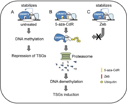
Activation-induced cytidine deaminase (AID) was originally identified as an inducer of somatic hypermutation (SHM) and class switch recombination (CSR) in immunoglobulin genes. However, AID can also cause mutations in host genes and contribute to cancer progression and drug resistance. In this study, molecular docking showed the interaction of free 5-aza-CdR and Zebularine (Zeb) with AID. However, only 5-aza-CdR-incorporated ssDNA bound to the active site of AID and inhibited AID expression through proteasomal degradation. 5-aza-CdR demonstrated cytotoxicity against AID-positive and -negative hematopoietic cancer cells. In contrast, Zeb exhibited a cytotoxic effect only in AID-negative cells due to its inability to inhibit AID expression. This differential effect might be due to the DNMT1 stabilization induced by AID, thus restricting the ability of Zeb to deplete DNMT1 and induce tumor suppressor genes (TSGs), such as p21, in AID-positive cells. Moreover, the in vivo anticancer effect of 5-aza-CdR but not Zeb in AID-positive hematopoietic cancer cells was demonstrated. The study not only displays the association of AID and DNMT1 and identifies a novel biological function of AID, but also provides novel information regarding the use of DNMT inhibitors to treat AID-positive hematopoietic cancers.


Figures

Figure 1
Figure 1. Molecular docking of DNMT inhibitors to AID
(A) Molecular structures of cytidine and its analogs. (B) The docking sites of DNMT inhibitors were analyzed using CDOCKER, and the best structures were selected based on the lowest CDOCKER_ INTERACTION_ENERGY for each ligand. (C) 5-aza-CdR and Zeb were incorporated into the DNA sequence (5'-AGCT-3') built with “Build and Edit Nucleic Acid” tools in Discovery Studio 2.55, and the binding modes were further analyzed. The top ten binding structures are shown. (D) The best insertion model is presented by a ribbon (left panel) and surface model (right panel).
Figure 2
Figure 2. 5-aza-CdR downregulated AID
Raji cells and SUP-B15 were treated with 5-aza-CdR (1-10 μM), Zeb (50-200 μM), or TSA (1 μM) for 4 days (A) or 5-aza-CdR (5 μM) for 24, 48, and 72 hrs (B). The protein expression levels of AID, DNMT1 and actin were analyzed through immunoblotting. (C) Raji cells were treated with 5-aza-CdR (1-10 μM) or Zeb (50-200 μM) for 4 days (left panel) or 5-aza-CdR (5 μM) for 24, 48, and 72 hrs (right panel). The mRNA levels of AICDA and actin were analyzed through RT-PCR. (D) Raji cells were treated with 5-aza-CdR (5-10 μM) or Zeb (100 μM) for 4 day. The relative mRNA levels of AICDA were analyzed through QRT-PCR
Figure 3
Figure 3. 5-aza-CdR reduced the protein stability of nuclear AID
(A) Upper-left panel: Raji cells were treated with 5-aza-CdR (1-10 μM) for 40 hrs, and MG132 (10 μM) was added for another 8 hrs. Upper-right panel: Raji cells were co-treated with 5-aza-CdR (5 μM) and MG132 (10 μM) for 8 hrs. The protein expression levels of AID and actin were analyzed through immunoblotting. Lower panel: Raji cells were pretreated with 5-aza-CdR (5 μM) for 16 hrs. Then, the cells were exposed to cycloheximide (20 μM) or cycloheximide/ MG132 (10 μM) for 4, 8, 10, and 12 hrs. The protein expression levels of AID and actin were analyzed through immunoblotting. (B) Left panel: Raji cells were pretreated with 5-aza-CdR (10 μM) for 19 hrs; then, the cells were exposed to MG132 (10 μM) for 5 hrs. AID was immunoprecipitated, and AID ubiquitination was examined through immunoblotting. Right panel: Raji cells were treated with 5-aza-CdR (5-10 μM) for 48 hrs, and the nuclear and cytosolic extracts were harvested. The protein expression levels of AID, tubulin, and lamin A/C were analyzed through immunoblotting. (C) Upper-left panel: K562AID4 cells were treated with 5-aza-CdR (10-50 μM) and Zeb (50 μM) for 48 hrs. Lower-left panel: K562AID4 cells were treated with 5-aza-CdR (10 μM) and Zeb (10 μM) for 48 hrs, and the nuclear and cytosolic extracts were harvested. The protein expression levels of flag-AID and actin were analyzed through immunoblotting. Right panel: K562AID4 cells were treated with 10 μM DNMT inhibitors for 4 days. The cells were fixed and stained with an anti-fag antibody and DAPI and subjected to confocal microscopy analysis. (D) K562AID4 cells were treated with 5-aza-CdR (10 μM), LMB (5 ng/ml), or both for 48 hrs. The cells were fixed and stained with an anti-fag antibody (yellow) and DAPI (blue) and subjected to confocal microscopy analysis. The images are shown at 1X and 3X magnification.
Figure 4
Figure 4. AID interferes with the cytotoxic effect of DNMT inhibitor in vitro
(A) K562, Raji, and SUP-B15 cells were treated with 5-aza-CdR (1-10 μM) or Zeb (50-200 μM) for 4 days, and cell viability was analyzed with the Alamar blue assay. Error bars indicate the mean ± SD of three independent experiments. (B) K562 and Raji cells were treated with 5-aza-CdR (5-10 μM) and Zeb (200 μM) for 0-6 days, and cell viability was analyzed with the trypan blue assay. (C) Left panel: Raji-shluc and Raji-shAIDH1 cells were treated with 5-aza-CdR (5-10 μM) or Zeb (50 μM) for 4 days, and cell viability was analyzed with the Alamar blue assay. **, p<0.01. Right panel: K562 and pooled K562AID cells were treated with 5-aza-CdR (10 μM) or Zeb (10 μM) for 4 days. Then, cell viability was analyzed with the Alamar blue assay. *, p<0.05. (D) Left panel: K562 and pooled K562AID cells were treated with Zeb (1-10 μM) for 4 days. Then, cell viability was analyzed with the Alamar blue assay. *, p<0.01. Right panel: K562 cells were transiently transfected with pCMV or pCMV-AID plasmids and exposed to Zeb (5 μM) for 3 days. The protein expression levels of flag-AID and the cell viability were examined through immunoblotting and the Alamar blue assay, respectively.
Figure 5
Figure 5. AID blocks DNMT inhibitor-induced degradation of DNMT1 and expression of TSG.s
(A) Left panel: K562 and Raji cells were treated with 5-aza-CdR (5-10 μM) or Zeb (200 μM) for 24 hrs. Right panel: Raji cells transduced with the shluc or shAID plasmid were treated with 5-aza-CdR (5-10 μM) or Zeb (50 μM) for 24 hrs. (B) Left panel: K562 and pooled K562AID cells were treated with 5-aza-CdR (5-10 μM) or Zeb (10 μM) for 24 hrs. Right panel: K562 and pooled K562AID cells were treated with 5-aza-CdR (10 μM) or Zeb (10 μM) for 96 hrs. The protein expression levels of DNMTs, p21, flag-AID, and actin were examined through immunoblotting. (C) Left panel: Raji-shluc, Raji-shAID, K562 and K562AID cells were treated with cycloheximide (20 μM) for 4, 8, and 12 hrs. Right panel: K562 and K562AID cells were pretreated with Zeb (10 μM) for 18 hrs and then exposed to cycloheximide (20 μM) for 1, 3, and 5 hrs. The protein expression levels of DNMT1, flag-AID, and actin were analyzed through immunoblotting. (D) Upper panel: AID and DNMT1 were co-immunoprecipitated from the total lysate of K562AID4 cells. Flag-AID was precipitated using the anti-fag antibody. Normal mouse IgG (mIgG) was used as the IP control. The protein expression levels of DNMT1 and AID were analyzed through immunoblotting. Lower panel: Raji cells were fixed and stained with anti-AID antibody (green), anti-DNMT1 antibody (red) and DAPI (blue) and subjected to confocal microscopy analysis. (E) Proteins were harvested from Raji-shluc and Raji-shAIDH1 cells, and protein expression levels of DNMT1, p21, Rassf1a, p53, AID, and tubulin were examined through immunoblotting.
Figure 6
Figure 6. The anti-cancer effect of DNMT inhibitors on tumor-engrafted mice
(A) KCL22 and KCL22+AID cells were treated with 5-aza-CdR (0.1-10 μM, left panel) and Zeb (10-100 μM, right panel) for 4 days, and cell viability was analyzed with the Alamar blue assay. The error bars represent the mean ± SD. **, p<0.01. (B) Left panel: KCL22 and KCL22+AID cells were treated with 5-aza-CdR (5-10 μM, left panel) and Zeb (10-25 μM, right panel) for 48 hrs. The protein expression levels of AID, DNMT1, and actin were analyzed through immunoblotting. Right panel: KCL22+AID cells were treated with 5-aza-CdR (5 and 10 μM) and Zeb (50 μM) for 4 days, and the nuclear extracts were harvested. The protein expression levels of AID and lamin A/C were examined through immunoblotting. (C) Left panel: Firefly luciferase-labeled KCL22+AID cells were i.t. injected into NOD/SCID recipient mice. After 21 days, tumor-bearing mice were selected and treated with PBS (n=5) or Zeb (500 mg/kg, n=5). Cell growth was examined using an in vivo imaging system (IVIS) at day 21, 37, and 43. Right panel: Firefly luciferase-labeled KCL22+AID cells were i.t. injected into NOD/SCID mice, and after 21 days, the mice were then treated with PBS (n=5) or 5-aza-CdR (0.5 mg/kg, n=5) for 21 days. Cell growth was examined using IVIS at day 21, 28, and 35. (D) Then the overall survival was depicted with Kaplan-Meier analysis.
Figure 7
Figure 7. A hypothetical model for the role of AID in the anticancer effect of DNMT inhibitors
(A) AID may co-localize with DNMT1 on DNA and enhance its stability in B-lymphoid malignancies. 5-aza-CdR and Zeb incorporate into DNA. (B) 5-aza-CdR targets the active site of AID and DNMT1 and then destabilizes AID and DNMT1 through the proteasome degradation pathway. Eventually, DNA is demethylated, and TSGs are induced. (C) Because Zeb is unable to bind to the active site of AID, DNMT1 is stabilized and avoids degradation by Zeb. Thus, the anticancer effect of Zeb is inhibited

Similar articles

Cited by

References

    1. Perez-Duran P, de Yebenes VG, Ramiro AR. Oncogenic events triggered by AID the adverse effect of antibody diversifcation. Carcinogenesis. 2007;28(12):2427–2433. - PubMed
    1. Honjo T, Muramatsu M, Fagarasan S. AID: how does it aid antibody diversity? Immunity. 2004;20(6):659–668. - PubMed
    1. Kinoshita K, Nonaka T. The dark side of activation-induced cytidine deaminase: relationship with leukemia and beyond. International journal of hematology. 2006;83(3):201–207. - PubMed
    1. Ramiro AR, Jankovic M, Callen E, Diflippantonio S, Chen HT, McBride KM, Eisenreich TR, Chen J, Dickins RA, Lowe SW, Nussenzweig A, Nussenzweig MC. Role of genomic instability and p53 in AID-induced c-myc-Igh translocations. Nature. 2006;440(7080):105–109. - PMC - PubMed
    1. Takizawa M, Tolarova H, Li Z, Dubois W, Lim S, Callen E, Franco S, Mosaico M, Feigenbaum L, Alt FW, Nussenzweig A, Potter M, Casellas R. AID expression levels determine the extent of cMyc oncogenic translocations and the incidence of B cell tumor development. The Journal of experimental medicine. 2008;205(9):1949–1957. - PMC - PubMed

Publication types

LinkOut - more resources