Two modes of exocytosis in an artificial cell
- PMID: 24457949
- PMCID: PMC3900996
- DOI: 10.1038/srep03847
Two modes of exocytosis in an artificial cell
Abstract
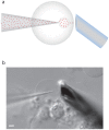
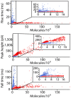
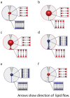
The details of exocytosis, the vital cell process of neuronal communication, are still under debate with two generally accepted scenarios. The first mode of release involves secretory vesicles distending into the cell membrane to release the complete vesicle contents. The second involves partial release of the vesicle content through an intermittent fusion pore, or an opened or partially distended fusion pore. Here we show that both full and partial release can be mimicked with a single large-scale cell model for exocytosis composed of material from blebbing cell plasma membrane. The apparent switching mechanism for determining the mode of release is demonstrated to be related to membrane tension that can be differentially induced during artificial exocytosis. These results suggest that the partial distension mode might correspond to an extended kiss-and-run mechanism of release from secretory cells, which has been proposed as a major pathway of exocytosis in neurons and neuroendocrine cells.
Figures


Similar articles
-
The dynamic nature of exocytosis from large secretory vesicles. A view from electrochemistry and imaging.Cell Calcium. 2023 Mar;110:102699. doi: 10.1016/j.ceca.2023.102699. Epub 2023 Jan 21. Cell Calcium. 2023. PMID: 36708611 Review.
-
Cdc42 controls the dilation of the exocytotic fusion pore by regulating membrane tension.Mol Biol Cell. 2014 Oct 15;25(20):3195-209. doi: 10.1091/mbc.E14-07-1229. Epub 2014 Aug 20. Mol Biol Cell. 2014. PMID: 25143404 Free PMC article.
-
Two distinct modes of exocytotic fusion pore expansion in large astrocytic vesicles.J Biol Chem. 2013 Jun 7;288(23):16872-16881. doi: 10.1074/jbc.M113.468231. Epub 2013 Apr 25. J Biol Chem. 2013. PMID: 23620588 Free PMC article.
-
Amperometric post spike feet reveal most exocytosis is via extended kiss-and-run fusion.Sci Rep. 2012;2:907. doi: 10.1038/srep00907. Epub 2012 Nov 30. Sci Rep. 2012. PMID: 23205269 Free PMC article.
-
Fusion pores, SNAREs, and exocytosis.Neuroscientist. 2013 Apr;19(2):160-74. doi: 10.1177/1073858412461691. Epub 2012 Sep 26. Neuroscientist. 2013. PMID: 23019088 Review.
Cited by
-
Mimicking subsecond neurotransmitter dynamics with femtosecond laser stimulated nanosystems.Sci Rep. 2014 Jun 23;4:5398. doi: 10.1038/srep05398. Sci Rep. 2014. PMID: 24954021 Free PMC article.
-
Mitochondrial Nanotunnels.Trends Cell Biol. 2017 Nov;27(11):787-799. doi: 10.1016/j.tcb.2017.08.009. Epub 2017 Sep 19. Trends Cell Biol. 2017. PMID: 28935166 Free PMC article. Review.
-
Mitochondrial fusion dynamics is robust in the heart and depends on calcium oscillations and contractile activity.Proc Natl Acad Sci U S A. 2017 Jan 31;114(5):E859-E868. doi: 10.1073/pnas.1617288114. Epub 2017 Jan 17. Proc Natl Acad Sci U S A. 2017. PMID: 28096338 Free PMC article.
-
Cholesterol Alters the Dynamics of Release in Protein Independent Cell Models for Exocytosis.Sci Rep. 2016 Sep 21;6:33702. doi: 10.1038/srep33702. Sci Rep. 2016. PMID: 27650365 Free PMC article.
-
Variations in Fusion Pore Formation in Cholesterol-Treated Platelets.Biophys J. 2016 Feb 23;110(4):922-9. doi: 10.1016/j.bpj.2015.12.034. Biophys J. 2016. PMID: 26910428 Free PMC article.
References
-
- Alvarez de Toledo G., Fernandez-Chacon R. & Fernandez J. M. Release of secretory products during transient vesicle fusion. Nature. 363, 554–558 (1993). - PubMed
-
- Wightman R. M. & Haynes C. L. Synaptic vesicles really do kiss and run. Nat Neurosci. 7, 321–322 (2004). - PubMed
-
- Fesce R., Grohovaz F., Valtorta F. & Meldolesi J. Neurotransmitter release: fusion or ‘kiss-and-run'? Trends Cell Biol. 4, 1–4 (1994). - PubMed
Publication types
MeSH terms
LinkOut - more resources
Full Text Sources
Other Literature Sources

