. 2014 Jan 23;5(1):e1016.
doi: 10.1038/cddis.2013.532.

Regulation of the proapoptotic functions of prostate apoptosis response-4 (Par-4) by casein kinase 2 in prostate cancer cells

Affiliations

Regulation of the proapoptotic functions of prostate apoptosis response-4 (Par-4) by casein kinase 2 in prostate cancer cells

A de Thonel et al. Cell Death Dis. .

Abstract

The proapoptotic protein, prostate apoptosis response-4 (Par-4), acts as a tumor suppressor in prostate cancer cells. The serine/threonine kinase casein kinase 2 (CK2) has a well-reported role in prostate cancer resistance to apoptotic agents or anticancer drugs. However, the mechanistic understanding on how CK2 supports survival is far from complete. In this work, we demonstrate both in rat and humans that (i) Par-4 is a new substrate of the survival kinase CK2 and (ii) phosphorylation by CK2 impairs Par-4 proapoptotic functions. We also unravel different levels of CK2-dependent regulation of Par-4 between species. In rats, the phosphorylation by CK2 at the major site, S124, prevents caspase-mediated Par-4 cleavage (D123) and consequently impairs the proapoptotic function of Par-4. In humans, CK2 strongly impairs the apoptotic properties of Par-4, independently of the caspase-mediated cleavage of Par-4 (D131), by triggering the phosphorylation at residue S231. Furthermore, we show that human Par-4 residue S231 is highly phosphorylated in prostate cancer cells as compared with their normal counterparts. Finally, the sensitivity of prostate cancer cells to apoptosis by CK2 knockdown is significantly reversed by parallel knockdown of Par-4. Thus, Par-4 seems a critical target of CK2 that could be exploited for the development of new anticancer drugs.


Figures

Figure 1
Figure 1
Both rodent and human Par-4 interact and are substrates of CK2. (a) COS cells were co-transfected with rat GFP-tagged Par-4 or empty GFP vector together with the CK2α and/or the CK2βHA-tagged subunits. In the left panel, immunoprecipitation of GFP vector, Par-4 (GFP) or CK2α subunits (HA). In right panel, immunoprecipitation of Par-4 (GFP) or CK2α and/or CK2β subunits (HA). The immunoprecipitations were followed by immunodetection of either CK2 (HA) or Par-4 (GFP). (b) Immunoprecipitation, from a same PC-3 cells extract, of endogenous human Par-4 (hPar-4) or CK2 subunits was followed by immunodetection of endogenous human CK2α, CK2β or Par-4. Inputs: proteins in total cell lysates. IP IgG: immunoprecipitation with a non-relevant antibody (IgG mouse). *HC, LC immunoglobulin heavy chain and light chain respectively. (c) The GFP-tagged CK2 kinase or GFP alone (as a control, Mock) were immunoprecipitated from transfected COS cells and used for an in vitro kinase assay in the presence of [γ−32P]ATP with either recombinant GST-Par-4 or GST alone, as substrates (left panel). Reaction products were resolved by SDS-PAGE on 10% gels, stained with Coomassie blue to verify that equal amounts of GST-Par-4 or GST alone were used in each reaction (lower panel), and autoradiography was performed (upper panel). In parallel, the amount of GFP (Mock) or CK2 subunits immunoprecipitated was analyzed by immunoblotting (right panel). (d) Recombinant GFP-tagged human Par-4 (GFP-hPar-4) was produced in vitro by the TNT rabbit reticulocyte lysate system (RRL), and lysates were subsequently immunoprecipitated using GFP antibody. Used as substrates, the immunoprecipitates were subjected to an in vitro kinase assay using recombinant CK2 (recCK2) in the presence of [γ−32P]ATP for 30 min. The CK2 inhibitor (TBB, 1 μM) was incubated 5 min before addition of CK2 recombinant (middle panel). After migration, phosphorylated Par-4 (32P-hPar-4) and autophosphorylation of recombinant CK2β (32P- CK2β) was detected by autoradiography. The amount of recombinant GFP-Par-4 produced by RRL was checked by western blotting (input, lower panel)
Figure 2
Figure 2
Identification of the CK2 phosphorylated sites S124 and S223 in rodent Par-4. (a) Two-dimensional phosphopeptide map analysis of the phosphorylated recombinant rat GST-Par-4. GST-Par-4 was phosphorylated by recombinant CK2 in the presence of [γ−32P]ATP. After migration, band corresponding to the radiolabeled phospho-Par-4 was excised from the membrane and digested by trypsin. Tryptic fragments were purified and resolved by two-dimensional thin layer electrophoresis and ascending chromatography followed by autoradiography to visualize the phosphopeptides. The separation origin is indicated as ‘0'. Three major phosphopeptides were revealed by autoradiography: P1, P1′, and P2. (b) Transfected wild-type Par-4 (rat) or 124A223A Par-4 mutants (GFP-tagged) were immunoprecipitated from COS cells and subjected to an in vitro kinase assay in the presence of recombinant CK2 and [γ−32P]ATP. Radiolabeled Par-4 was visualized upon autoradiography. Immunoblot analysis confirmed that equal amounts of GFP-Par-4 and GFP-124A223A were immunoprecipitated from each cell lysate (inputs, lower panel)
Figure 3
Figure 3
Phosphorylation of rodent Par-4 by CK2 blocks Par-4 proapoptotic functions. (a–c) PC-3 cells were transfected with empty GFP vector control (Mock), rat GFP-tagged Par-4, GFP-tagged 124A223A mutant (mimicking non-phosphorylated Par-4 form) or 124D223D mutant (mimicking phosphorylated Par-4 form) and then treated with recombinant death ligand TRAIL (500 ng/ml, 3 h). (a) Green fluorescent (transfected) cells were gated (represent around 20% of total cell) and further analyzed for apoptosis by Hoechst staining. Bars represent the mean±S.D. of at least four independent experiments. (b) FACS analysis of the caspase-3 activity was assessed by red fluorescent signal FLICA (Fluorogenic inhibitors of caspase-3 activation). Bars represent the mean±S.D. of at least four independent experiments. (c) Immunoblot analysis of caspase-8 and PARP cleavage (included both GFP-positive and -negative cells) at the indicated times. *P<0.05, **P<0.01. (d) PC-3 cells, transfected as above, were treated 8 h with 200 nM of Paclitaxel, and then apoptosis was determined by detection of caspases 8 and PARP cleavage. Hsp90 and 14-3-3 were used as a loading control
Figure 4
Figure 4
Rodent Par-4 phosphorylation by CK2 prevents caspase-mediated Par-4 cleavage. (a) PC-3 cells transfected with the GFP-tagged constructs wild-type rat Par-4, 124A/D, 223A/D, 124A223A or 124D223D mutants of Par-4 were treated with TRAIL (500 ng/ml, 3 h) in the presence or absence of Z-VAD (15 μM). Par-4 cleavage was determined by western blotting using anti-GFP antibody. Hsp90 was used as a loading control. (b) Wild-type Par-4, 124A/D, 223A/D, 124D223D and 124A223A mutant proteins (GFP tagged) were produced in vitro by a TNT RRL system. Recombinant caspase-8 (upper panel) or caspase-3 and -7 (Casp3, Casp7, lower panel) were incubated with the indicated Par-4 proteins for 3 h at 37 °C, and Par-4 cleavage was assessed by western blotting using GFP antibody. (c) Caspase assay was performed on pre-phosphorylated rat GST-Par-4 (in vitro kinase assay using RecCK2 as described above Figure 2), and cleaved Par-4 was detected by immunoblot. *NS: non-specific bands are probably due to the presence of recombinant Par-4 degraded forms. To note: cleaved GST Par-4 migrates at lower molecular weight than cleaved GFP-Par-4, given GST tag is located in the N-terminus of Par-4 and not on C-terminus as the GFP-tag. (d–f) PC-3 cells transfected with Par-4 wild-type or FLAG-tagged Par-4 (124–332) were treated as above with TRAIL and analyzed for apoptosis by Hoechst staining (d) or FACS analysis of the caspase-3 activity (FLICA) (e) and by immunoblotting analysis of caspase-8 and PARP cleavage (f). Bars represent the mean±S.D. of at least three independent experiments. Flag-Par-4 (124–332) and GFP were revealed in the same membrane using corresponding antibodies (lower panel). Endogenous Par-4 was used as a loading control. *P<0.05, **P<0.01
Figure 5
Figure 5
Human Par-4 is phosphorylated both in vitro and in vivo on S231 (ortholog of rodent S223) by CK2 and this phosphorylation impairs apoptosis. (a) Conservation of the CK2 recognition motif (including the serine 231 of human Par-4) was evaluated in different species. (b) Recombinant Myc-tagged human Par-4 (hPar-4) or 231D mutant proteins were produced with TNT RRL system. An in vitro kinase assay was performed with the immunoprecipitates as substrates, in the presence of recombinant CK2 and [γ−32P]ATP. Phosphorylated Par-4 was detected by autoradiography. Production of recombinant Myc tagged proteins were checked by western blotting (right panel). (c) Phosphorylation of endogenous Par-4 was detected by western blotting (left panel), using the human anti-phosphoserine231-Par-4 antibody (Ph231 hPar-4), in PC-3 cells treated (+) or not (−) with TRAIL for 9 h, and densitometry analysis was done. The blocking peptide was used in order to test the specificity of the phospho-antibody. In parallel, the percentage of apoptosis induced by TRAIL (9 h) was assessed by Hoetsch staining. Bars represent the mean±S.D. of at least three independent experiments (right panel). (d–f) HCT116 cells were transfected with empty Myc vector control (Mock), human Myc-tagged Par-4, 231A or 231D mutant Par-4 (mimicking unphosphorylated and phosphorylated hPar-4, respectively) and then treated with recombinant TRAIL (150 ng/ml, 3 h). Expression of the different constructs was assessed by western blotting using Myc antibody, and apoptosis was monitored by immunoblot analysis of caspase-8 and PARP cleavage (d) and by DAPI staining (e and f). Bar graph shows semi-quantified densitometry from PARP and Caspase-8 western blotting analysis. Bars represent the mean±S.D. of at least four independent experiments. Hsp90 was used as a loading control. Bar=10 μm, magnification × 63; *P<0.05. a.u, arbitrary unit
Figure 6
Figure 6
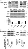
Par-4 is highly phosphorylated on S231 (ortholog of rodent S223) in human prostate cancer cells compared with normal counterparts. (a) Upper panel, phosphorylation of Par-4 on S231 was studied by western blotting in prostate cancer cells (PC-3, LnCap cells) and in normal prostate cells (PrCE, PNT2C2). In parallel, endogenous expression of Par-4 and CK2 subunits in the different cell lines was determined (middle panel). Hsp90 was used as a loading control. Lower panel, analysis of CK2 activity in the different prostate cells studied using Cyclex CK2 screening kit. Bars represent the mean±S.D. of at least two independent experiments. (b) PC-3 cells were transfected with CK2 siRNA (siCK2T, ThermoFischer) or scrambled siRNA fluorescently labeled with FAM (Scr siRNA) for 48 h and then treated or not with TRAIL (500 ng/ml, 3 h). Endogenous phosphorylated human Par-4 was detected by western blotting using the anti-phospho231 Par-4 antibody (Ph231-hPar-4) (left panel). Par-4 expression (left panel) and downregulation of CK2α protein (right panel) were evaluated by immunoblotting using the corresponding antibodies. Bar graph shows semi-quantified densitometry from western blotting analysis
Figure 7
Figure 7
Anti-apoptotic role of CK2 is dependent on Par-4 in human prostate cancer cells. (a–d) PC-3 cells were transfected with different CK2 siRNAs (siCK2T from ThermoFischer (a) or siCK2AB from Ambion (b)) together with scrambled siRNA fluorescently labeled with FAM (Scr siRNA) or different Par-4 siRNA (siPar-4T from ThermoFisher or siPar-4sc from Santa Cruz) for 48 h. Then, cells were treated or not with TRAIL (500 ng/ml, 3 h). The downregulation of Par-4 and CK2α proteins was confirmed by immunoblot using the corresponding antibodies. Apoptosis was monitored (a and b) by immunoblot analysis of caspase-8 and PARP (long and short cleaved PARP forms) cleavage and (c and d) by DAPI staining. Graphs represent semi-quantified densitometry analysis from PARP and caspase-8 western blotting analysis of panel (a). See also Supplementary Figure S10A for the densitometric analysis of panel (b). Bars represent the mean±S.D. of at least four independent experiments. Hsp90 was used as a loading control. Bar=10 μm, magnification × 63; *P<0.05. a.u, arbitrary unit

Similar articles

Cited by

References

    1. Chakraborty M, Qiu SG, Vasudevan KM, Rangnekar VM. Par-4 drives trafficking and activation of Fas and Fasl to induce prostate cancer cell apoptosis and tumor regression. Cancer Res. 2001;61:7255–7263. - PubMed
    1. El-Guendy N, Rangnekar VM. Apoptosis by Par-4 in cancer and neurodegenerative diseases. Exp Cell Res. 2003;283:51–66. - PubMed
    1. Nalca A, Qiu SG, El-Guendy N, Krishnan S, Rangnekar VM. Oncogenic Ras sensitizes cells to apoptosis by Par-4. J Biol Chem. 1999;274:29976–29983. - PubMed
    1. Sells StephenF, Wood DP, Joshi-barve SS, Muthukumar S, Jacob RJ, Crist SA, et al. Commonality of apoptosis of the gene programs in androgen-dependent prostate cells. Cell Growth Differs. 1994;5:457–466. - PubMed
    1. Mattson MP, Duan W, Chan SL, Camandola S. Par-4: an emerging pivotal player in neuronal apoptosis and neurodegenerative disorders. J Mol Neurosci. 1999;13:17–30. - PubMed

Publication types

Substances