. 2014 Jan 23;5(1):e1019.
doi: 10.1038/cddis.2013.559.

c-Jun N-terminal kinase has a key role in Alzheimer disease synaptic dysfunction in vivo

Affiliations

c-Jun N-terminal kinase has a key role in Alzheimer disease synaptic dysfunction in vivo

A Sclip et al. Cell Death Dis. .

Abstract

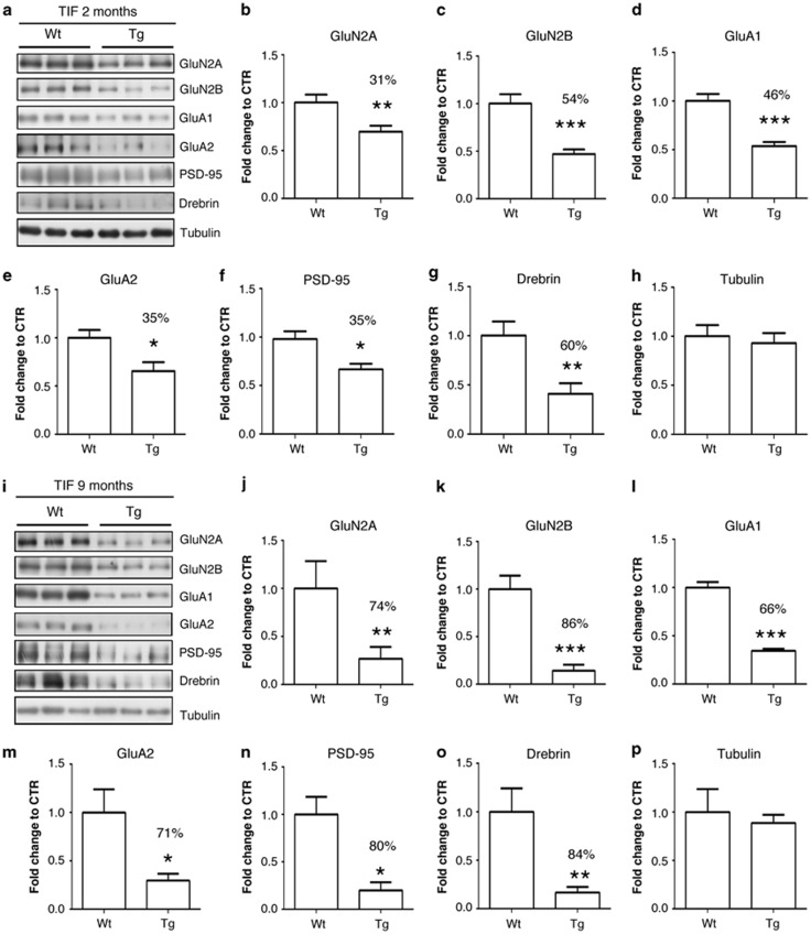
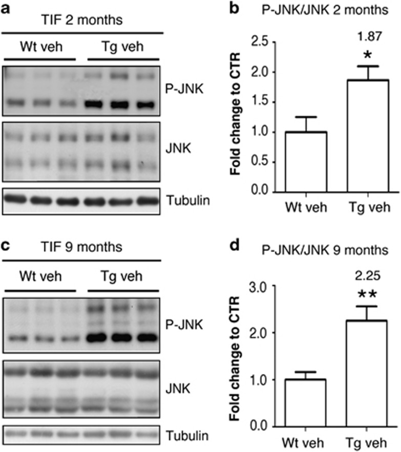
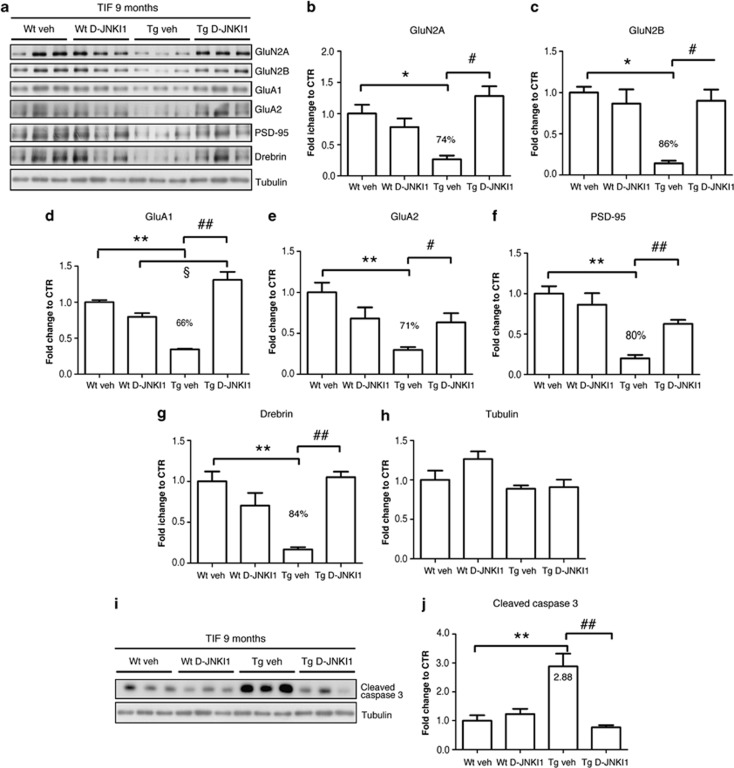
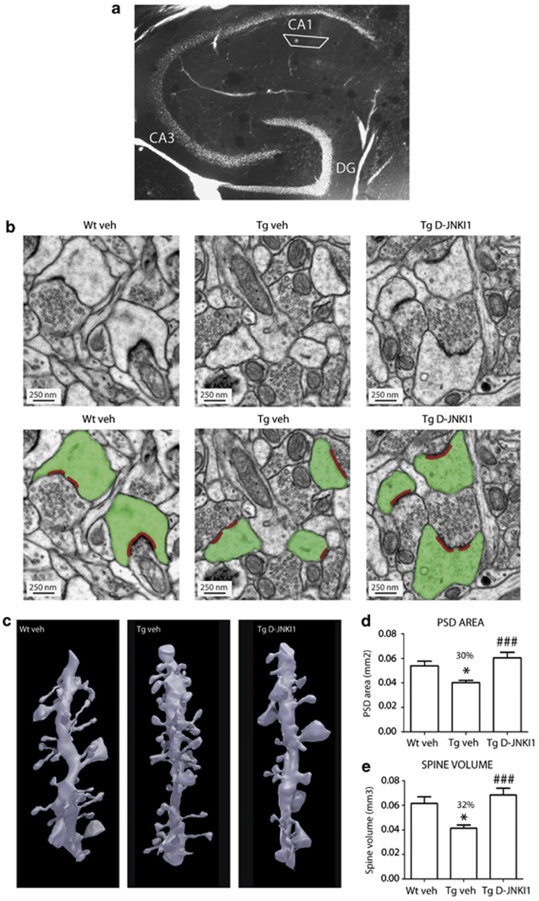
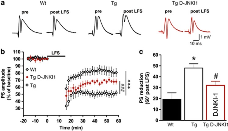
Altered synaptic function is considered one of the first features of Alzheimer disease (AD). Currently, no treatment is available to prevent the dysfunction of excitatory synapses in AD. Identification of the key modulators of synaptopathy is of particular significance in the treatment of AD. We here characterized the pathways leading to synaptopathy in TgCRND8 mice and showed that c-Jun N-terminal kinase (JNK) is activated at the spine prior to the onset of cognitive impairment. The specific inhibition of JNK, with its specific inhibiting peptide D-JNKI1, prevented synaptic dysfunction in TgCRND8 mice. D-JNKI1 avoided both the loss of postsynaptic proteins and glutamate receptors from the postsynaptic density and the reduction in size of excitatory synapses, reverting their dysfunction. This set of data reveals that JNK is a key signaling pathway in AD synaptic injury and that its specific inhibition offers an innovative therapeutic strategy to prevent spine degeneration in AD.


Figures

Figure 1
Figure 1
Synaptopathy occurred in TgCRND8 mice, already at an early stage of AD pathology. (ah) Western blot and relative quantification performed on the TIF fraction of 2-months-old Wt and TgCRND8 mice. Tg mice showed a significant reduction in the PSD levels of GluN2A (31%) (b) and of GluN2B (54%) (c) subunits of NMDAr, as well as reduction of GluA1 (46%) (d) and of GluA2 (35%) (e) subunits of AMPAr, of PSD-95 (35%) (f) and of drebrin (60%) (g) if compared with age-matched Wt mice (Student's t-test, *P<0.05, **P<0.01, ***P<0.001, n=6). Tubulin levels were not affected (h) (Student's t-test, P>0.05, n=6). (ip) Western blot and relative quantification performed on the TIF fraction of 9-months-old Wt and TgCRND8 mice. Tg mice showed a severe reduction in the PSD levels of GluN2A (74%) (j) and of GluN2B (86%) (k) subunits of NMDAr, as well as reduction of GluA1 (66%) (l) and of GluA2 (71%) (m) subunits of AMPAr, of PSD-95 (80%) (n) and of drebrin (84%) (o) if compared with age-matched Wt mice (Student's t-test, *P<0.05, **P<0.01, ***P<0.001, n=6). Tubulin levels were not affected (p) (Student's t-test, P>0.05, n=6)
Figure 2
Figure 2
JNK and caspase-3 pathways were activated in the postsynaptic compartment in TgCRND8 mice. (a and b) Western blot and relative quantification showing P-JNK and JNK levels in the TIF fraction of 2-months-old Wt and TgCRND8 mice. P-JNK/JNK ratio was increased by 1.87-fold in TgCRND8 mice if compared with the age-matched Wt mice (Student's t-test, *P<0.05, n=6). (c and d) Western blot and relative quantification showing P-JNK and JNK levels in the TIF fraction of 9-months-old Wt and TgCRND8 mice. P-JNK/JNK ratio was increased by 2.25-fold in TgCRND8 mice if compared with the age-matched Wt mice (Student's t-test, **P<0.01, n=6)
Figure 3
Figure 3
D-JNKI1 prevented Aβ-oligomers-induced loss of postsynaptic proteins from the PSD in TgCRND8 mice. (ah) Western blot and relative quantification of TIF fraction obtained from hippocampus homogenate of 9-months-old Wt and TgCRND8 mice, chronically treated with vehicle (water) or D-JNKI1 (22 mg/kg for 5 months). TgCRND8 mice treated with vehicle showed a strong loss of GluN2A (b) and GluN2B (c) subunits of NMDAr, GluA1 (d) and GluA2 (e) subunits of AMPAr, PSD-95 (f) and drebrin (g) from the PSD. D-JNKI1 chronic treatment completely prevented these alterations in Tg mice, whereas it did not affect protein levels in Wt mice (two-way analysis of variance (ANOVA), Bonferroni post-hoc test, *P<0.05, **P<0.01 Wt veh versus Tg veh; #P<0.05, ##P<0.01 Tg veh versus Tg D-JNKI1; §P<0.05 Wt D-JNKI1 versus Tg D-JNKI1, n=6). Tubulin levels (h) were not affected by the treatment or by mice genotype. (i and j) Western blot and relative quantification showing cleaved caspase-3 levels in the TIF fraction of 9-months-old Wt and TgCRND8 mice treated with vehicle (water) or D-JNKI1 (22 mg/kg for 5 months). D-JNKI1 chronic treatment completely prevented activation of caspase-3 in TgCRND8 mice (Two-way ANOVA, Bonferroni post-hoc test, **P<0.01 Wt veh versus Tg veh; ##P<0.01 Tg veh versus Tg D-JNKI1, n=6)
Figure 4
Figure 4
D-JNKI1 prevented morphological alterations in dendritic spines observed in TgCRND8 mice. Alteration in spine morphology was assessed by performing serial sectioning electron microscope analysis. (a) Micrograph of a section through the hippocampus showing the position of the neutrophil sampled at the level of the stratum radiatum of the hippocampus. The black box shows the position of the block whereas the asterisk represent the site of the analysis. (b) Electron micrographs showing excitatory synapses on spines in TgCRND8 mice treated with vehicle (Tg veh) or with 22 mg/kg D-JNKI1 (Tg D-JNKI1). D-JNKI1 treatment induced an increase in the PSD (red in figure) area and in the postsynaptic spine (green in figure) volume if compared with vehicle-treated TgCRND8 mice. (c) Three-dimensional EM reconstruction illustrating dendritic segments from TgCRND8 mice treated with vehicle or D-JNKI1; note the appearance of large spines in the D-JNKI1-treated case. (d and e) Graphs showing the effect of D-JNKI1 treatment on PSD area (d) and in spine volume (e) in TgCRND8 mice. TgCRND8 mice show a significant reduction in the PSD area (30%) (d) and spine volume (32%) (e). D-JNKI1 treatment restores normal sizes of spines and PSD area (one-way ANOVA, Bonferroni post-hoc test, *P<0.05 Wt veh versus Tg veh; ###P<0.001 Tg veh versus Tg D-JNKI1, Wt veh n=122 spines, Tg veh n=168 spines; TgCRND8 D-JNKI1 n=178 spines)
Figure 5
Figure 5
D-JNKI1 restored synaptic function in TgCRND8 mice. Electrophysiological analysis. (a) Example pairs of traces of field potentials showing population spikes (PS) evoked before and after the induction of LTD by a LFS protocol in hippocampal slices cut from Wt (left) and TgCRND8 mice (center) and in hippocampal slices from Tg mice incubated with D-JNKI1 (3 μM, 2 h) (right). (b) Time-course graph of population spike (PS) amplitudes showing LTD induced by LFS protocol in slices from Wt mice (diamonds), Tg mice (open circles) and in slices from Tg mice incubated with D-JNKI1 (red-filled circles) (two-way ANOVA, ***P<0.001 Wt versus Tg, ###P<0.001 Tg versus Tg D-JNKI1; Wt n=11, Tg n=8, Tg D-JNKI1 n=4). (c) Histogram summarizing the reduction of the PS amplitude measured in control slices from Tg (white bar) compared with Wt mice (black bar) (Student's t-test, *P<0.05), and in slices from Tg mice incubated with D-JNKI1 (red bar) compared with the non-treated control slices from Tg mice (Student's t-test, #P<0.05)

Similar articles

Cited by

References

    1. Scheff SW, Price DA, Schmitt FA, Mufson EJ. Hippocampal synaptic loss in early Alzheimer's disease and mild cognitive impairment. Neurobiol Aging. 2006;27:1372–1384. - PubMed
    1. Crews L, Masliah E. Molecular mechanisms of neurodegeneration in Alzheimer's disease. Hum Mol Genet. 2010;19:R12–R20. - PMC - PubMed
    1. Gomez-Isla T, Price JL, McKeel DW, Jr, Morris JC, Growdon JH, Hyman BT. Profound loss of layer II entorhinal cortex neurons occurs in very mild Alzheimer's disease. J Neurosci. 1996;16:4491–4500. - PMC - PubMed
    1. Lambert MP, Barlow AK, Chromy BA, Edwards C, Freed R, Liosatos M, et al. Diffusible, nonfibrillar ligands derived from Abeta1-42 are potent central nervous system neurotoxins. Proc Natl Acad Sci USA. 1998;95:6448–6453. - PMC - PubMed
    1. Lesne S, Koh MT, Kotilinek L, Kayed R, Glabe CG, Yang A, et al. A specific amyloid-beta protein assembly in the brain impairs memory. Nature. 2006;440:352–357. - PubMed

Publication types