. 2014 Jan 23;5(1):e1022.
doi: 10.1038/cddis.2013.539.

Stress-induced RNASET2 overexpression mediates melanocyte apoptosis via the TRAF2 pathway in vitro

Affiliations

Stress-induced RNASET2 overexpression mediates melanocyte apoptosis via the TRAF2 pathway in vitro

Q Wang et al. Cell Death Dis. .

Abstract

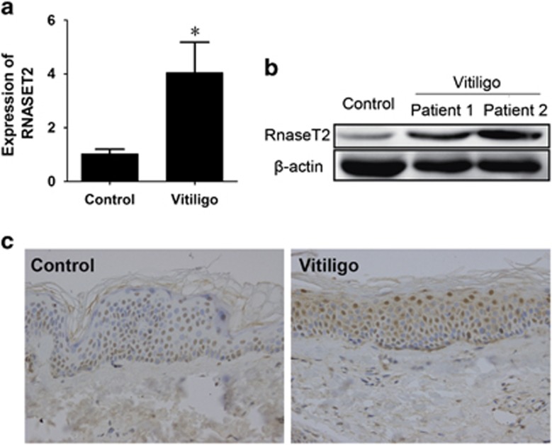
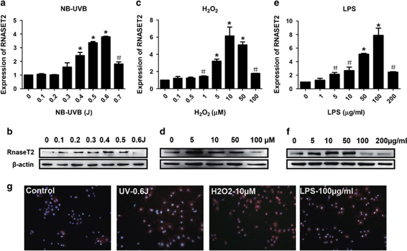
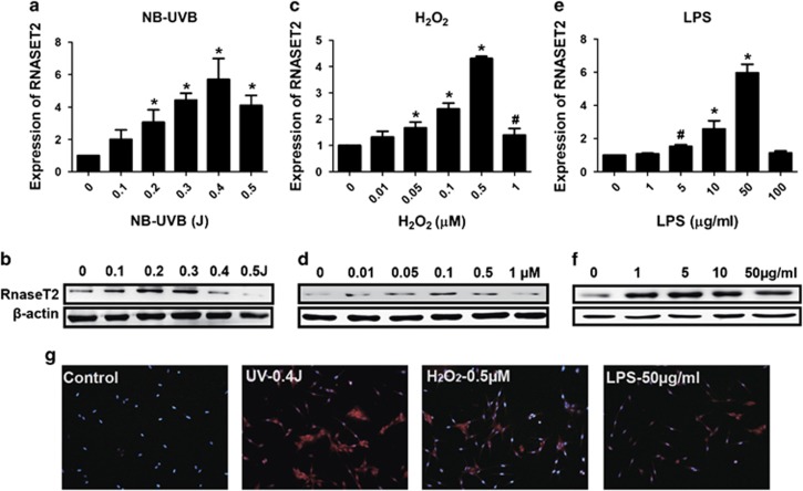
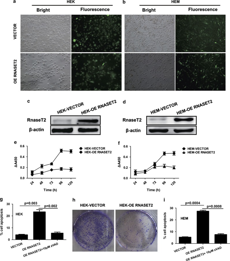
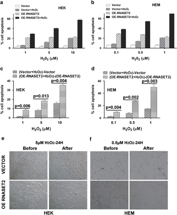
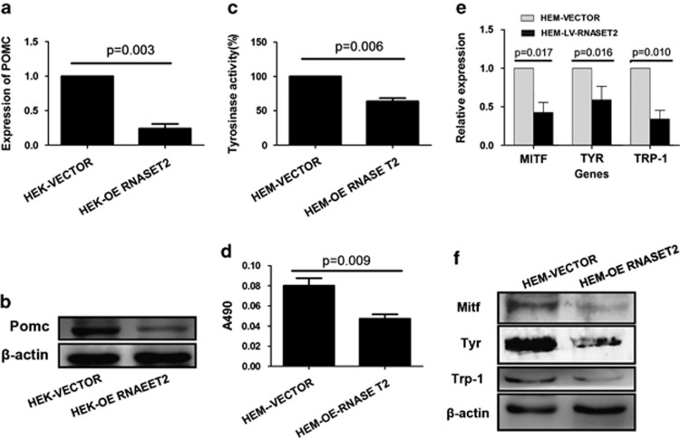
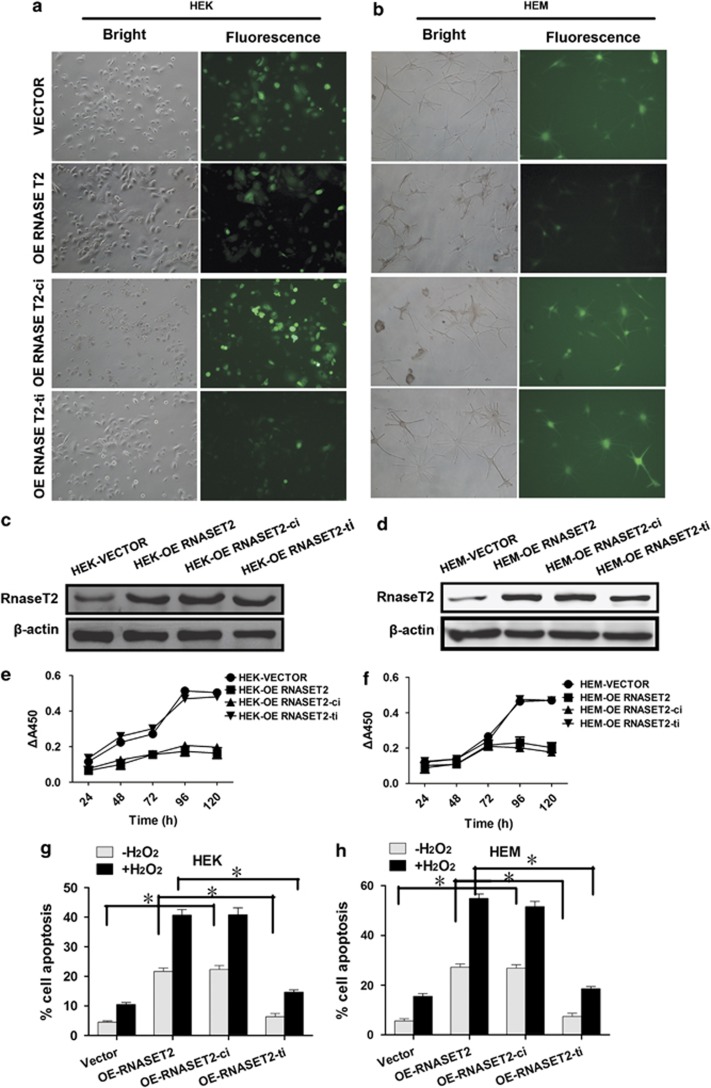
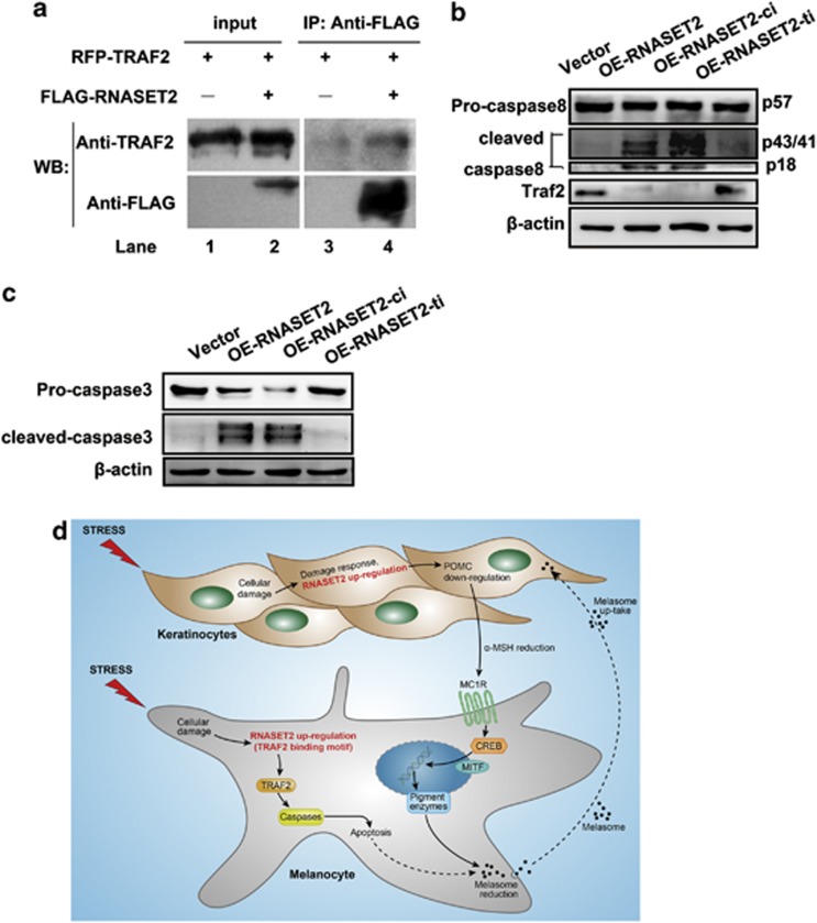
The recent genome-wide association study identified a link between vitiligo and genetic variants in the ribonuclease T2 (RNASET2) gene; however, the functional roles of RNASET2 in vitiligo pathogenesis or in melanocyte apoptosis have yet to be determined. The current study was designed to investigate the vitiligo-related expression pattern of RNASET2 and its molecular function involving apoptosis-related signaling proteins and pathways. The results showed overexpression of RNASET2 in epidermis specimens from 40 vitiligo patients compared with that from matched healthy controls. In addition, in vitro analyses indicated that overexpression of RNASET2 was inducible in cultured primary human melanocytes and keratinocytes by stress conditions, that is, exposure to UV irradiation, hydrogen peroxide, and inflammatory factors, respectively, and led to increased cell apoptosis via the tumor necrosis factor receptor-associated factor 2 (TRAF2)-caspases pathway through the physical interaction of RNASET2 with TRAF2. Thus, RNASET2 may contribute to vitiligo pathogenesis by inhibiting TRAF2 expression and, as such, RNASET2 may represent a potential therapeutic target of vitiligo.


Figures

Figure 1
Figure 1
RNASET2 is overexpressed in clinical specimens of epithelial lesions from vitiligo patients. (a) RNASET2 mRNA expression in 40 vitiligo specimens and 40 healthy tissues detected by qRT-PCR and plotted as mean±S.D. from three independent experiments. *P<0.01. (b) RNASET2 protein expression in two representative vitiligo specimens and one healthy specimen detected by western blotting. (c) RNASET2 protein expression in representative healthy and vitiligo tissues detected by immunohistochemistry. Magnification: × 400
Figure 2
Figure 2
Stress-induced RNASET2 expression in HEKs. (a and b) Excessive UV irradiation. (c and d) H2O2. (e and f) LPS. (a, c, and e) Plots of β-actin-normalized RNASET2 mRNA expression detected by qRT-PCR after 24 h under stress conditions. Data are expressed as the mean±S.D. of values from three independent experiments. #P<0.05, *P<0.01 versus control group, by one-way ANOVA with Dunnett's multiple comparison test. (b, d, and f) Western blotting detection of RNASET2 protein expression after 48 h under stress conditions. (g) Indirect immunofluorescence detection of RNASET2 protein expression in HEKs after 48 h under stress conditions. Magnification: × 200. Western blotting and immunofluorescence experiments were repeated at least three times and representative results are shown
Figure 3
Figure 3
Stress-induced RNASET2 expression in HEMs. (a and b) Excessive UV irradiation. (c and d) H2O2. (e and f) LPS. (a, c, and e) Plots of β-actin-normalized RNASET2 mRNA expression detected by qRT-PCR after 24 h under stress conditions. Data are expressed as the mean±S.D. of values from three independent experiments. #P<0.05, *P<0.01 versus control group, by one-way ANOVA with Dunnett's multiple comparison test. (b, d, and f) Western blotting detection of RNASET2 protein expression after 48 h under stress conditions. (g) Indirect immunofluorescence detection of RNASET2 protein expression in HEKs after 48 h under stress conditions. Magnification: × 200. Western blot and immunofluorescence experiments were repeated at least three times and representative results are shown
Figure 4
Figure 4
Lentivirus-mediated RNASET2 overexpression perturbs growth of HEKs and HEMs in vitro. At 72 h post transfection, (a and b) image of HEKs (magnification: × 100) and HEMs ( × 200) transfected with empty vector (VECTOR) or wild-type RNASET2-overexpressing vector (OE RNASET2), and (c and d) overexpression of RNASET2 in HEKs and HEMs confirmed by western blotting. Transfected cells were collected and seeded for further assays. (e and f) Plots of growth curves over time as indicated for HEKs and HEMs. (g and i) Analyses of HEKs and HEMs undergoing apoptosis in the absence or presence of 10 μM zVAD-fmk for 24 h at day 3 after seeding. (eg and i) Data are expressed as the mean±S.D. of values from three independent experiments. (h) Colony-forming assay of HEKs was conducted at day 14 after seeding. Experiments were repeated at least three times and representative results are shown
Figure 5
Figure 5
HEKs and HEMs overexpressing RNASET2 are hypersensitive to oxidative stress. Transfected cells were collected and seeded for further assays. All the experiments were conducted at day 3 after seeding. (a and b) Analyses of apoptosis with or without different H2O2 concentration for 24 h in HEKs (1, 5, and 10 μM) and HEMs (0.1, 0.5, and 1 μM). (c and d) Comparison of net cell apoptosis of a and b induced by H2O2 in HEKs/HEMs-VECTOR and HEKs/HEMs-OE RNASET2. Paired t-test was used to determine significance of between-group differences. (e and f) Changes in cell morphology before and during oxidative stress. HEKs-VECTOR and HEKs-OE RNASET2 were observed after a 24-h exposure to 5 μM H2O2 (left), whereas HEMs-VECTOR and HEMs-OE RNASET2 were observed after a 24-h exposure to 0.5 μM H2O2 (right). Experiments were repeated at least three times; representative examples are shown
Figure 6
Figure 6
Overexpression of RNASET2 in HEKs and HEMs leads to reduced melanin synthesis. Transfected cells were collected and seeded for further assays. All the experiments were conducted at day 3 after seeding. (a and b) In HEKs, OE RNASET2 significantly decreased the expression of POMC as detected by (a) qRT-PCR and (b) western blotting. (cf) In HEMs, OE of RNASET2 significantly decreased melanin synthesis, as shown by (c) TYR activity assay and (d) melanin quantification. (e and f) In HEMs, OE of RNASET2 significantly decreased the expression of MITF, TYR, and TRP-1 as detected by (e) qRT-PCR and (f) western blotting. Quantitative data are presented as mean±S.D. from three independent experiments. Qualitative experiments were repeated at least three times and representative results are shown
Figure 7
Figure 7
Contribution of the catalytic activity and TRAF2-binding site to cell growth effects mediated by RNASET2 overexpression in HEKs and HEMs. At 72 h post transfection, (a and b) image of HEKs (magnification: × 100) and HEMs ( × 200) transfected with empty vector (VECTOR), wild-type RNASET2-overexpressing vector (OE RNASET2), catalytically inactive RNASET2-overexpressing vector (OE RNASET2-ci), or TRAF2-binding site mutant RNASET2-overexpressing vector (OE RNASET2-ti), and (c and d) RNASET2 overexpression in HEKs and HEMs confirmed by western blotting. Transfected cells were collected and seeded for further assays. (e and f) Plots of growth curves over time as indicated for HEKs and HEMs. (g and h) Apoptosis assays of HEKs with or without exposure to 5 μM H2O2 (left) and HEMs with or without exposure to 0.5 μM H2O2 (right) for 24 h at day 3 after seeding. Data are expressed as the mean±S.D. of values from three independent experiments. *P<0.01
Figure 8
Figure 8
RNASET2 promotes apoptosis via the TRAF2–caspase pathway. (a) RNASET2 immunoprecipitation with TRAF2. pmRFP-TRAF2 was transfected into H1299 cells along with GV287-RNASET2-FLAG (lanes 2 and 4) or alone (lanes 1 and 3). Western blotting membrane probed with an antibody against TRAF2 (upper) and against FLAG (lower) is shown. (b) Western blotting detection of TRAF2 and caspase-8 expression in HEMs. OE of RNASET2 and OE of RNASET2–ci produced proteolytic activation of caspase-8 and low TRAF2 expression compared with VECTOR and OE RNASET2-ti. (c) Western blotting detection of caspase-3 expression. (d) Hypothesized contribution of RNASET2 protein overexpression in the pathogenesis of vitiligo. RNASET2 may promote cell apoptosis and hypersensitization to stress via the TRAF2–caspase pathway

Similar articles

Cited by

References

    1. Taieb A, Picardo M. Clinical practice. Vitiligo. N Engl J Med. 2009;360:160–169. - PubMed
    1. Schaffer JV, Bolognia JL. The treatment of hypopigmentation in children. Clin Dermatol. 2003;21:296–310. - PubMed
    1. Westerhof W, D'Ischia M. Vitiligo puzzle: the pieces fall in place. Pigment Cell Res. 2007;20:345–359. - PubMed
    1. Quan C, Ren YQ, Xiang LH, Sun LD, Xu AE, Gao XH, et al. Genome-wide association study for vitiligo identifies susceptibility loci at 6q27 and the MHC. Nat Genet. 2010;42:614–618. - PubMed
    1. Acquati F, Nucci C, Bianchi MG, Gorletta T, Taramelli R. Molecular cloning, tissue distribution, and chromosomal localization of the human homolog of the R2/Th/Stylar ribonuclease gene family. Methods Mol Biol. 2001;160:87–101. - PubMed

Publication types

MeSH terms