. 2014;8(3):222-9.
doi: 10.4161/chan.27662.

Upregulation of KCNQ1/KCNE1 K+ channels by Klotho

Upregulation of KCNQ1/KCNE1 K+ channels by Klotho

Ahmad Almilaji et al. Channels (Austin). 2014.

Abstract

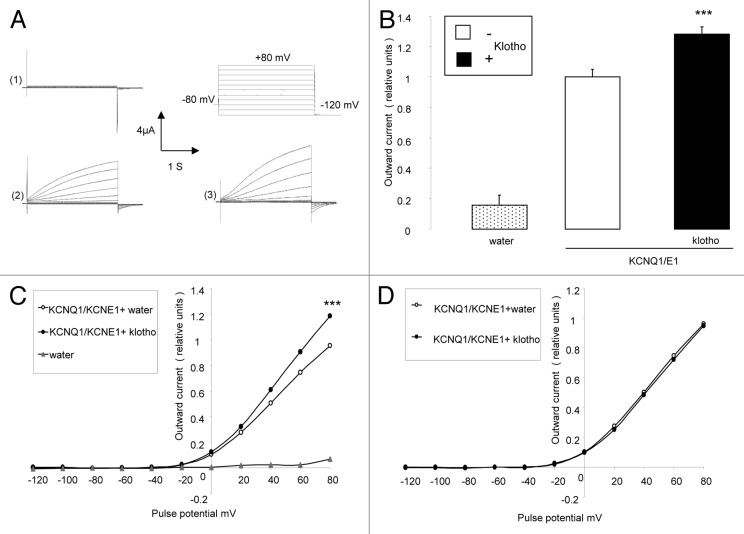
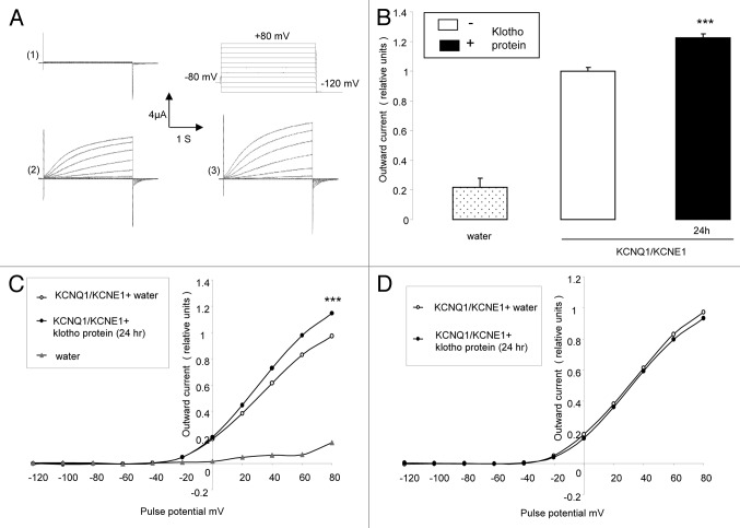
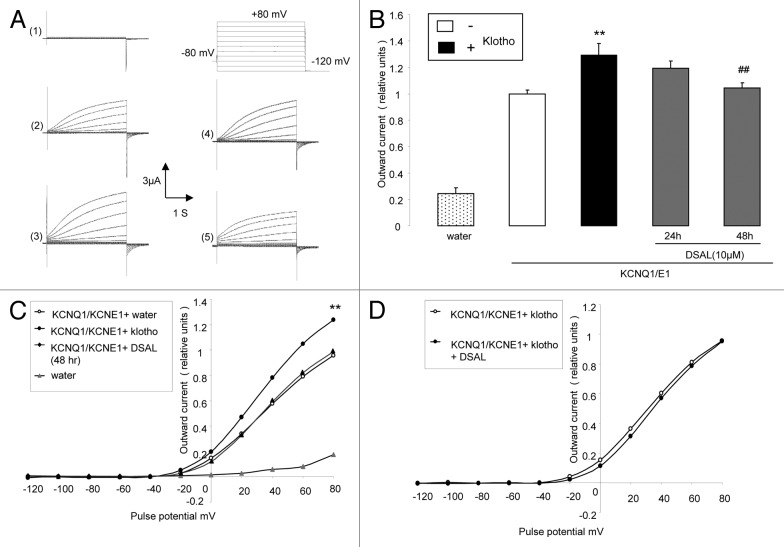
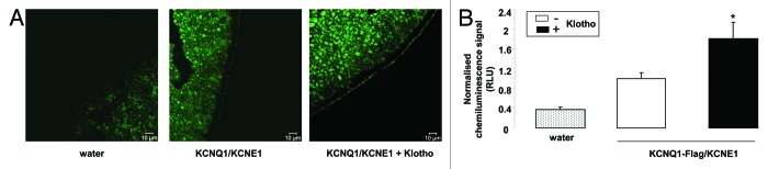
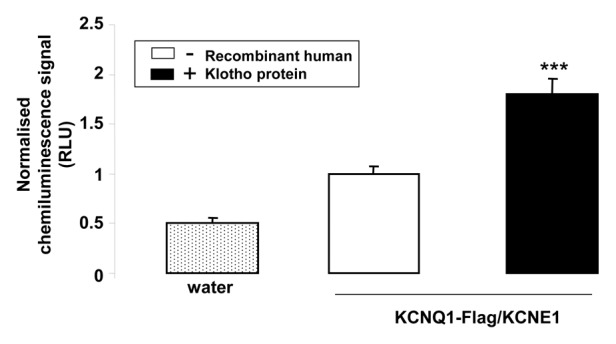
Klotho is a transmembrane protein expressed primarily in kidney, parathyroid gland, and choroid plexus. The extracellular domain could be cleaved off and released into the systemic circulation. Klotho is in part effective as β-glucuronidase regulating protein stability in the cell membrane. Klotho is a major determinant of aging and life span.Overexpression of Klotho increases and Klotho deficiency decreases life span. Klotho deficiency may further result in hearing loss and cardiac arrhythmia. The present study explored whether Klotho modifies activity and protein abundance of KCNQ1/KCNE1, a K(+) channel required for proper hearing and cardiac repolarization. To this end, cRNA encoding KCNQ1/KCNE1 was injected in Xenopus oocytes with or without additional injection of cRNA encoding Klotho. KCNQ1/KCNE1 expressing oocytes were treated with human recombinant Klotho protein (30 ng/mL) for 24 h. Moreover, oocytes which express both KCNQ1/KCNE1 and Klotho were treated with 10 μM DSA L (D-saccharic acid-1,4-lactone), a β-glucuronidase inhibitor. The KCNQ1/KCNE1 depolarization-induced current (I(Ks)) was determined utilizing dual electrode voltage clamp, while KCNQ1/KCNE1 protein abundance in the cell membrane was visualized utilizing specific antibody binding and quantified by chemiluminescence. KCNQ1/KCNE1 channel activity and KCNQ1/KCNE1 protein abundance were upregulated by coexpression of Klotho. The effect was mimicked by treatment with human recombinant Klotho protein (30 ng/mL) and inhibited by DSA L (10 μM). In conclusion, Klotho upregulates KCNQ1/KCNE1 channel activity by “mainly” enhancing channel protein abundance in the plasma cell membrane, an effect at least partially mediated through the β-glucuronidase activity of Klotho protein.


Figures

None
Figure 1. Effect of Klotho coexpression on current in KCNQ1/KCNE1 expressing Xenopus oocytes. (A) Original tracings demonstrating outward K+ currents activated by depolarization from -120 to +80 mV in 20 mV steps from a holding potential of -80 mV in Xenopus oocytes injected with water (1), injected with cRNA encoding KCNQ1/KCNE1 (2) and in Xenopus oocytes injected with cRNA encoding KCNQ1/KCNE1 and Klotho (3). (B) Arithmetic means ± SEM (n = 16–57) of the normalized depolarization-induced K+ current at +80 mV in Xenopus oocytes injected with water (dotted bar), with cRNA encoding KCNQ1/KCNE1(white bar) or with cRNA encoding KCNQ1/KCNE1 and Klotho (black bar). *** indicates statistically significant (P < 0.001) difference of KCNQ1/KCNE1 and Klotho expressing Xenopus oocytes from Xenopus oocytes expressing KCNQ1/KCNE1 alone. (C) Arithmetic means ± SEM (n = 16–57) of the normalized depolarization-induced K+ current as a function of voltage in Xenopus oocytes injected with water (gray triangles), with cRNA encoding KCNQ1/KCNE1 (white circles) or with cRNA encoding KCNQ1/KCNE1 and Klotho (black circles). *** indicates statistically significant (P < 0.001) difference of KCNQ1/KCNE1 and Klotho expressing Xenopus oocytes from Xenopus oocytes expressing KCNQ1/KCNE1 alone. (D) Arithmetic means ± SEM (n = 56–57) of the normalized depolarization-induced K+ current to the maximum peak current of each respective group as a function of voltage in Xenopus oocytes injected with cRNA encoding KCNQ1/KCNE1 (white circles) or with cRNA encoding KCNQ1/KCNE1 and Klotho (black circles).
None
Figure 2. Effect of treatment with recombinant Klotho protein on current in KCNQ1/KCNE1 expressing Xenopus oocytes. (A) Original tracings demonstrating outward K+ currents activated by depolarization from -120 to +80 mV in 20 mV steps from a holding potential of -80 mV in Xenopus oocytes injected with water (1),or injected with cRNA encoding KCNQ1/KCNE1 without (2) or with (3) a 24 h pretreatment with recombinant Klotho protein (30 ng/ml). (B) Arithmetic means ± SEM (n = 4–17) of the normalized depolarization-induced K+ current at +80 mV in Xenopus oocytes injected with water (dotted bar), or with cRNA encoding KCNQ1/KCNE1 without (white bar) or with (black bar) a 24 h pretreatment with recombinant Klotho protein (30 ng/ml). *** indicates statistically significant (P < 0.001) difference of Klotho treated KCNQ1/KCNE1 expressing Xenopus oocytes from untreated KCNQ1/KCNE1 expressing oocytes. (C) Arithmetic means ± SEM (n = 4–17) of the normalized depolarization-induced K+ current as a function of voltage in Xenopus oocytes injected with water (gray triangles), or with cRNA encoding KCNQ1/KCNE1 without (white circles) or with (black circles) a 24 h pretreatment with recombinant Klotho protein (30 ng/ml). *** indicates statistically significant (P < 0.001) difference of Klotho treated KCNQ1/KCNE1 expressing Xenopus oocytes from untreated KCNQ1/KCNE1 expressing Xenopus oocytes. (D) Arithmetic means ± SEM (n = 15–17) of the depolarization-induced K+ current (normalized to the maximum peak current of each respective group) as a function of voltage in Xenopus oocytes injected with cRNA encoding KCNQ1/KCNE1 without (white circles) or with (black circles) a 24 h pretreatment with recombinant Klotho protein (30 ng/ml).
None
Figure 3. Effect of β-glucuronidase inhibitor (DSAL) on current in KCNQ1/KCNE1 and Klotho expressing Xenopus oocytes. (A) Original tracings demonstrating outward K+ currents activated by depolarization from -120 to +80 mV in 20 mV steps from a holding potential of -80 in Xenopus oocytes injected with water (1), injected with cRNA encoding KCNQ1/KCNE1 alone (2) and in Xenopus oocytes injected with cRNA encoding both KCNE1/KCNQ1 and Klotho in the absence of β-glucuronidase inhibitor (3), or in the presence of DSAL for 24 h (4) or 48 h (5). (B) Arithmetic means ± SEM (n = 9–23) of the normalized depolarization-induced K+ current at +80 mV in Xenopus oocytes injected with water (dotted bar), injected with cRNA encoding KCNQ1/KCNE1 alone (white bar) or injected with cRNA encoding both KCNQ1/KCNE1 and Klotho in the absence of β-glucuronidase inhibitor DSAL (black bar), or in the presence of DSAL for 24 h (first gray bar) or 48 h (second gray bar). ** indicates statistically significant (P < 0.01) difference of Klotho and KCNQ1/KCNE1 expressing Xenopus oocytes from Xenopus oocytes expressing KCNQ1/KCNE1 alone. ## indicates statistically significant (P < 0.01) difference of DSAL-treated from untreated KCNQ1/KCNE1 and Klotho expressing Xenopus oocytes. (C) Arithmetic means ± SEM (n = 9–23) of the normalized depolarization-induced K+ current as a function of voltage in Xenopus oocytes injected with water (gray triangles), injected with cRNA encoding KCNQ1/KCNE1 alone (white circles) and in Xenopus oocytes injected with cRNA encoding both KCNQ1/KCNE1 and klotho in the absence of β-glucuronidase inhibitor DSAL (black circles), or in the presence of DSAL for 48 h (black triangles). ** indicates statistically significant (P < 0.01) difference of Klotho and KCNQ1/KCNE1 expressing Xenopus oocytes from Xenopus oocytes expressing KCNQ1/KCNE1 alone. (D) Arithmetic means ± SEM (n = 18–23) of the depolarization-induced K+ current (normalized to the maximum peak current of each group) as a function of voltage in Xenopus oocytes injected with cRNA encoding KCNQ1/KCNE1 and Klotho without treatment (white circles) or treated for 48 h with DSAL (black circles).
None
Figure 4. Effect of Klotho coexpression on KCNQ1/KCNE1 protein abundance in the cell membrane of Xenopus oocytes. (A) Confocal microscopy of the KCNQ1/KCNE1 protein abundance in Xenopus oocytes injected with water (left), injected with cRNA encoding KCNQ1/KCNE1 alone (middle) or expressing KCNQ1/KCNE1 together with Klotho (right). The images are representative for 3 independent experiments. (B) Arithmetic means ± SEM (n = 55–76) of the chemiluminescence of KCNQ1-Flag/KCNE1 protein abundance in Xenopus oocytes injected with water (dotted bar), injected with cRNA encoding KCNQ1-Flag/KCNE1 alone (white bar), or expressing KCNQ1-Flag/KCNE1 with Klotho (black bar). * (P < 0.05) indicates statistically significant difference from the protein abundance in Xenopus oocytes expressing KCNQ1-Flag/KCNE1 alone.
None
Figure 5. Effect of treatment with recombinant Klotho protein on KCNQ1/KCNE1 protein abundance in the cell membrane of Xenopus oocytes. Arithmetic means ± SEM (n = 70–72) of the chemiluminescence of KCNQ1-Flag/KCNE1 protein abundance in Xenopus oocytes injected without (dotted bar) or with cRNA encoding KCNQ1-Flag/KCNE1 without (white bar) or with (black bar) a 24 h pretreatment with recombinant Klotho protein (30 ng/ml). *** (P < 0.001) indicates statistically significant difference from the protein abundance in Xenopus oocytes expressing KCNQ1-Flag/KCNE1 alone.

Comment in

References

    1. Takeshita K, Fujimori T, Kurotaki Y, Honjo H, Tsujikawa H, Yasui K, Lee JK, Kamiya K, Kitaichi K, Yamamoto K, et al. Sinoatrial node dysfunction and early unexpected death of mice with a defect of klotho gene expression. Circulation. 2004;109:1776–82. doi: 10.1161/01.CIR.0000124224.48962.32. - DOI - PubMed
    1. Tsujikawa H, Kurotaki Y, Fujimori T, Fukuda K, Nabeshima Y. Klotho, a gene related to a syndrome resembling human premature aging, functions in a negative regulatory circuit of vitamin D endocrine system. Mol Endocrinol. 2003;17:2393–403. doi: 10.1210/me.2003-0048. - DOI - PubMed
    1. Kuro-o M, Matsumura Y, Aizawa H, Kawaguchi H, Suga T, Utsugi T, Ohyama Y, Kurabayashi M, Kaname T, Kume E, et al. Mutation of the mouse klotho gene leads to a syndrome resembling ageing. Nature. 1997;390:45–51. doi: 10.1038/36285. - DOI - PubMed
    1. Kuro-o M, M K-o Klotho. Pflugers Arch. 2010;459:333–43. doi: 10.1007/s00424-009-0722-7. - DOI - PubMed
    1. Imura A, Iwano A, Tohyama O, Tsuji Y, Nozaki K, Hashimoto N, Fujimori T, Nabeshima Y. Secreted Klotho protein in sera and CSF: implication for post-translational cleavage in release of Klotho protein from cell membrane. FEBS Lett. 2004;565:143–7. doi: 10.1016/j.febslet.2004.03.090. - DOI - PubMed

Publication types

LinkOut - more resources