

Safety and effects of the vector for the Leber hereditary optic neuropathy gene therapy clinical trial
- PMID: 24457989
- PMCID: PMC4266107
- DOI: 10.1001/jamaophthalmol.2013.7630
Safety and effects of the vector for the Leber hereditary optic neuropathy gene therapy clinical trial
Abstract
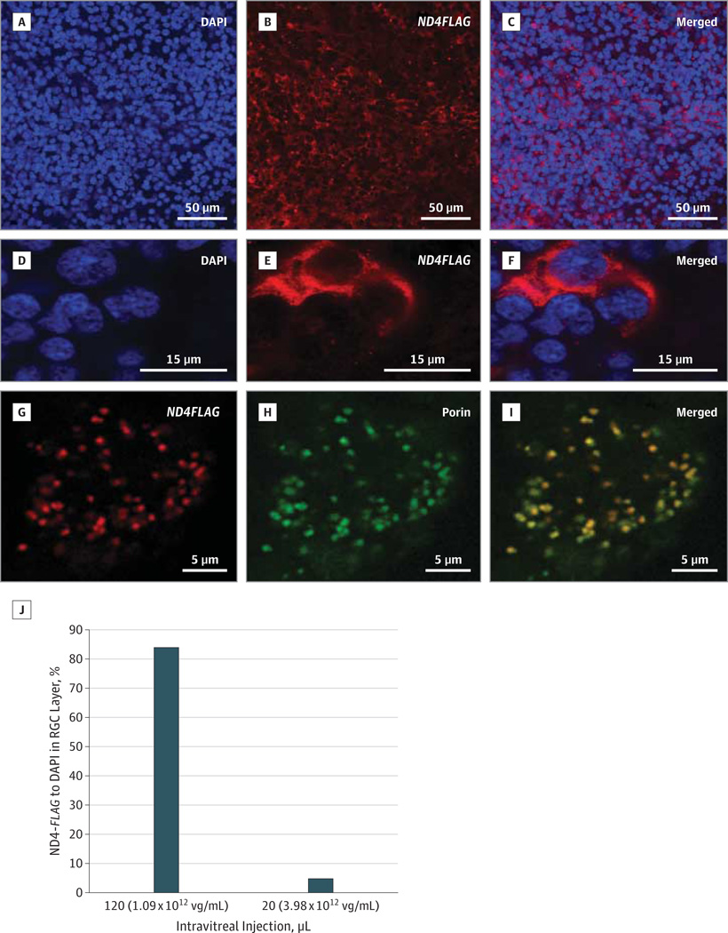
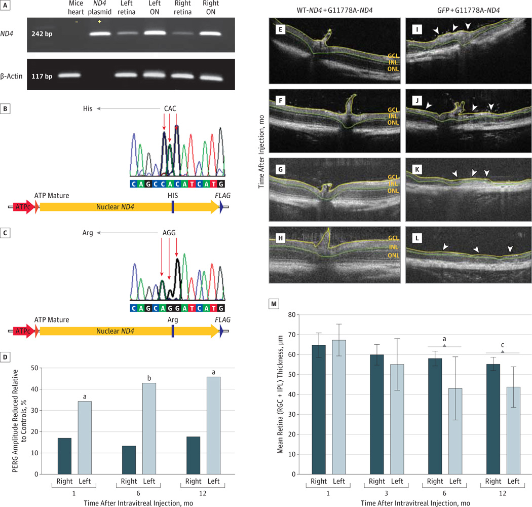
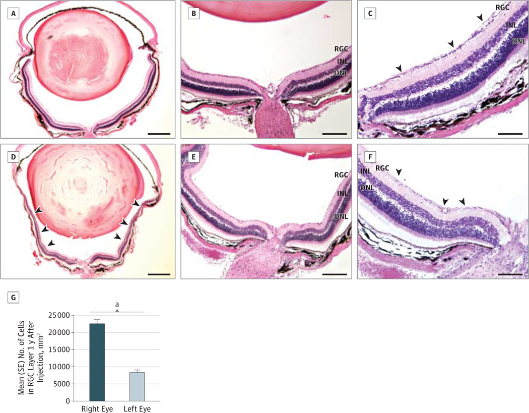
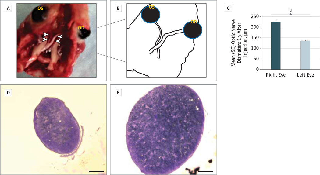
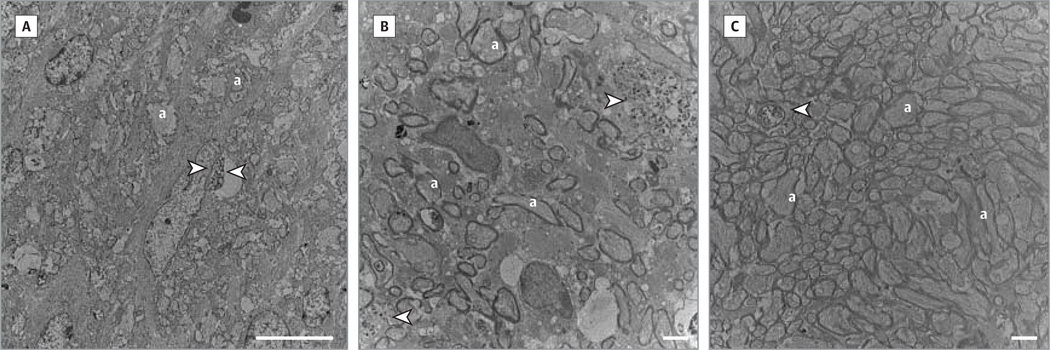
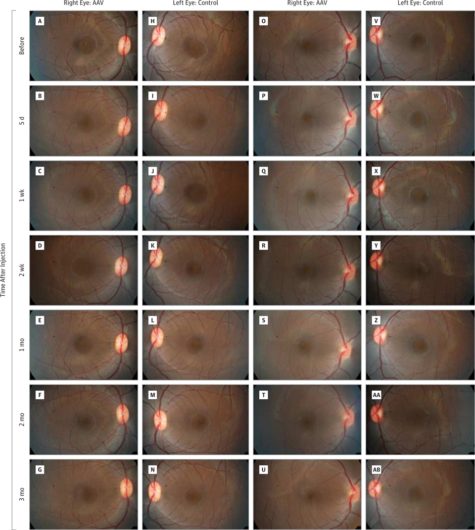
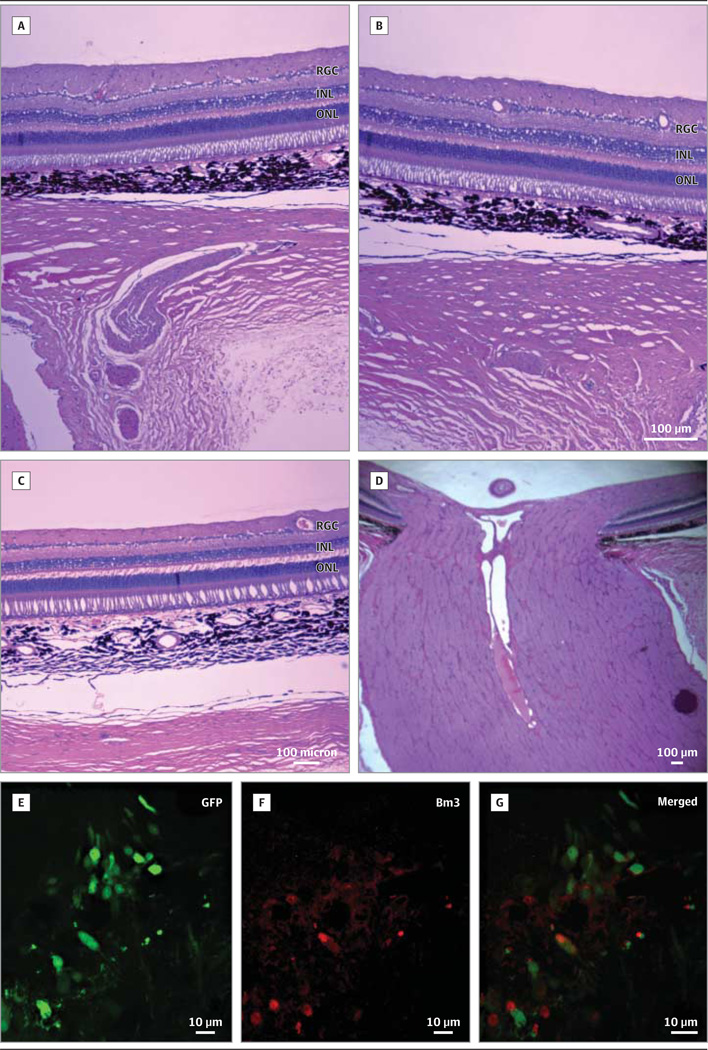
IMPORTANCE We developed a novel strategy for treatment of Leber hereditary optic neuropathy (LHON) caused by a mutation in the nicotinamide adenine dinucleotide dehydrogenase subunit IV (ND4) mitochondrial gene. OBJECTIVE To demonstrate the safety and effects of the gene therapy vector to be used in a proposed gene therapy clinical trial. DESIGN AND SETTING In a series of laboratory experiments, we modified the mitochondrial ND4 subunit of complex I in the nuclear genetic code for import into mitochondria. The protein was targeted into the organelle by agency of a targeting sequence (allotopic expression). The gene was packaged into adeno-associated viral vectors and then vitreally injected into rodent, nonhuman primate, and ex vivo human eyes that underwent testing for expression and integration by immunohistochemical analysis and blue native polyacrylamide gel electrophoresis. During serial follow-up, the animal eyes underwent fundus photography, optical coherence tomography, and multifocal or pattern electroretinography. We tested for rescue of visual loss in rodent eyes also injected with a mutant G11778A ND4 homologue responsible for most cases of LHON. EXPOSURE Ocular infection with recombinant adeno-associated viral vectors containing a wild-type allotopic human ND4 gene. MAIN OUTCOMES AND MEASURES Expression of human ND4 and rescue of optic neuropathy induced by mutant human ND4. RESULTS We found human ND4 expressed in almost all mouse retinal ganglion cells by 1 week after injection and ND4 integrated into the mouse complex I. In rodent eyes also injected with a mutant allotopic ND4, wild-type allotopic ND4 prevented defective adenosine triphosphate synthesis, suppressed visual loss, reduced apoptosis of retinal ganglion cells, and prevented demise of axons in the optic nerve. Injection of ND4 in the ex vivo human eye resulted in expression in most retinal ganglion cells. Primates undergoing vitreal injection with the ND4 test article and followed up for 3 months had no serious adverse reactions. CONCLUSIONS AND RELEVANCE Expression of our allotopic ND4 vector in the ex vivo human eye, safety of the test article, rescue of the LHON mouse model, and the severe irreversible loss of visual function in LHON support clinical testing with mutated G11778A mitochondrial DNA in our patients.
Conflict of interest statement
Figures
Similar articles
-
LHON gene therapy vector prevents visual loss and optic neuropathy induced by G11778A mutant mitochondrial DNA: biodistribution and toxicology profile.Invest Ophthalmol Vis Sci. 2014 Oct 23;55(12):7739-53. doi: 10.1167/iovs.14-15388. Invest Ophthalmol Vis Sci. 2014. PMID: 25342621 Free PMC article.
-
Mutant NADH dehydrogenase subunit 4 gene delivery to mitochondria by targeting sequence-modified adeno-associated virus induces visual loss and optic atrophy in mice.Mol Vis. 2012;18:1668-83. Epub 2012 Jun 20. Mol Vis. 2012. PMID: 22773905 Free PMC article.
-
Trial end points and natural history in patients with G11778A Leber hereditary optic neuropathy : preparation for gene therapy clinical trial.JAMA Ophthalmol. 2014 Apr 1;132(4):428-36. doi: 10.1001/jamaophthalmol.2013.7971. JAMA Ophthalmol. 2014. PMID: 24525545 Free PMC article.
-
Leber hereditary optic neuropathy gene therapy.Curr Opin Ophthalmol. 2024 May 1;35(3):244-251. doi: 10.1097/ICU.0000000000001028. Epub 2023 Dec 20. Curr Opin Ophthalmol. 2024. PMID: 38117686 Free PMC article. Review.
-
Gene therapy for Leber hereditary optic neuropathy.Expert Opin Biol Ther. 2024 Jun;24(6):521-528. doi: 10.1080/14712598.2024.2359015. Epub 2024 Jun 28. Expert Opin Biol Ther. 2024. PMID: 38939999 Review.
Cited by
-
Gene therapy reforms photoreceptor structure and restores vision in NPHP5-associated Leber congenital amaurosis.Mol Ther. 2021 Aug 4;29(8):2456-2468. doi: 10.1016/j.ymthe.2021.03.021. Epub 2021 Mar 27. Mol Ther. 2021. PMID: 33781914 Free PMC article.
-
Mechanisms of mesenchymal stem/stromal cell function.Stem Cell Res Ther. 2016 Aug 31;7(1):125. doi: 10.1186/s13287-016-0363-7. Stem Cell Res Ther. 2016. PMID: 27581859 Free PMC article. Review.
-
Fundus imaging of retinal ganglion cells transduced by retrograde transport of rAAV2-retro.Exp Eye Res. 2022 Jun;219:109084. doi: 10.1016/j.exer.2022.109084. Epub 2022 Apr 20. Exp Eye Res. 2022. PMID: 35460667 Free PMC article.
-
Retinal gene therapy using adeno-associated viral vectors: multiple applications for a small virus.Hum Gene Ther. 2014 Aug;25(8):671-8. doi: 10.1089/hum.2014.2530. Hum Gene Ther. 2014. PMID: 25136913 Free PMC article. No abstract available.
-
Is there treatment for Leber hereditary optic neuropathy?Curr Opin Ophthalmol. 2015 Nov;26(6):450-7. doi: 10.1097/ICU.0000000000000212. Curr Opin Ophthalmol. 2015. PMID: 26448041 Free PMC article. Review.
References
-
- Leber T. Über hereditare und congenital-angelegete Sehnerverleiden. Graefes Archiv Klin Experimentelle Opthalmol. 1871;7:249–271.
-
- Gray RE, Law RH, Devenish RJ, Nagley P. Allotopic expression of mitochondrial ATP synthase genes in nucleus of Saccharomyces cerevisiae. Methods Enzymol. 1996;264:369–389. - PubMed
-
- Neupert W. Protein import into mitochondria. Annu Rev Biochem. 1997;66:863–917. - PubMed
Publication types
MeSH terms
Substances
Grants and funding
LinkOut - more resources
Full Text Sources
Other Literature Sources
Medical
