

Chorioamnionitis-induced fetal gut injury is mediated by direct gut exposure of inflammatory mediators or by lung inflammation
- PMID: 24458021
- PMCID: PMC3949018
- DOI: 10.1152/ajpgi.00260.2013
Chorioamnionitis-induced fetal gut injury is mediated by direct gut exposure of inflammatory mediators or by lung inflammation
Abstract
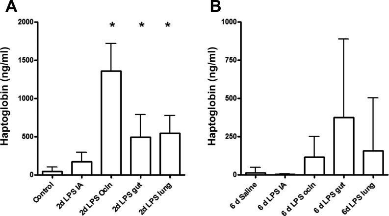
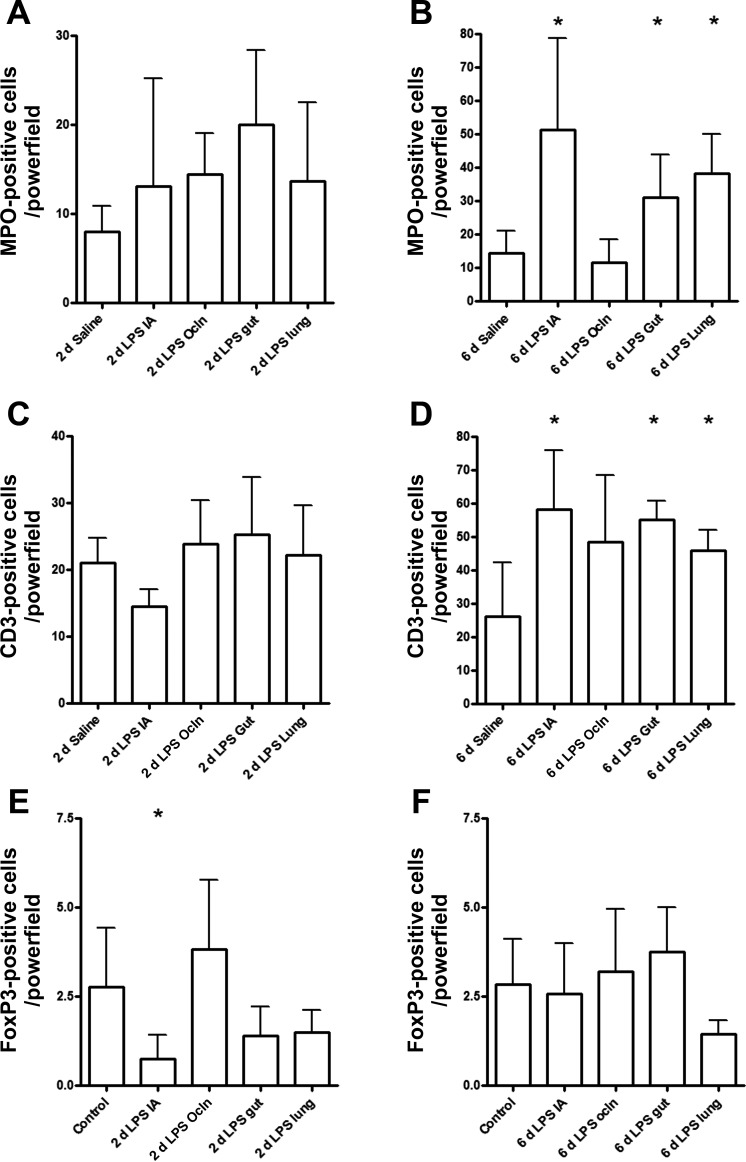
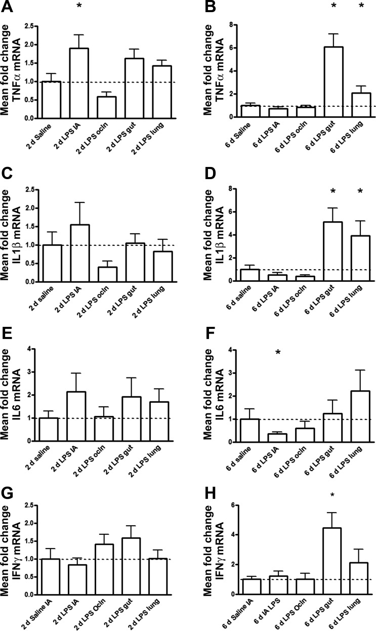
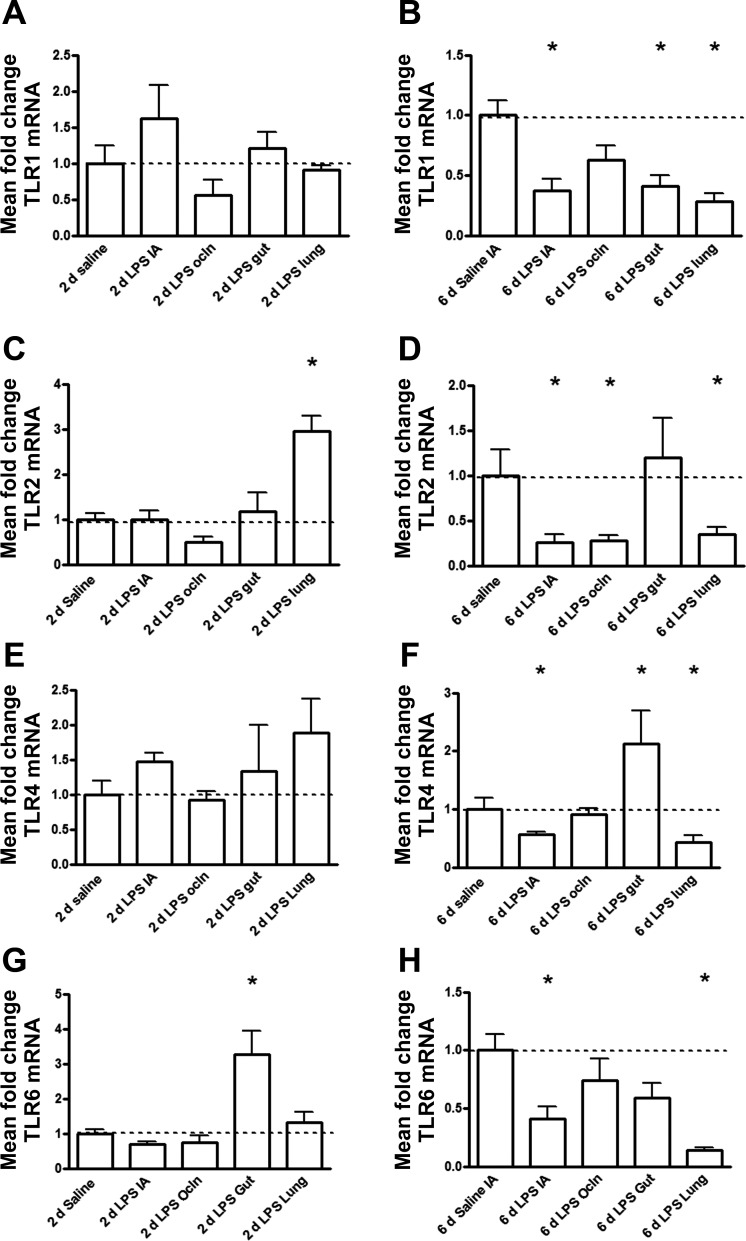
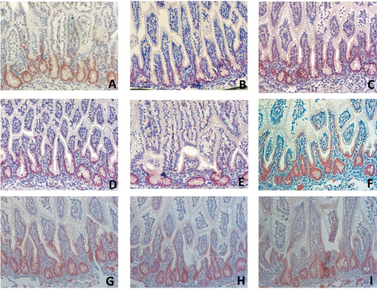
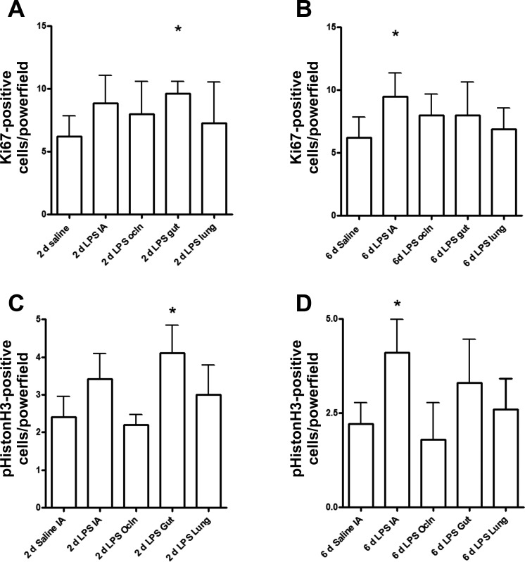
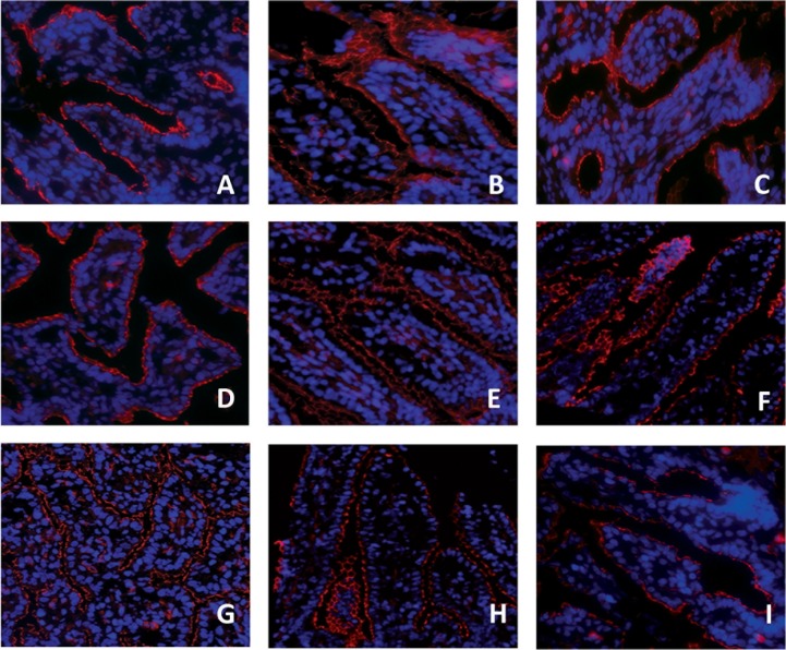
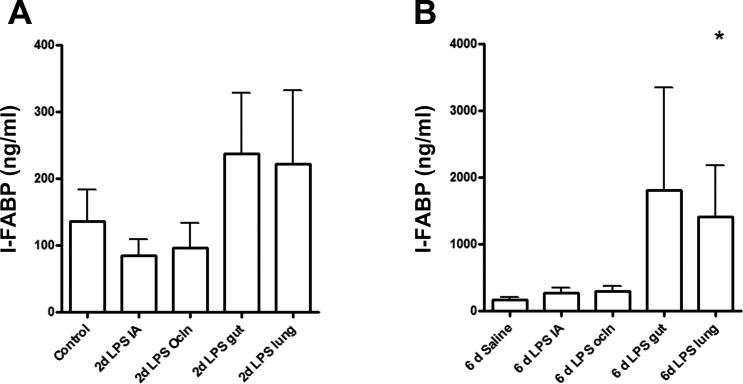
Intra-amniotic exposure to proinflammatory agonists causes chorioamnionitis and fetal gut inflammation. Fetal gut inflammation is associated with mucosal injury and impaired gut development. We tested whether this detrimental inflammatory response of the fetal gut results from a direct local (gut derived) or an indirect inflammatory response mediated by the chorioamnion/skin or lung, since these organs are also in direct contact with the amniotic fluid. The gastrointestinal tract was isolated from the respiratory tract and the amnion/skin epithelia by fetal surgery in time-mated ewes. Lipopolysaccharide (LPS) or saline (controls) was selectively infused in the gastrointestinal tract, trachea, or amniotic compartment at 2 or 6 days before preterm delivery at 124 days gestation (term 150 days). Gastrointestinal and intratracheal LPS exposure caused distinct inflammatory responses in the fetal gut. Inflammatory responses could be distinguished by the influx of leukocytes (MPO(+), CD3(+), and FoxP3(+) cells), tumor necrosis factor-α, and interferon-γ expression and differential upregulation of mRNA levels for Toll-like receptor 1, 2, 4, and 6. Fetal gut inflammation after direct intestinal LPS exposure resulted in severe loss of the tight junctional protein zonula occludens protein 1 (ZO-1) and increased mitosis of intestinal epithelial cells. Inflammation of the fetal gut after selective LPS instillation in the lungs caused only mild disruption of ZO-1, loss in epithelial cell integrity, and impaired epithelial differentiation. LPS exposure of the amnion/skin epithelia did not result in gut inflammation or morphological, structural, and functional changes. Our results indicate that the detrimental consequences of chorioamnionitis on fetal gut development are the combined result of local gut and lung-mediated inflammatory responses.
Keywords: endotoxin; fetal inflammatory response; necrotizing enterocolitis; sheep.
Figures
Similar articles
-
Selective IL-1α exposure to the fetal gut, lung, and chorioamnion/skin causes intestinal inflammatory and developmental changes in fetal sheep.Lab Invest. 2016 Jan;96(1):69-80. doi: 10.1038/labinvest.2015.127. Epub 2015 Oct 26. Lab Invest. 2016. PMID: 26501868
-
Prophylactic Interleukin-2 Treatment Prevents Fetal Gut Inflammation and Injury in an Ovine Model of Chorioamnionitis.Inflamm Bowel Dis. 2015 Sep;21(9):2026-38. doi: 10.1097/MIB.0000000000000455. Inflamm Bowel Dis. 2015. PMID: 26002542
-
Intra-amniotic LPS and antenatal betamethasone: inflammation and maturation in preterm lamb lungs.Am J Physiol Lung Cell Mol Physiol. 2012 Feb 15;302(4):L380-9. doi: 10.1152/ajplung.00338.2011. Epub 2011 Dec 9. Am J Physiol Lung Cell Mol Physiol. 2012. PMID: 22160306 Free PMC article.
-
Chorioamnionitis - new ideas from experimental models.Neonatology. 2011;99(4):320-5. doi: 10.1159/000326620. Epub 2011 Jun 23. Neonatology. 2011. PMID: 21701204 Review.
-
Fetal immune response to chorioamnionitis.Semin Reprod Med. 2014 Jan;32(1):56-67. doi: 10.1055/s-0033-1361823. Epub 2014 Jan 3. Semin Reprod Med. 2014. PMID: 24390922 Free PMC article. Review.
Cited by
-
Perinatal Hyperoxia and Developmental Consequences on the Lung-Brain Axis.Oxid Med Cell Longev. 2022 Feb 24;2022:5784146. doi: 10.1155/2022/5784146. eCollection 2022. Oxid Med Cell Longev. 2022. PMID: 35251477 Free PMC article. Review.
-
Pulmonary Consequences of Prenatal Inflammatory Exposures: Clinical Perspective and Review of Basic Immunological Mechanisms.Front Immunol. 2020 Jun 19;11:1285. doi: 10.3389/fimmu.2020.01285. eCollection 2020. Front Immunol. 2020. PMID: 32636848 Free PMC article. Review.
-
CD161 contributes to prenatal immune suppression of IFNγ-producing PLZF+ T cells.J Clin Invest. 2019 May 30;129(9):3562-3577. doi: 10.1172/JCI125957. J Clin Invest. 2019. PMID: 31145102 Free PMC article.
-
Recent advances in the prevention of preterm birth.F1000Res. 2017 Jul 18;6:F1000 Faculty Rev-1139. doi: 10.12688/f1000research.11385.1. eCollection 2017. F1000Res. 2017. PMID: 28781747 Free PMC article. Review.
-
Understanding Host-Pathogen Interactions in Acute Chorioamnionitis Through the Use of Animal Models.Front Cell Infect Microbiol. 2021 Jul 27;11:709309. doi: 10.3389/fcimb.2021.709309. eCollection 2021. Front Cell Infect Microbiol. 2021. PMID: 34386434 Free PMC article. Review.
References
-
- Adams-Chapman I. Long-term impact of infection on the preterm neonate. Semin Perinatol 36: 462–470, 2012 - PubMed
-
- Alam M, Midtvedt T, Uribe A. Differential cell kinetics in the ileum and colon of germfree rats. Scand J Gastroenterol 29: 445–451, 1994 - PubMed
-
- Andrews WW, Goldenberg RL, Faye-Petersen O, Cliver S, Goepfert AR, Hauth JC. The Alabama Preterm Birth study: polymorphonuclear and mononuclear cell placental infiltrations, other markers of inflammation, and outcomes in 23- to 32-week preterm newborn infants. Am J Obstet Gynecol 195: 803–808, 2006 - PubMed
-
- Been JV, Lievense S, Zimmermann LJ, Kramer BW, Wolfs TG. Chorioamnionitis as a risk factor for necrotizing enterocolitis: a systematic review and meta-analysis. J Pediatr 162: 236–242, 2013 - PubMed
Publication types
MeSH terms
Substances
Grants and funding
LinkOut - more resources
Full Text Sources
Other Literature Sources
Medical
Research Materials
Miscellaneous