. 2014 Jan 24:4:3854.
doi: 10.1038/srep03854.

The fidelity of stochastic single-molecule super-resolution reconstructions critically depends upon robust background estimation

Affiliations

The fidelity of stochastic single-molecule super-resolution reconstructions critically depends upon robust background estimation

Eelco Hoogendoorn et al. Sci Rep. .

Abstract

The quality of super resolution images obtained by stochastic single-molecule microscopy critically depends on image analysis algorithms. We find that the choice of background estimator is often the most important determinant of reconstruction quality. A variety of techniques have found use, but many have a very narrow range of applicability depending upon the characteristics of the raw data. Importantly, we observe that when using otherwise accurate algorithms, unaccounted background components can give rise to biases on scales defeating the purpose of super-resolution microscopy. We find that a temporal median filter in particular provides a simple yet effective solution to the problem of background estimation, which we demonstrate over a range of imaging modalities and different reconstruction methods.


Figures

Figure 1
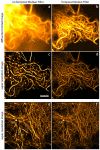
Figure 1. Background and foreground estimation by temporal median filtering.
Panels in the left column (a,d,g) show raw data frames from LifeAct-mEos3.2, MyosinIIa-Alexa532 and MyosinIIa-Alexa647 data sets. Middle column (b,e,h) shows the background estimated for that frame using the temporal median filter (window size of 101 frames with 10 frame interpolation). Right column (c,f,i) shows the foreground calculated by subtracting the estimated background from the raw frames. For display purposes the values were clipped at zero in order to only show the fluorescence that is higher than the estimated background. Panel j shows the raw fluorescence trace (MyosinIIa Alexa 647 data set) at two adjacent pixels (arrow in panel h) and the corresponding background estimate.
Figure 2
Figure 2. Application of a temporal median filter prior to localization analysis improves fidelity of two-color GSDIM data.
GSDIM imaging of myosinIIa independently labeled with Alexa532 (a–c) and Alexa647 (e–g). Without the utilization of the temporal median filter, the RapidSTORM reconstruction of the Alexa532 data set shows localizations that are skewed towards regions of high fluorescence (b) and exhibit poor co-localization with the Alexa647 (f) based on Pearson's cross-correlation analysis (f, inset). Use of the temporal median filter prior to running the localization eliminates these artifacts in the Alexa532 reconstruction (c) and shows higher correlation with the Alexa647 reconstruction (g, inset). Intensity traces (d, h) are normalized to the area under the trace. Similar analysis for alternative reconstruction methods can be found in supplemental Figs S2,3. Scale bar: 3 μm.
Figure 3
Figure 3. Reconstructions for LifeAct-mEos3.2 HeLa cell using RapidSTORM, QuickPALM and deconvolution with and without the temporal median filter applied.
An area that shows a high-degree of structured (heterogeneous) background fluorescence indicated with an arrow in Fig S1a-b leads to a spurious structure when using RapidSTORM (median smoothing 5 px setting) or deconvolution if the temporal median was not applied. When the temporal median filter prior to running the reconstruction analysis was applied (b, e, h) the effect of structured background is greatly reduced in the reconstruction such that the intensity profiles are now in much closer agreement for the different methods (c, f, i). This illustrates the relative importance of background estimation in the overall reconstruction process. Scale bar: 3 μm.
Figure 4
Figure 4
GSDIM data of two HeLA cells with LifeAct-Venus Panel a shows the sum of all frames in the data sets; panel b, shows the sum of all background subtracted frames, both represent a diffraction limited image. Panels c and d show the RapidSTORM reconstruction obtained without and with application of the temporal median filter. The analysis of these data sets revealed that temporal median filtering reduces the presence of strong foci at filament crossings, which appear to induce deformations that are not apparent in the diffraction limited images. Panels e and f show the RapidSTORM reconstruction obtained without and with application of the temporal median filter of another HeLa cell with an intricate F-actin network. The used threshold for all reconstructions was the same and the Gaussian smoothing filter was selected in this case (1 sigma). Color scale for images in panels c,d and e,f were chosen to be equal (scale bar 3 μm).
Figure 5
Figure 5
Thin filaments at different inter-filament distances (a) simulated with structured background (b). The structured background leads both artifacts and distorted structures (c), which are mitigated by the utilization of a temporal median filter prior to performing the localization analysis (d), resulting in a more accurate rendering of the structures. (e) Quantification of synthetic filaments shown in c-d. Pairs of filaments, each with thickness 10 nm (black bar). The profiles shown in red and green represent the quantified mean profile of the reconstructed filaments measured from the midline outward. Reconstructions were obtained from RapidSTORM, with settings median smoothing (5 px) and threshold 100 and rendered using the obtained amplitude blurred with a Gaussian with a SEM of 2 nm. Prior to reconstruction by RapidSTORM the background was accounted for using the temporal median filter, with a filter size of 101 frames and 10 frame interpolation.
Figure 6
Figure 6. RapidSTORM reconstructions of a ring with radius 75 nm with a threshold of 10 photons and using the standard spatial median filter without (a) and with (b) application of the temporal median filter prior to reconstruction.
Background corrected reconstruction no longer show artifacts in the reconstructions and in all cases reliably reconstruct the ring. For these simulations the event cycle amplitude A = [200, 800, 3200] was varied and the structured background, where the peak of the structured background was varied using the values bs = [0, 10, 20, 30, 40, 50] photons using the same event list for each case with simulations settings as described in the supplementary information. The radial profiles of the rings were calculated for each panel and are shown together with the original ring in the right hand side column. The intensity scale in each panel was adjusted to show the full intensity range independent of the other panels. The localizations were obtained from RapidSTORM with settings median smoothing (5 px) and threshold 100 and rendered using the obtained amplitude blurred with a Gaussian with a SEM of 2 nm. Prior to reconstruction by RapidSTORM the background was accounted for using the temporal median filter, with a filter size of 101 frames and 10 frame interpolation.
Figure 7
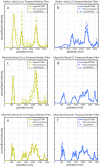
Figure 7. Line scan profiles for the LifeAct-mEos3.2, MyosinIIa-Alexa532 and MyosinIIa-Alexa647 data sets from (Fig. 2, 3,S2, S3) put side by side, using three reconstruction methods without and with application of the temporal median filter.
The panels a, c and e reveal that the different methods give different results for the same data set when no temporal median filter is applied. The panels b, d and f reveals that application of the temporal median filter yields results that are in close agreement for the three reconstruction methods used.

Comment in

References

    1. Gould T. J., Hess S. T. & Bewersdorf J. Optical Nanoscopy: From Acquisition to Analysis. Annual Review of Biomedical Engineering 14, 231–254 (2012). - PMC - PubMed
    1. Ewers H. 4. Nano Resolution Optical Imaging Through Localization Microscopy. 1–20 (2012). 10.1016/b978-0-12-385872-6.00004-0.
    1. Wolter S., Löschberger A., Holm T. & Aufmkolk S. rapidSTORM: accurate, fast open-source software for localization microscopy. Nature (2012). - PubMed
    1. Henriques R. et al. QuickPALM: 3D real-time photoactivation nanoscopy image processing in ImageJ. Nature Methods 7, 339–340 (2010). - PubMed
    1. Mukamel E., Babcock H. & Zhuang X. Statistical Deconvolution for Superresolution Fluorescence Microscopy. Biophysical Journal 102, 2391–2400 (2012). - PMC - PubMed

Publication types