. 2014 Jun;63(6):2097-113.
doi: 10.2337/db12-1459. Epub 2014 Jan 23.

Glycogen synthase kinase-3β inhibition ameliorates cardiac parasympathetic dysfunction in type 1 diabetic Akita mice

Affiliations

Glycogen synthase kinase-3β inhibition ameliorates cardiac parasympathetic dysfunction in type 1 diabetic Akita mice

Yali Zhang et al. Diabetes. 2014 Jun.

Abstract

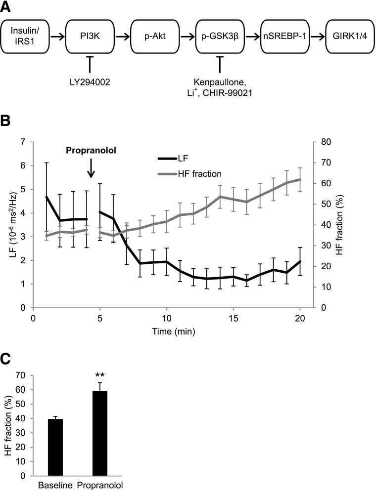
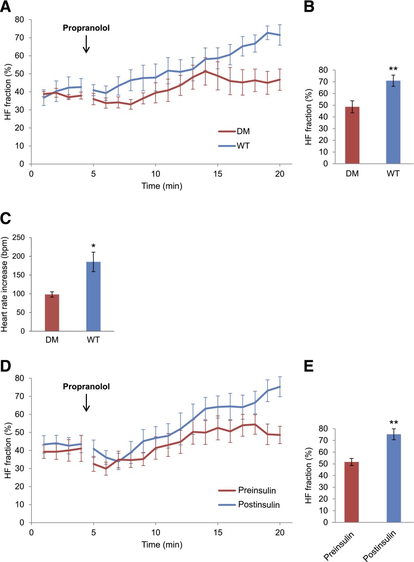
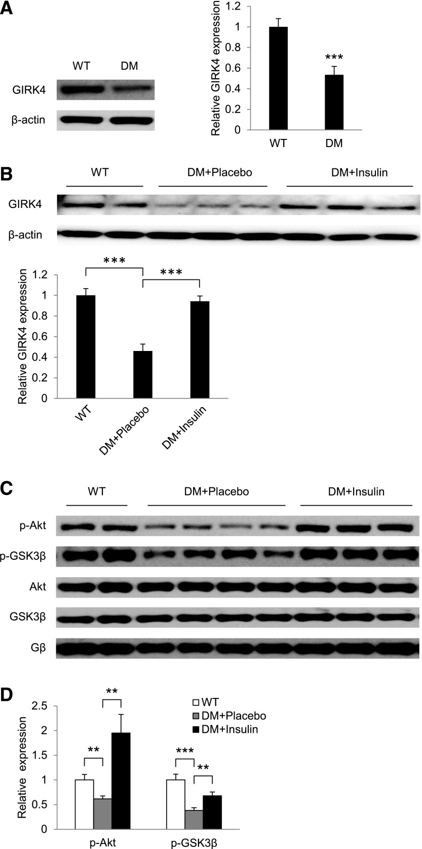
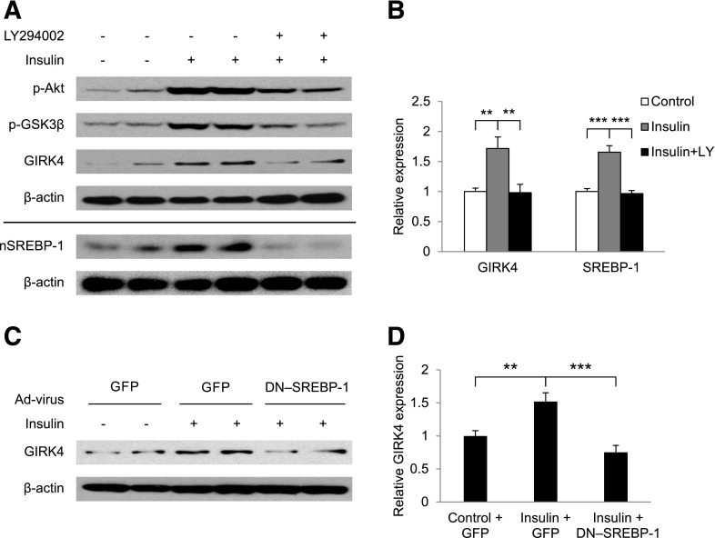
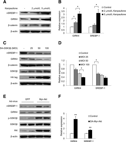
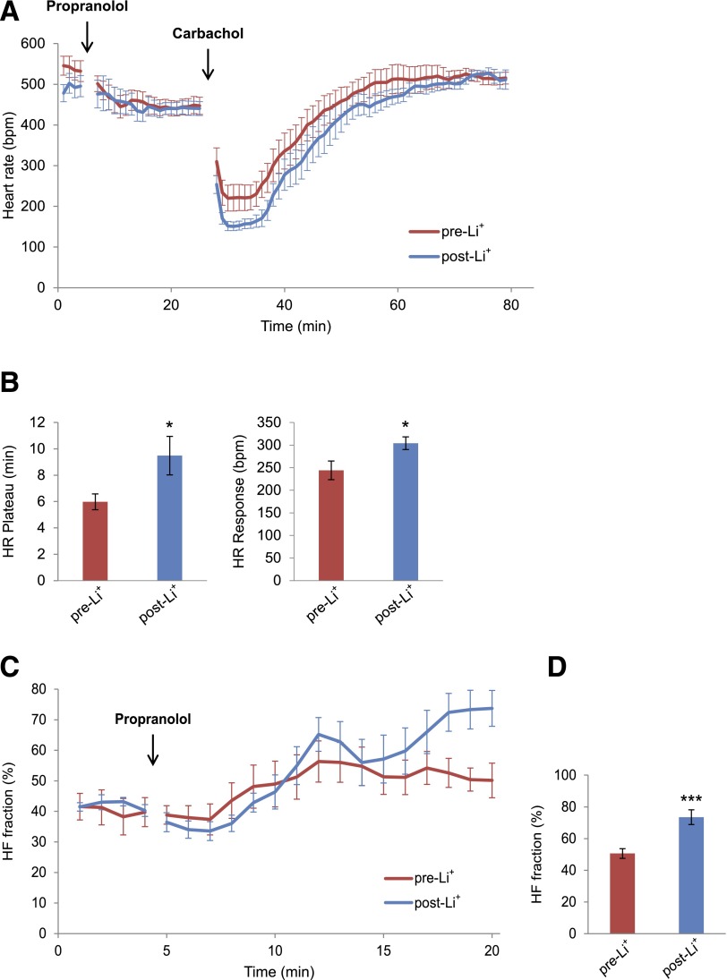
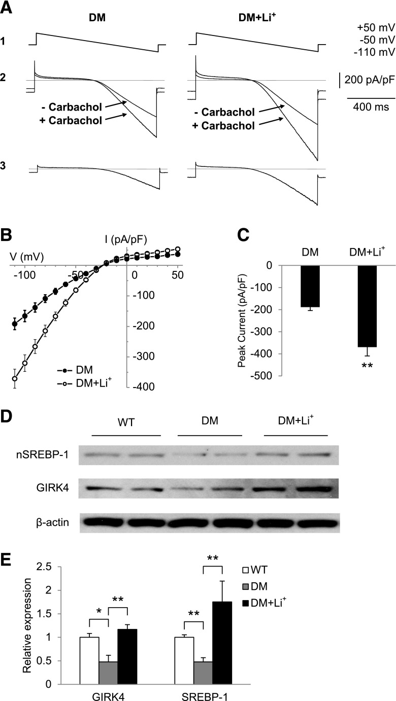
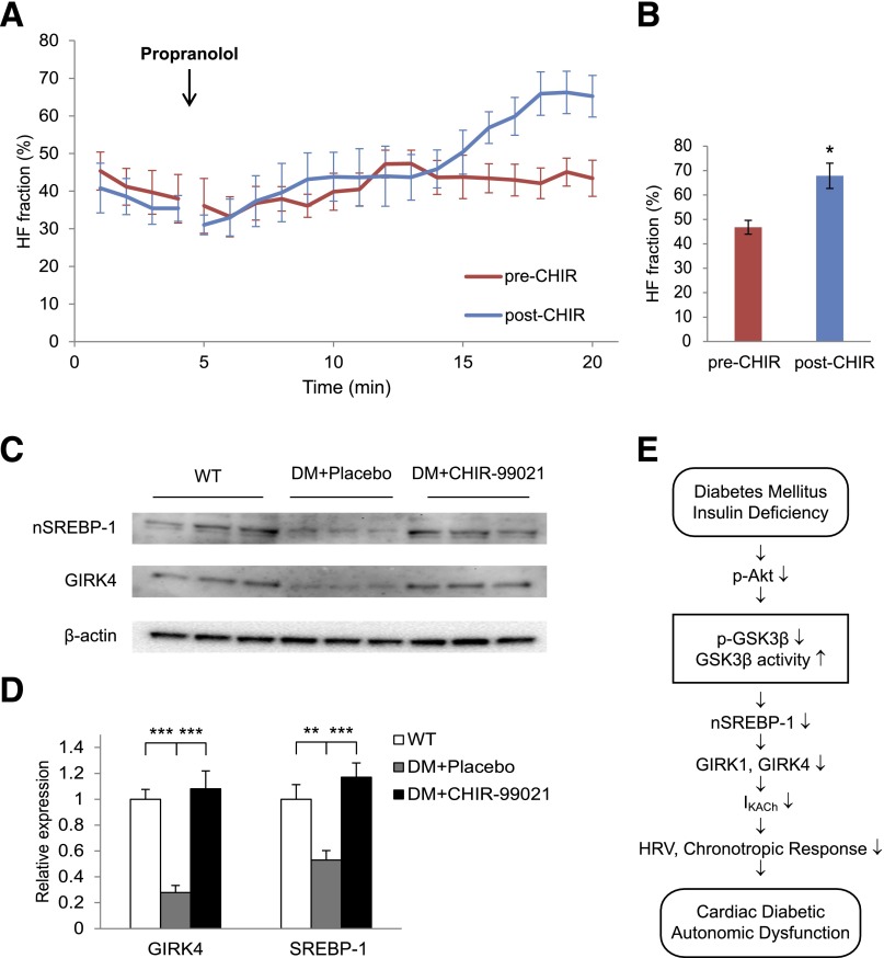
Decreased heart rate variability (HRV) is a major risk factor for sudden death and cardiovascular disease. We previously demonstrated that parasympathetic dysfunction in the heart of the Akita type 1 diabetic mouse was due to a decrease in the level of the sterol response element-binding protein (SREBP-1). Here we demonstrate that hyperactivity of glycogen synthase kinase-3β (GSK3β) in the atrium of the Akita mouse results in decreased SREBP-1, attenuation of parasympathetic modulation of heart rate, measured as a decrease in the high-frequency (HF) fraction of HRV in the presence of propranolol, and a decrease in expression of the G-protein coupled inward rectifying K(+) (GIRK4) subunit of the acetylcholine (ACh)-activated inward-rectifying K(+) channel (IKACh), the ion channel that mediates the heart rate response to parasympathetic stimulation. Treatment of atrial myocytes with the GSK3β inhibitor Kenpaullone increased levels of SREBP-1 and expression of GIRK4 and IKACh, whereas a dominant-active GSK3β mutant decreased SREBP-1 and GIRK4 expression. In Akita mice treated with GSK3β inhibitors Li(+) and/or CHIR-99021, Li(+) increased IKACh, and Li(+) and CHIR-99021 both partially reversed the decrease in HF fraction while increasing GIRK4 and SREBP-1 expression. These data support the conclusion that increased GSK3β activity in the type 1 diabetic heart plays a critical role in parasympathetic dysfunction through an effect on SREBP-1, supporting GSK3β as a new therapeutic target for diabetic autonomic neuropathy.


Figures

Figure 1
Figure 1
A: Schematic representation of the insulin-signaling cascade. Insulin binding to the insulin receptor results in phosphorylation of insulin receptor substrate (IRS1/2), which in turn activates PI3K, which converts Akt to the activated phosphorylated form. p-Akt phosphorylates and inactivates GSK3β, whose role in regulation of SREBP-1 and GIRK4 is suggested. LY294002 is a PI3K inhibitor; Kenpaullone, Li+, and CHIR-99021 are GSK3β inhibitors. B: Time course of changes in HF fraction and the LF power in response to propranolol. ECGs were monitored in 4-month-old WT male mice and recorded continuously for 5 min before an intraperitoneal injection of 1 mg/kg propranolol and continued for 15 min. Composite plots of HF fraction and LF power were computed as described in research design and methods. C: Quantitation of HF fraction at baseline and 15 min after propranolol. Results are the mean ± SEM. **P < 0.01.
Figure 2
Figure 2
Comparison of HF fraction of HRV in WT, Akita, and insulin-treated Akita mice, as well as heart rate change after atropine injection in WT and Akita mice. ECGs were monitored in 4-month-old male mice before the intraperitoneal injection of 1 mg/kg propranolol and continued for 15 min as described in research design and methods. A: Comparison of groupwise averaged (± SEM) composite plots of the HF fraction before and over the duration of the propranolol phase in WT and Akita diabetic mice (DM). B: Quantitation of HF fraction at 15 min after propranolol injection. C: Comparison of the increase in heart rate averaged for 3 min before and 3 min after the injection of atropine in Akita and WT mice. D: Comparison of composite plots of the response of Akita mice to propranolol before (preinsulin) and 10 days after insulin treatment (postinsulin). Each mouse served as its own control. E: Quantitation of HF fraction at 15 min after propranolol injection before and after insulin treatment. Results are reported as mean ± SEM. Statistical comparisons were by Student t test throughout the figure. *P < 0.05, **P < 0.01.
Figure 3
Figure 3
GIRK4 expression and insulin signaling are decreased in atria of Akita diabetic mice (DM). Western blot analysis of GIRK4 expression in atria of WT and Akita (DM) mice (A) and WT mice and placebo- and insulin-treated Akita mice (B). The bar graphs represent densitometry analysis of Western blots normalized to β-actin. C: Western blot analysis of p-Akt, Akt, p-GSK3β, and GSK3β in atria of age-matched WT and placebo- and insulin-treated Akita mice. D: Bar graphs of levels of p-Akt and p-GSK3β in placebo (DM+Placebo) and insulin-treated Akita mice normalized to the expression of the β subunit of Gαi2. **P < 0.01, ***P < 0.001.
Figure 4
Figure 4
Insulin stimulation of GIRK4 expression depends on PI3K and SREBP-1. Embryonic chick atrial myocytes were incubated for 16 h with vehicle or 100 nmol/L insulin, with or without 10 μmol/L LY294002. A: Effect of LY294002 on expression of GIRK4, p-Akt, p-GSK3β, and nSREBP-1 as determined by Western blot analysis. B: Densitometric analysis of GIRK4 and nSREBP-1 expression from experiments similar to those in panel A normalized to β-actin. C: Effect of infection of embryonic chick atrial myocytes with Ad-GFP or GFP–DN–SREBP-1 on insulin-stimulated GIRK4 expression. D: Densitometric analysis of GIRK4 expression from experiments similar to those in panel C normalized to β-actin. **P < 0.01, ***P < 0.001.
Figure 5
Figure 5
GSK3β regulation of nSREBP-1 and GIRK4 levels in chick atrial myocytes. A: Western blots demonstrate dose dependence of Kenpaullone inhibition of GSK3β on GIRK4 and nSREBP-1 levels. B: Densitometric analysis of Western blots similar to those in A, normalized to β-actin. C: Western blot analysis of levels of GIRK4 and nSREBP-1 protein in atrial myocytes infected with increasing multiplicity of infection (MOI) of an adenovirus expressing an HA-tagged DA-GSK3β. D: Densitometric analysis of Western blots similar to those in C. E: Effect of adenoviral expression of Myr-Akt on the phosphorylation of GSK3β and levels of nSREBP-1 and GIRK4 proteins. Chick atrial myocytes were infected with Ad-Myr-Akt or Ad-GFP at an MOI of 50 pfu/cell for 3 h, followed by incubation for 48 h in fresh medium. Cells were harvested, and levels of nSREBP-1, GIRK4, and p-GSK3β were determined. F: Densitometric analysis of Western blots similar to those in E. Data are normalized to β-actin. *P < 0.05, **P < 0.01.
Figure 6
Figure 6
Li+ treatment of the Akita mouse increases both the negative chronotropic response of the heart to the parasympathetic receptor agonist carbamylcholine and the HF fraction of HRV. A: Negative chronotropic response of 4-month-old male Akita mice to carbamylcholine before and after a 7-day treatment with Li+. Mice were pretreated with intraperitoneal (i.p.) propranolol, 1 mg/kg, to block the β-adrenergic reflex response to carbamylcholine, followed 20 min later by 0.2 mg/kg i.p. carbamylcholine. Heart rate was recorded as described in research design and methods. Data are the composite mean heart rates obtained from moving average beat data of 11 Akita mice before Li+ (pre-Li+) and 7 days after Li+ (post-Li+). B, Left panel: Duration of bradycardia after carbamylcholine injection defined as the elapsed time from the carbamylcholine-induced bradycardia until the initiation of recovery given here as the heart rate plateau. Right panel: Magnitude of the negative chronotropic response to carbamylcholine. The difference between baseline heart rate immediately before carbamylcholine injection and the lowest heart rate after carbamylcholine injection were used to compute the heart rate response. C: Composite plots of the averaged (± SEM) time course of the increase in HF fraction after injection of propranolol in mice pre-Li+ and post-Li+. D: Comparison of the magnitude of HF fraction pre-Li+ and post-Li+ computed 15 min after propranolol injection. For these experiments, each mouse served as its own control. *P < 0.05, ***P < 0.01
Figure 7
Figure 7
Li+ treatment increases IKACh in atrial myocytes and increases levels of nSREBP-1 and GIRK4 expression in the Akita atrium. IKACh was determined as described in research design and methods, and I-V plots were constructed. A: I-V relationship of the carbamylcholine-induced whole-cell currents elicited from a 1-s voltage ramp with a continuously changing voltage from +50 to −110 mV (1); current from a typical atrial myocyte with and without 20 µmol/L carbamylcholine (2), and current generated by subtracting the trace obtained before and after the addition of carbamylcholine (3). B: I-V plots constructed from a series of data points as in A3. Data are the mean ± SEM of 12 recordings each from cells from four untreated Akita mice and four Li+-treated Akita mice. C: Quantitation of peak inward currents from A. **P = 0.006 compared with control. D: Levels of nSREBP-1 and GIRK4 in atria from WT, DM and Li+-treated Akita diabetic mice (DM+Li+) determined by Western blot analysis of atrial extracts of 2 mice in each group. E: Densitometric analysis of nSREBP-1 and GIRK4; 5 mice in each group determined as in D. Data were normalized to the expression of β-actin. *P < 0.05, **P < 0.01.
Figure 8
Figure 8
The GSK3β inhibitor CHIR-99021 increases HF fraction and levels of expression of nSREBP-1 and GIRK4 in Akita mice. A: Composite plots of the averaged (± SEM) time course of the increase in HF fraction after injection of propranolol in mice before and after treatment with CHIR-99021. B: Comparison of the magnitude of HF fraction pre–CHIR-99021 and post–CHIR-99021 computed 15 min after propranolol injection. CHIR-99021 had no effect on baseline HF fraction (data not shown). For these experiments, each mouse served as its own control. C: Levels of nSREBP-1 and GIRK4 in atria from WT, placebo (DM + Placebo), and CHIR-99021–treated Akita mice (DM + CHIR-99021) determined by Western blot analysis. D: Densitometric analysis of nSREBP-1 and GIRK4 from C. Data were normalized to the expression of β-actin. E: Schematic representation of the proposed effect of hypoinsulinemia on GSK3β and the development of parasympathetic dysfunction. *P < 0.05, **P < 0.01, ***P < 001.

Comment in

References

    1. Aronson D, Rayfield EJ, Chesebro JH. Mechanisms determining course and outcome of diabetic patients who have had acute myocardial infarction. Ann Intern Med 1997;126:296–306 - PubMed
    1. Brown DW, Giles WH, Greenlund KJ, Valdez R, Croft JB. Impaired fasting glucose, diabetes mellitus, and cardiovascular disease risk factors are associated with prolonged QTc duration. Results from the Third National Health and Nutrition Examination Survey. J Cardiovasc Risk 2001;8:227–233 - PubMed
    1. Allessie MA, Lammers WJ, Bonke IM, Hollen J. Intra-atrial reentry as a mechanism for atrial flutter induced by acetylcholine and rapid pacing in the dog. Circulation 1984;70:123–135 - PubMed
    1. Aronson D. Pharmacologic modulation of autonomic tone: implications for the diabetic patient. Diabetologia 1997;40:476–481 - PubMed
    1. Valensi P, Paries J, Attali JR; French Group for Research and Study of Diabetic Neuropathy. Cardiac autonomic neuropathy in diabetic patients: influence of diabetes duration, obesity, and microangiopathic complications–the French multicenter study. Metabolism 2003;52:815–820 - PubMed

Publication types

MeSH terms

Substances