

Osteoblast-chondrocyte interactions in osteoarthritis
- PMID: 24458429
- PMCID: PMC3933767
- DOI: 10.1007/s11914-014-0192-5
Osteoblast-chondrocyte interactions in osteoarthritis
Abstract
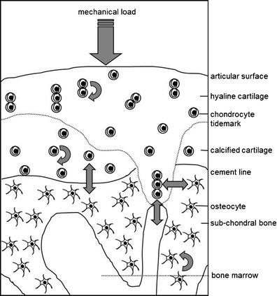
There is now general agreement that osteoarthritis (OA) involves all structures in the affected joint, culminating in the degradation of the articular cartilage. It is appropriate to focus particularly on the subchondral bone because characteristic changes occur in this tissue with disease progression, either in parallel, or contributing to, the loss of cartilage volume and quality. Changes in both the articular cartilage and the subchondral bone are mediated by the cells in these two compartments, chondrocytes and cells of the osteoblast lineage, respectively, whose primary roles are to maintain the integrity and function of these tissues. In addition, altered rates of bone remodeling across the disease process are due to increased or decreased osteoclastic bone resorption. In the altered mechanical and biochemical environment of a progressively diseased joint, the cells function differently and show a different profile of gene expression, suggesting direct effects of these external influences. There is also ex vivo and in vitro evidence of chemical crosstalk between the cells in cartilage and subchondral bone, suggesting an interdependence of events in the two compartments and therefore indirect effects of, for example, altered loading of the joint. It is ultimately these cellular changes that explain the altered morphology of the cartilage and subchondral bone. With respect to crosstalk between the cells in cartilage and bone, there is evidence that small molecules can transit between these tissues. For larger molecules, such as inflammatory mediators, this is an intriguing possibility but remains to be demonstrated. The cellular changes during the progression of OA almost certainly need to be considered in a temporal and spatial manner, since it is important when and where observations are made in either human disease or animal models of OA. Until recently, comparisons have been made with the assumption, for example, that the subchondral bone is behaviorally uniform, but this is not the case in OA, where regional differences of the bone are evident using magnetic resonance imaging (MRI). Nevertheless, an appreciation of the altered cell function during the progression of OA will identify new disease modifying targets. If, indeed, the cartilage and subchondral bone behave as an interconnected functional unit, normalization of cell behavior in one compartment may have benefits in both tissues.
Conflict of interest statement
D.M. Findlay declares that he has no conflicts of interest. G.J. Atkins declares that he has no conflicts of interest.
Figures
References
-
- Principles of osteoarthritis - its definition, character, derivation, and modality-related recognition. Croatia: InTech; 2012.
-
- Pesesse L, Sanchez C, Delcour JP, Bellahcene A, Baudouin C, Msika P, et al. Consequences of chondrocyte hypertrophy on osteoarthritic cartilage: potential effect on angiogenesis. Osteoarthritis Cartilage. 2013. - PubMed
-
- Dell'accio F, De Bari C, Eltawil NM, Vanhummelen P, Pitzalis C. Identification of the molecular response of articular cartilage to injury, by microarray screening: Wnt-16 expression and signaling after injury and in osteoarthritis. Arthritis Rheum. 2008;58:1410–21. doi: 10.1002/art.23444. - DOI - PubMed
Publication types
MeSH terms
LinkOut - more resources
Full Text Sources
Other Literature Sources
Medical
