. 2014 Jan 24;343(6169):432-7.
doi: 10.1126/science.1247606.

Adaptation of innate lymphoid cells to a micronutrient deficiency promotes type 2 barrier immunity

Affiliations

Adaptation of innate lymphoid cells to a micronutrient deficiency promotes type 2 barrier immunity

S P Spencer et al. Science. .

Abstract

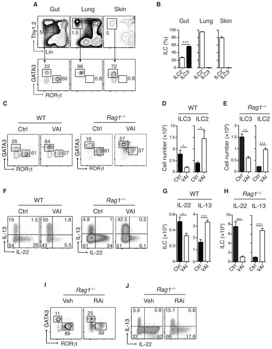
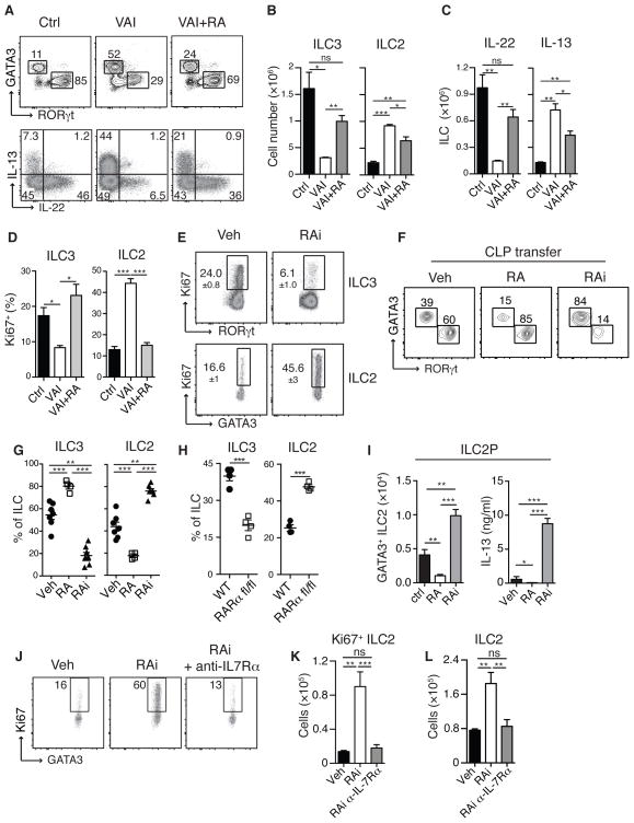
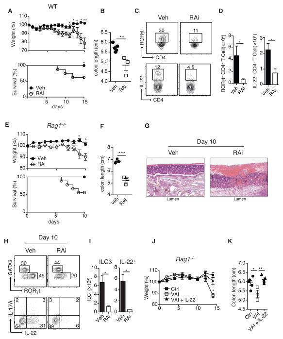
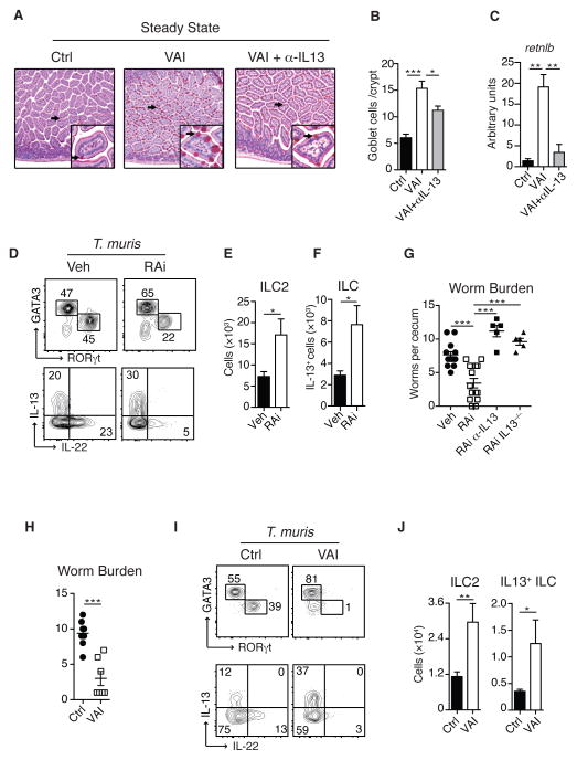
How the immune system adapts to malnutrition to sustain immunity at barrier surfaces, such as the intestine, remains unclear. Vitamin A deficiency is one of the most common micronutrient deficiencies and is associated with profound defects in adaptive immunity. Here, we found that type 3 innate lymphoid cells (ILC3s) are severely diminished in vitamin A-deficient settings, which results in compromised immunity to acute bacterial infection. However, vitamin A deprivation paradoxically resulted in dramatic expansion of interleukin-13 (IL-13)-producing ILC2s and resistance to nematode infection in mice, which revealed that ILCs are primary sensors of dietary stress. Further, these data indicate that, during malnutrition, a switch to innate type 2 immunity may represent a powerful adaptation of the immune system to promote host survival in the face of ongoing barrier challenges.


Figures

Figure 1
Figure 1. ILC3 are enriched in the GI tract and depend on retinoic acid
A) Flow cytometric analysis of cells isolated from small intestinal lamina propria (gut), lung and skin of naïve C57Bl/6 mice. Upper panel represents live CD45+ cells stained with Thy1.2 and lineage (lin) markers (NK1.1, TCRβ, TCRγδ, CD11b, CD11c, CD4, CD8a, CD8b, CD19, GR-1, DX5, Ter119). Lower panel displays cells gated on Lin and Thy1.2 expression (ILC), stained for RORgt (ILC3) and GATA3 (ILC2). B) Frequencies of ILC2 and ILC3 in gut, lung and skin. C) Small intestinal lamina propria (SiLP) cells from control (Ctrl) or vitamin A insufficient (VAI) WT or Rag1−/− mice, gated on Lin, Thy1.2+ cells and analyzed for GATA3 and RORγt expression. D–E) Total numbers of ILC3 (RORγt+) and ILC2 (GATA3+) cells in the SiLP of WT and Rag1−/− mice. F) Intracellular IL-13 and IL-22 expression in Lin Thy1.2+ cells following stimulation with PMA and ionomycin and G–H) Total numbers of IL-22 and IL-13 producing ILC in the SiLP. I) SiLP ILC2 and ILC3 from Rag1−/− mice treated with vehicle control (Veh) or retinoic acid receptor inhibitor (RAi) for 8 days and J) Intracellular IL-13 and IL-22 expression in ILC following stimulation with PMA and ionomycin. Results are representative of at least three independent experiments with 3–5 mice in each experimental group. All graphs display means ±SEM.
Figure 2
Figure 2. Retinoic acid dynamically regulates developmental balance between ILC subsets
SiLP cells from WT Ctrl and VAI or VAI Rag1−/− mice treated with all-trans retinoic acid every three days for 12 days (VAI + RA). A) GATA3 and RORγt expression in Lin, Thy1.2+ cells (upper panel). Intracellular IL-13 and IL-22 expression in Lin Thy1.2+ cells following stimulation with PMA and ionomycin (lower panel). B) Total numbers of ILC3 and ILC2 cells in SiLP and C) total numbers of IL-22 and IL-13 producing ILC in the SiLP. D) Frequencies of Ki67 expression in ILC3 and ILC2 isolated from SiLp of Ctrl, VAI or VAI +RA Rag1−/− mice. E) SiLP cells from Rag1−/− mice treated with RAi or Veh for 8 days stained for intracellular Ki67 in ILC3 (upper panel) cells and ILC2 (lower panel). F) 100,000 CD45.1+ common lymphoid progenitors (CLP) were transferred into congenic CD45.2+ Rag2−/−/gc−/− mice and treated either with vehicle (Veh), RA or RAi. 14 Days after transfer, SiLP cells were isolated, stained for GATA3 and RORγt and gated on CD45.1+, Lin and Thy1.2+ cells. G) Quantification of relative proportion of ILC3 and ILC2 in recipient mice and H) frequencies of ILC2 and ILC3 from RARafl/fl mice. Transferred cells were gated on GFP+ cells I) ILC2 progenitors (ILC2P) were sort purified from bone marrow and cultured in vitro with IL-7 and SCF in the presence of Veh, RA and RAi.for 7 days. Total numbers of Thy1.2+ GATA3+ cells (left panel) and IL-13 production in the culture supernatant (right panel). J) Ki67 expression in small intestinal ILC2 from mice treated with RAi or Veh and anti-IL7Ra. K) Total number of Ki67+ ILC2 and L) total number of ILC2. Data are representative of three (A–E, G–H) or two (I–L) independent experiments with 3–4 mice in each experimental group or at least two independent experiments with 3 experimental groups of cells isolated from 2 mice each (F). (G) displays pooled data from three independent experiments. All graphs display means ±SEM.
Figure 3
Figure 3. Vitamin A deficiency results in impaired immunity to bacterial infections
WT (A–D) and Rag1−/− (E–K) mice treated with Veh or RAi were infected with C. rodentium. A) Percentile change of original body weight and frequency of surviving animals and B) colon length of WT mice treated with RAi or Veh. C) Large intestine lamina propria (LiLP) cells isolated from Veh or RAI treated WT mice 10 days after infection with C. rodentium, gated on CD4+ and TCRb+ cells and analyzed for RORγt (upper panel) and IL-22 expression (lower panel). D) Total numbers of RORγt+ and IL-22+ CD4+ T cells. E) Percentile change of original body weight and frequency of surviving Rag1−/− mice treated with Veh or RAi and infected with C. rodentium. F) Colon length and G) representative H&E histological sections of colonic tissue analyzed 10 days post-infection. H) LILP ILC2 and ILC3 from Veh or RAI treated Rag1−/− mice 10 days after infection with C. rodentium (upper panel) and intracellular IL-17A and IL-22 expression in ILC following stimulation with PMA and ionomycin (lower panel). I) Total numbers of RORγt expressing ILC and total numbers of IL-22 producing ILC per colon. J) Weight loss of Ctrl, VAI or VAI Rag1−/− mice treated with IL-22 (VAI+IL-22) and infected with C. rodentium. K) Colon length analyzed 13 days post-infection. Data represent at least two independent experiments with 3–5 mice in each experimental group. All graphs display means ±SEM.
Figure 4
Figure 4. Vitamin A deficiency increases ILC2 mediated immunity to helminth infections
A). Small intestine histologic sections of Ctrl, VAI or VAI Rag1−/− mice treated with anti-IL13 antibody (VAI+αIL13) stained with PAS to visualize goblet cells. B) Total numbers of PAS-positive goblet cells per crypt and C) Retnlb (Relm-β) gene expression in the small intestine of Ctrl, VAI or VAI+αIL13 Rag1−/− mice. D) Lamina propria ILC2 and ILC3 isolated from the cecum of Ctrl or VAI WT mice 13 days after oral infection with T. muris (upper panel) and intracellular IL-13 and IL-22 expression in ILC after stimulation with PMA and ionomycin (lower panel). E) Total numbers of ILC2 and F) total numbers of IL-13 producing ILC in the cecum. G) Worm burden in the cecum of Veh, RAi, RAi treated IL-13−/− (RAi IL-13−/−) and RAi mice treated with neutralizing anti-IL13 antibody (RAi a-IL13) 12 days after infection. H) Number of worms in Ctrl and VAi Rag1−/− mice 12 days after infection and I) intracellular GATA3 and RORγt (upper panel) and IL-13 and IL-22 expression (lower panel) in cecal ILC. J) Total numbers of ILC2 and IL-13+ ILC in the cecum of T. muris infected mice. Data represents at least two (A–B, H–J) or three (D–G) independent experiments with 3–5 mice in each experimental group. Data in G) Veh and RAi is pooled from three experiments, RAi a-IL13 and RAi IL-13−/− represent one experiment each. All graphs display means ±SEM.

Comment in

References

    1. Ley RE, Peterson DA, Gordon JI. Ecological and evolutionary forces shaping microbial diversity in the human intestine. Cell. 2006;124:837–848. - PubMed
    1. WHO. Soil-transmitted helminth infections (factsheet) World Health Organization; 2013.
    1. Kiss EA, et al. Natural aryl hydrocarbon receptor ligands control organogenesis of intestinal lymphoid follicles. Science. 2011;334:1561–1565. - PubMed
    1. Lee JS, et al. AHR drives the development of gut ILC22 cells and postnatal lymphoid tissues via pathways dependent on and independent of Notch. Nature immunology. 2012;13:144–151. - PMC - PubMed
    1. Li Y, et al. Exogenous stimuli maintain intraepithelial lymphocytes via aryl hydrocarbon receptor activation. Cell. 2011;147:629–640. - PubMed

Publication types