Review
. 2014 Feb 24;53(9):2312-30.
doi: 10.1002/anie.201307761. Epub 2014 Jan 23.

Small-molecule control of intracellular protein levels through modulation of the ubiquitin proteasome system

Affiliations
Review

Small-molecule control of intracellular protein levels through modulation of the ubiquitin proteasome system

Dennis L Buckley et al. Angew Chem Int Ed Engl. .

Abstract

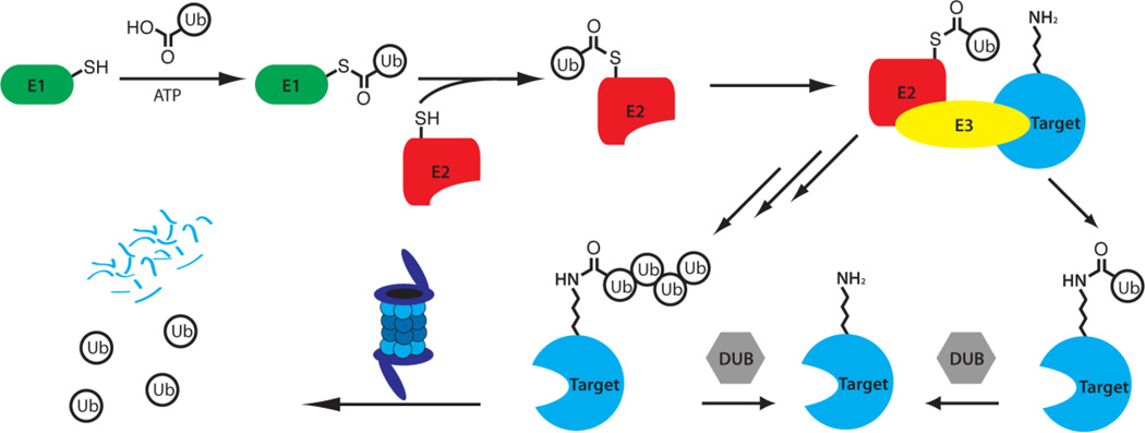
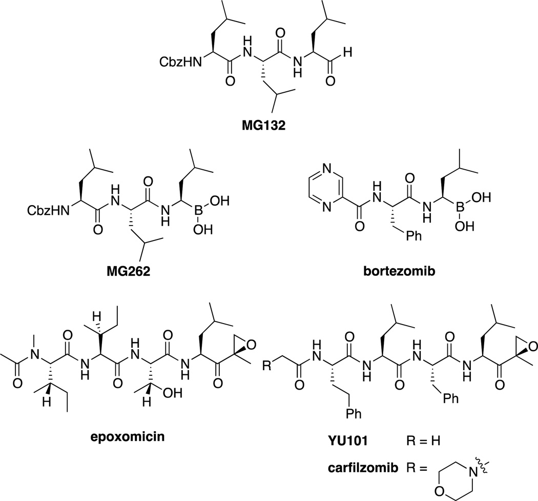
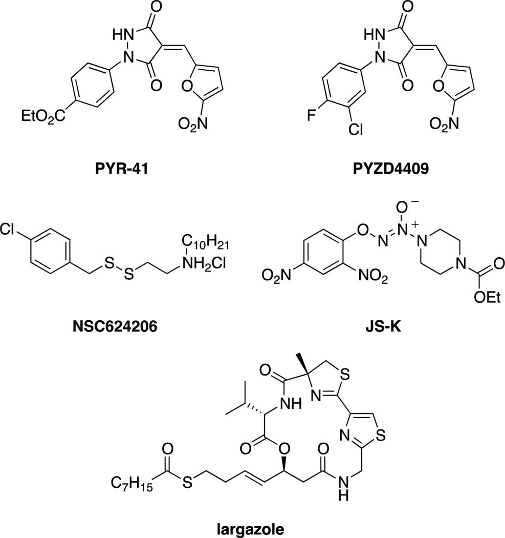
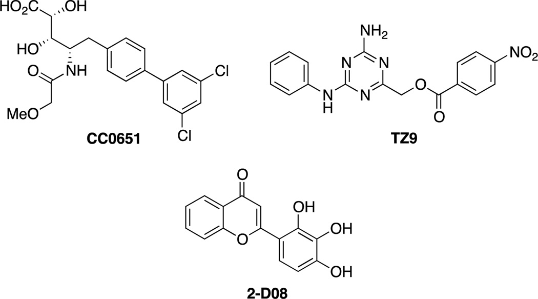
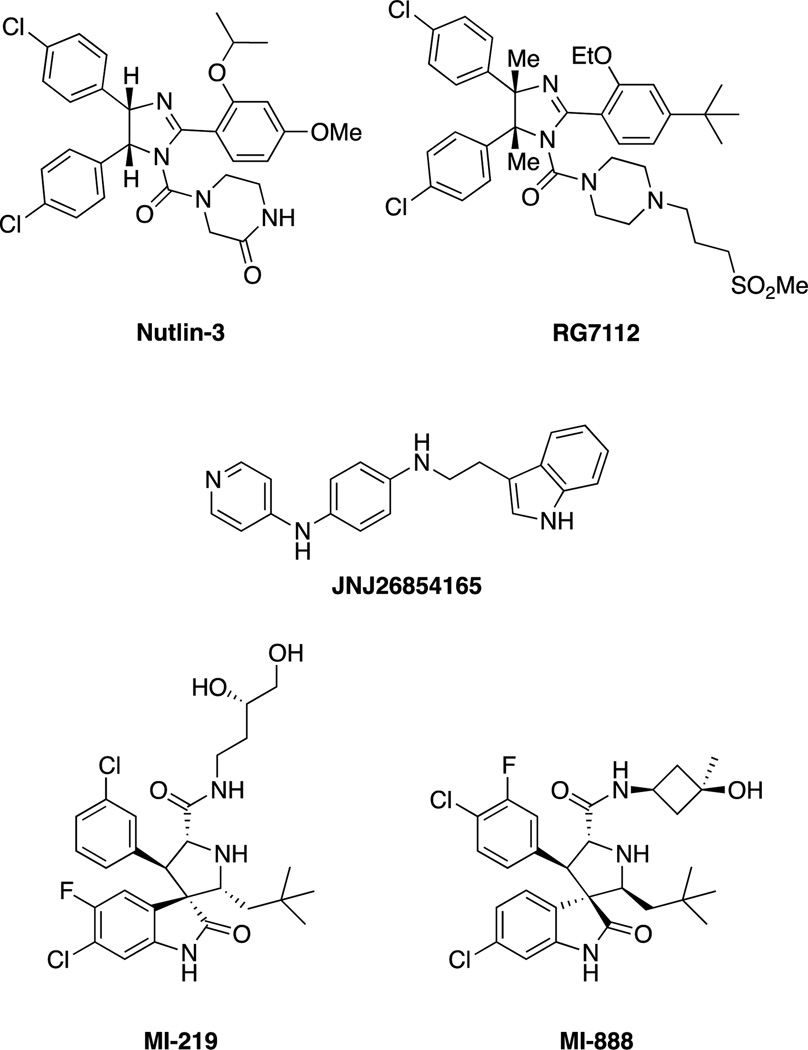
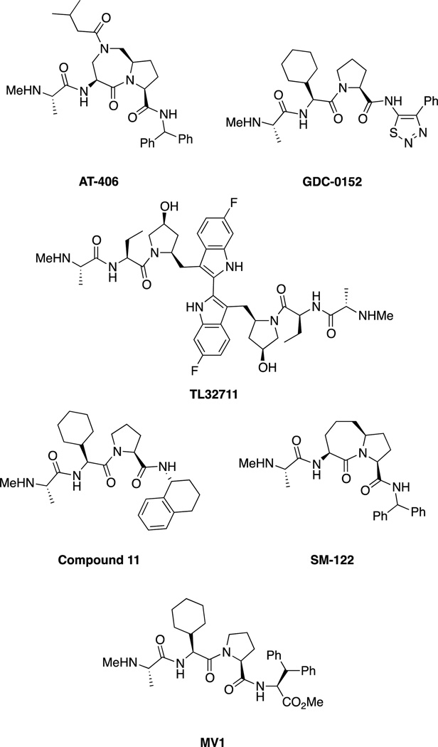
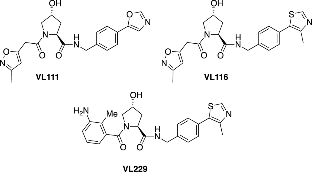
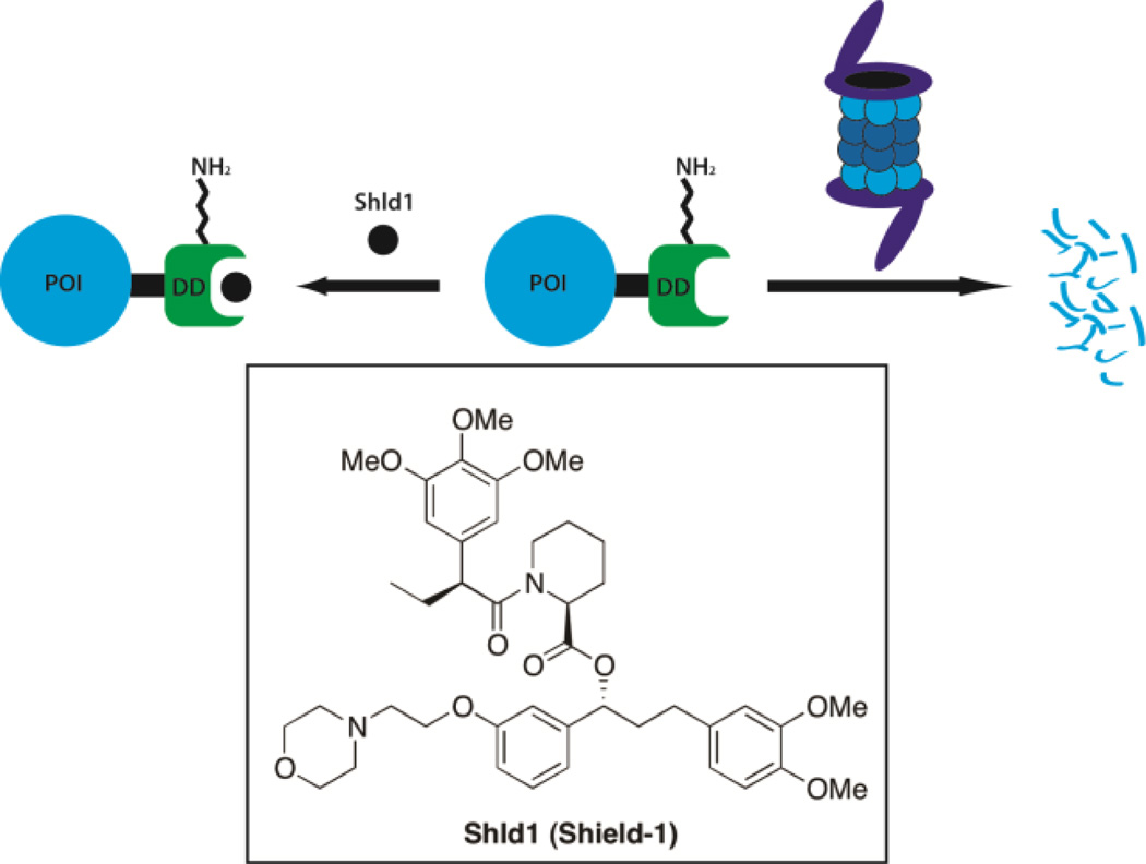
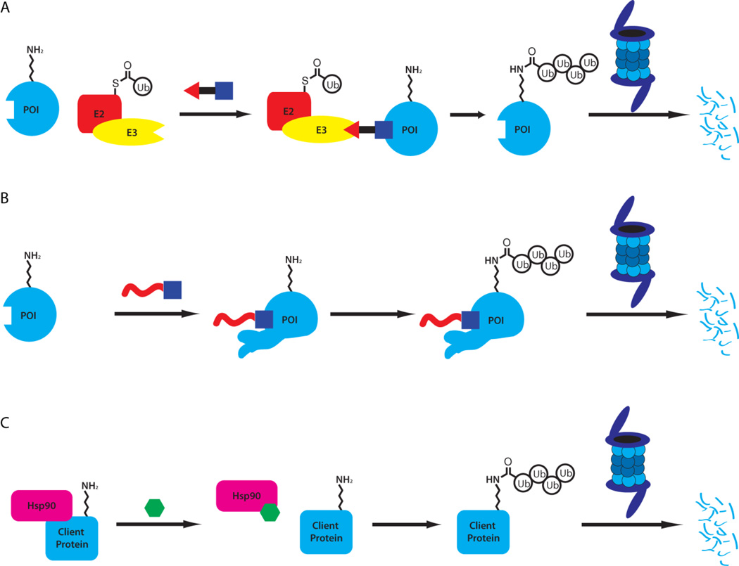
Traditionally, biological probes and drugs have targeted the activities of proteins (such as enzymes and receptors) that can be readily controlled by small molecules. The remaining majority of the proteome has been deemed "undruggable". By using small-molecule modulators of the ubiquitin proteasome, protein levels, rather than protein activity, can be targeted instead, thus increasing the number of druggable targets. Whereas targeting of the proteasome itself can lead to a global increase in protein levels, the targeting of other components of the UPS (e.g., the E3 ubiquitin ligases) can lead to an increase in protein levels in a more targeted fashion. Alternatively, multiple strategies for inducing protein degradation with small-molecule probes are emerging. With the ability to induce and inhibit the degradation of targeted proteins, small-molecule modulators of the UPS have the potential to significantly expand the druggable portion of the proteome beyond traditional targets, such as enzymes and receptors.

Keywords: drug design; inhibitors; proteasome; protein degradation; ubiquitin.


Figures

Figure 1
Figure 1
Summary of the Ubiquitin Proteasome Pathway (UPS). Ubiquitin is activated by the E1 ubiquitin-activating enzyme and transferred to an E2 ubiquitinating conjugating enzyme. The E2 then transfers the ubiquitin to a target protein with the assistance of an E3 ubiquitin ligase that recognizes the target protein. The process may then be repeated to form polyubiquitin chains, which bind to the regulatory particle of the 26S proteasome, leading to the degradation of the target protein and the recycling of the ubiquitin units.[4a, 5]
Figure 2
Figure 2
Summary of proteasome inhibitors, including the widely used probe compound MG132, as well as bortezomib/Velcade™ and carfilzomib/Kyprolis™, FDA approved drugs for the treatment of multiple myeloma.
Figure 3
Figure 3
Small molecule inhibitors of E1 ubiquitin-activating enzyme.
Figure 4
Figure 4
Small molecule inhibitors of E2 ligases. CC0651 inhibits hCdc34;[46a] TZ9 inhibits Rad6;[47] 2-D08 inhibits the SUMO E2, UBc-9.[48]
Figure 5
Figure 5
Nutlin, the first MDM2 inhibitor and other selected MDM2 inhibitors.
Figure 6
Figure 6
Summary of IAP inhibitors including AT-406 (developed by Ascenta Therapeutics and the University of Michigan),[62] which is administered orally in Phase 1 trials for solid tumors and lymphoma, Genentech/Roche’s GDC-0152 which is administered intravenously and is in Phase I trials for metastatic malignancies,[6b, 63] and the bivalent TL32711 (administered intravenously) developed by Tetralogics Pharma.[6b, 58] LCL161 (Novartis), AEG35156 and AEG40826 (Aegera), and YM155 (Astellas Pharma) are also in clinical trials but are not shown.[6b, 58] Selected SMAC mimics such as SM-122 and MV1 are also shown but are not in clinical trials.
Figure 7
Figure 7
A) Depiction of the SCF E3 ligase complex. The complex contains a cullin (which is NEDDylated when active) which binds a RING domain as well as the adaptor Skp1. Skp1 also binds various F box proteins (such asβTrCP, Cdc4 and Skp2) which function as the substrate interaction motif, binding target proteins which are ubiquitinated. B) Inhibitors of SCF E3 ligases. The specific E3 inhibited is shown in parentheses.
Figure 8
Figure 8
Structures of hydroxyproline-based molecules capable of inhibiting the interaction between VHL and a peptide derived from HIF in vitro.
Figure 9
Figure 9
Shld1 is used to stabilize mutant FKBP proteins (as well as FKBP fusion proteins).[74]
Figure 10
Figure 10
Strategies for induced protein degradation include direct recruitment of an E3 ligase with A) PROTACs, B) induced protein misfolding (or mimicking misfolding) with hydrophobic tags (or ligand-mediated degradation) and C) the inhibition of chaperones such as Hsp90.
Figure 11
Figure 11
PROTACs are heterobifunctional molecules that combine an E3 ligase ligand (shown on the right) with ligands for various proteins of interest (shown on the left). This recruits the E3 ligase to the protein of interest, leading to ubiquitination and degradation. Peptidic ligands have been used to target E3 ligases SCFβTrCP and VHL; small molecule ligands have been used to target MDM2 and cIAP1.
Figure 12
Figure 12
Structures of HyT13 and HyT36 and their ability to degrade HaloTag-GFP fusion proteins at 10 μM.[101]
Figure 13
Figure 13
Structures of Boc3Arg containing degraders of GST and eDHFR.
Figure 14
Figure 14
Despite being designed as traditional antagonists or inhibitors, fulvestrant and CI-1033 were discovered to induce the degradation of the ER and ErbB2 respectively.
Figure 15
Figure 15
Geldanamycin and analogs.
Figure 16
Figure 16
Selected synthetic Hsp90 inhibitors: PU3,[115] PU-H71[116], NVP-AUY922[117] and SNX-5422[118].
Figure 17
Figure 17
Inhibitors of DUBs. The specific DUB inhibited is shown in parentheses.
Figure 18
Figure 18
Inhibitors of 19S regulatory particle associated DUBs. IU1 inhibits USP14 while b-AP15 inhibits USP14 and UCHL5.
Scheme 1
Scheme 1
Proposed mechanism for the inactivation of the proteasome by epoxomicin. Initial hemiketal formation is followed by epoxide opening by the terminal amine, to form a stable morpholine adduct, which has been observed through x-ray crystallography.[29]
Scheme 2
Scheme 2
MLN4924 is able to react covalently with the activated NEDD8-NAE intermediate to form a non-hydrolysable covalent bond with NEDD8. The MLN4924-NEDD8 adduct then acts as a mimic of the adenylated-NEDD8 substrate, competitively inhibiting NAE.[44]

Similar articles

Cited by

References

    1. Raina K, Crews CM. J. Biol. Chem. 2010;285:11057–11060. - PMC - PubMed
    2. Schneekloth JS, Jr, Crews CM. ChemBioChem. 2005;6:40–46. - PMC - PubMed
    1. Crews CM. Chem. Biol. 2010;17:551–555. - PMC - PubMed
    1. Arkin MR, Wells JA. Nat. Rev. Drug Discov. 2004;3:301–317. - PubMed
    2. Wells JA, Mcclendon CL. Nature. 2007;450:1001–1009. - PubMed
    1. Hershko A. Angew. Chem. Int. Ed. 2005;44:5932–5943. - PubMed
    2. Hershko A, Ciechanover A. Annu. Rev. Biochem. 1992;61:761–807. - PubMed
    1. Kravtsova-Ivantsiv Y, Sommer T, Ciechanover A. Angew. Chem. Int. Ed. 2013;52:192–198. - PubMed

MeSH terms