. 2014 Mar 7;289(10):6404-6414.
doi: 10.1074/jbc.M113.501205. Epub 2014 Jan 23.

Polycystin-1 negatively regulates Polycystin-2 expression via the aggresome/autophagosome pathway

Affiliations

Polycystin-1 negatively regulates Polycystin-2 expression via the aggresome/autophagosome pathway

Valeriu Cebotaru et al. J Biol Chem. .

Abstract

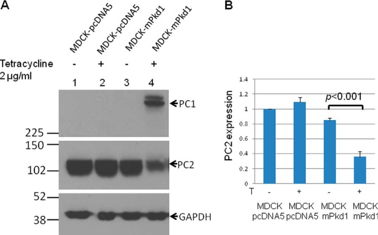
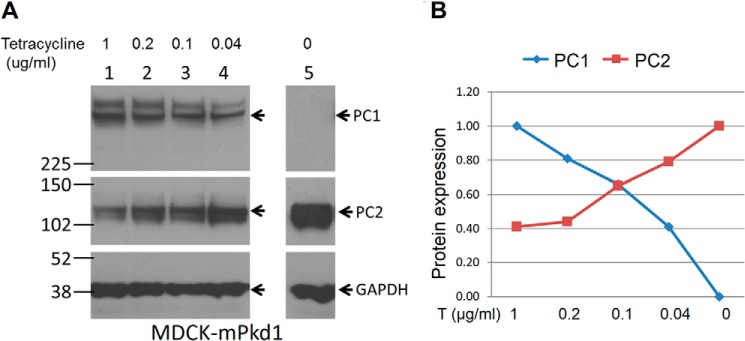
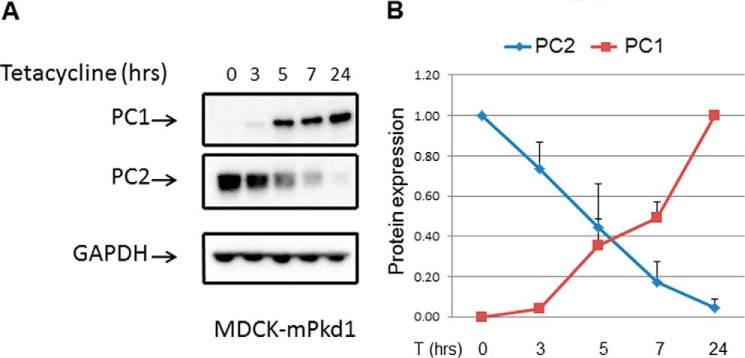
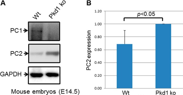
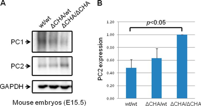
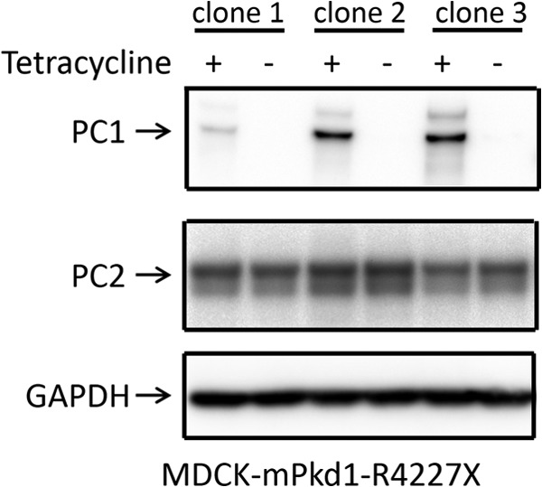
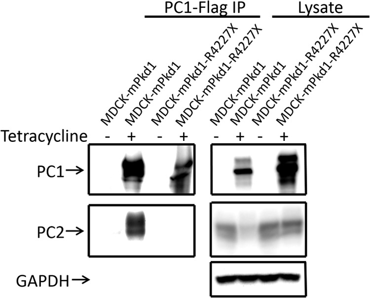
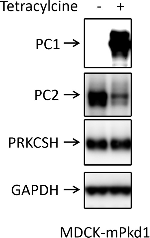
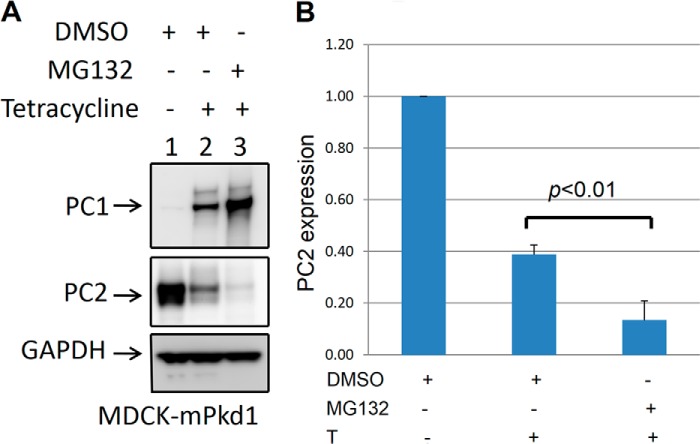
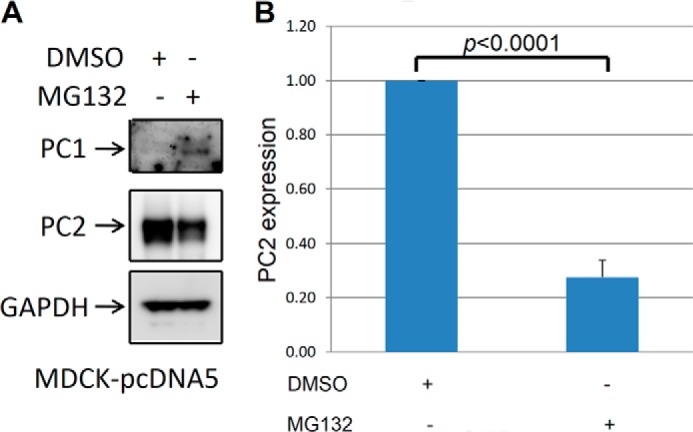
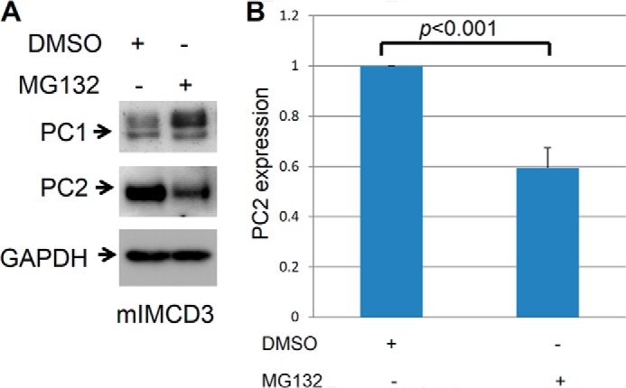
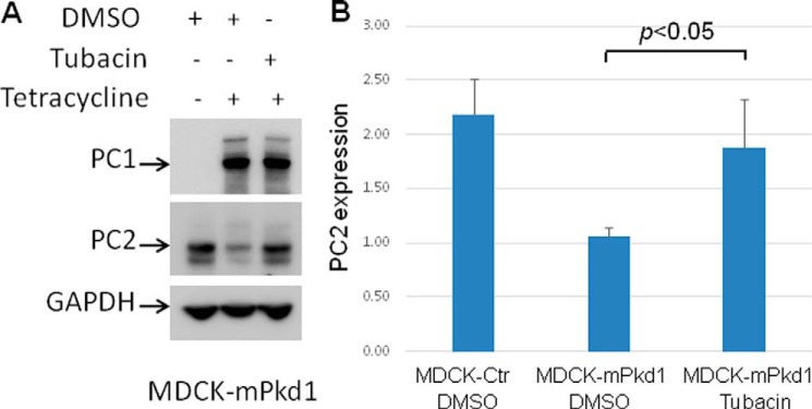
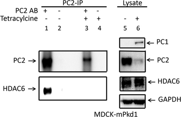
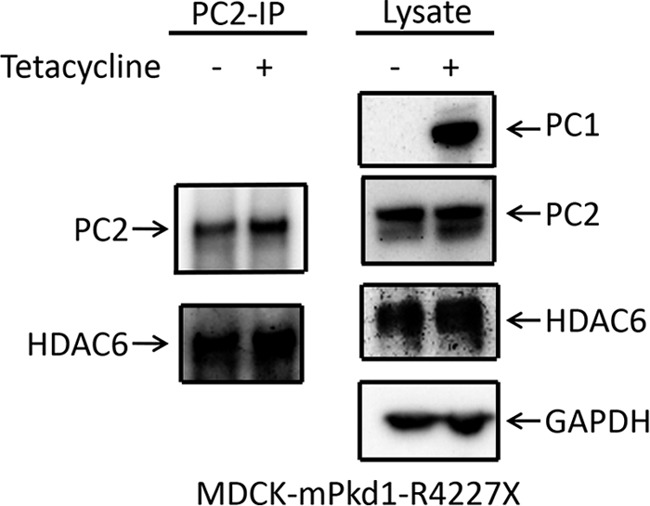
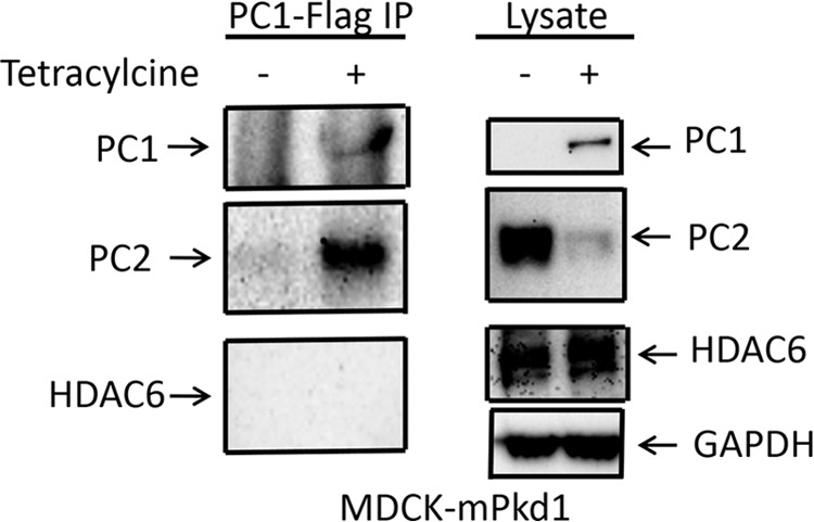
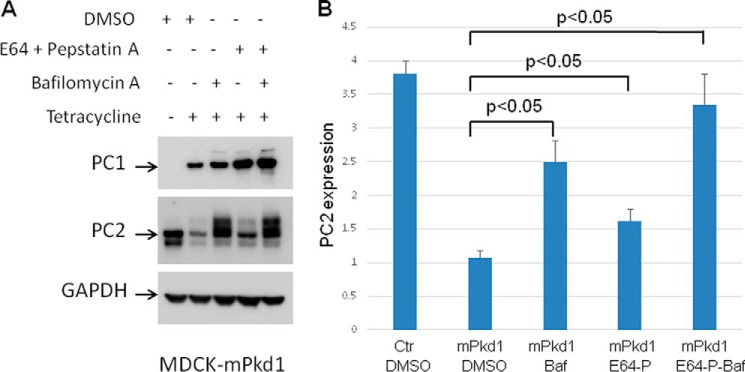
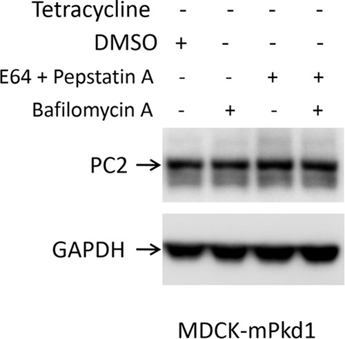
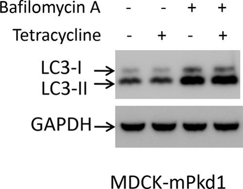
Mutations of the PKD1 and PKD2 genes, encoding polycystin-1 (PC1) and polycystin-2 (PC2), respectively, lead to autosomal dominant polycystic kidney disease. Interestingly, up-regulation or down-regulation of PKD1 or PKD2 leads to polycystic kidney disease in animal models, but their interrelations are not completely understood. We show here that full-length PC1 that interacts with PC2 via a C-terminal coiled-coil domain regulates PC2 expression in vivo and in vitro by down-regulating PC2 expression in a dose-dependent manner. Expression of the pathogenic mutant R4227X, which lacks the C-terminal coiled-coil domain, failed to down-regulate PC2 expression, suggesting that PC1-PC2 interaction is necessary for PC2 regulation. The proteasome and autophagy are two pathways that control protein degradation. Proteins that are not degraded by proteasomes precipitate in the cytoplasm and are transported via histone deacetylase 6 (HDAC6) toward the aggresomes. We found that HDAC6 binds to PC2 and that expression of full-length PC1 accelerates the transport of the HDAC6-PC2 complex toward aggresomes, whereas expression of the R4227X mutant fails to do so. Aggresomes are engulfed by autophagosomes, which then fuse with the lysosome for degradation; this process is also known as autophagy. We have now shown that PC1 overexpression leads to increased degradation of PC2 via autophagy. Interestingly, PC1 does not activate autophagy generally. Thus, we have now uncovered a new pathway suggesting that when PC1 is expressed, PC2 that is not bound to PC1 is directed to aggresomes and subsequently degraded via autophagy, a control mechanism that may play a role in autosomal dominant polycystic kidney disease pathogenesis.

Keywords: Calcium Channels; Physiology; Protein Degradation; Protein Processing; Trafficking.


Figures

FIGURE 1.
FIGURE 1.
PC1 down-regulates PC2 expression. Full-length mPkd1 was stably transfected into MDCK cells using a Flp-In T-REx core kit (Invitrogen). mPkd1 has an N-terminal Halo Tag and a C-terminal FLAG tag. The transfected MDCK cells were grown on Transwell plates for 72 h, and PC1 expression was induced with tetracycline (2 μg/ml) for 24 h. Lanes 1 and 2 are Western blot lysates from MDCK cells stably transfected with an empty plasmid. Lanes 3 and 4 are Western blot lysates from MDCK cells stably transfected with an mPkd1 inducible plasmid. A, PC1 expression induced by tetracycline (top panel, lane 4) led to down-regulation of PC2 (middle panel, lane 4). This experiment was repeated more than 10 times. It is noteworthy that when the blot is exposed for long time, PC1 presents as a double band. Qian et al. (38) showed that PC1 undergoes cleavage at the G protein-coupled receptor proteolytic site into an N-terminal fragment and C-terminal fragment. PC1 antibodies that we used were made against the N-terminal fragment, and hence the top band is full-length PC1, and the bottom band is the N-terminal fragment. B, summary of PC2 expression. We used MultiGauge software to measure density of PC2 bands, shown as averages of three experiments and standard deviation. In each experiment the most intense band was considered 100%, and the rest of the bands were compared with this band. PC1 and PC2 bands were normalized to GAPDH. We used Student's t test to compare PC2 expression in MDCK-mPkd1 cells when PC1 is uninduced versus induced.
FIGURE 2.
FIGURE 2.
PC1 down-regulates PC2 expression in a dose-dependent manner. A, the tetracycline dose was gradually decreased to decrease PC1 expression. We found that PC1 down-regulates PC2 expression in a dose-dependent manner: the higher the PC1 expression, the lower the PC2 expression. B, summary of PC2 expression after PC1 titration.
FIGURE 3.
FIGURE 3.
PC1 expression was induced with tetracycline (2 μg/ml) for different time points. A, PC1 was induced at the same time in a set of MDCK cells, and proteins were harvested at different time points (3, 5, 7, and 24 h). This experiment again demonstrated that PC2 down-regulation by PC1 is dose-dependent. B, summary of PC2 expression when PC1 is titrated.
FIGURE 4.
FIGURE 4.
PC1 regulates PC2 expression in vivo. A, PC2 expression was decreased in wild-type mouse embryos (Wt, 14.5 days after conception) when compared with Pkd1 knock-out embryos (ko). This is a Western blot, and the entire experiment was repeated three times. B, summary of PC2 expression in wild-type and Pkd1 knock-out embryos.
FIGURE 5.
FIGURE 5.
PC1 regulates PC2 expression in vivo. A, PC2 expression was decreased in wild-type mouse embryos (15.5 days after conception) when compared with Pkd1ΔCHACHA embryos (17). This is a Western blot, and the entire experiment was repeated three times. Pkd1ΔCHACHA lacks the C-terminal tail of PC1 and has a HA tag added to the truncated C-terminal end of PC1. Pkd1ΔCHACHA knock-out is embryonically lethal, similar to Pkd1 knock-out. B, summary of PC2 expression in Pkd1wt/wt, Pkd1ΔCHA/wt, and Pkd1ΔCHACHA embryos.
FIGURE 6.
FIGURE 6.
The mPkd1-R4227X mutant does not affect PC2 expression. To determine whether an interaction between PC1 and PC2 is necessary for the regulation of PC2 expression, we generated a plasmid that includes a truncated mouse Pkd1 (mPkd1-R4227X) lacking the last 76 amino acids, which cannot interact with PC2. Like the full-length mPkd1, this mPkd1-R4227X had an N-terminal HaloTag and a C-terminal FLAG tag. The mPkd1-R4227X plasmid was stably transfected into MDCK cells using a Flp-In T-REx core kit. MDCK cells were grown for 72 h, and expression of full-length mPkd1 and mPkd1-R4227X was induced with tetracycline (2 μg/ml) for 24 h. Overexpression of the mPkd1-R4227X mutant in MDCK cells (top panel) had no effect on PC2 expression (middle panel). Note that there are three different clones. This is a Western blot.
FIGURE 7.
FIGURE 7.
The mPkd1-R4227X mutant does not interact with PC2. We tested the mutant in a co-immunoprecipitation assay to confirm that the mPkd1-R4227X protein does not interact with PC2. Full-length mPkd1 was used as a positive control for the co-immunoprecipitation of PC2. This a Western blot, and the entire experiment was repeated three times.
FIGURE 8.
FIGURE 8.
PC1 expression does not affect PRKCSH expression. Tetracycline treatment induced PC1 expression in MDCK cells but had no effect on PRKCSH expression. This is a Western blot, and the entire experiment was repeated three times.
FIGURE 9.
FIGURE 9.
The proteasome inhibitor MG132 increases the expression of recombinant PC1 and leads to further down-regulation of PC2 expression in MDCK cells. For this experiment, we used MDCK cells that were stably transfected with an inducible mPkd1 plasmid (see details in Fig. 1 legend). Cells were grown on Transwell plates for 72 h, and PC1 expression was induced with tetracycline (2 μg/ml) for 72 h. Cells were treated with MG132 (1 μm/ml) for 16 h before harvesting. Control cells were treated with vehicle (DMSO). A, treatment with MG-132 led to increased PC1 expression and further down-regulation of PC2. This is a Western blot, and the entire experiment was repeated more than five times. B, summary of PC2 expression after PC1 induction and proteasome treatment in MDCK cells.
FIGURE 10.
FIGURE 10.
The proteasome inhibitor MG132 increases endogenous PC1 expression and decreases PC2 expression in MDCK cells. A, MDCK cells stably transfected with an empty plasmid were treated with MG132 (1 μm/ml) for 16 h before harvesting, and control cells were treated with vehicle (DMSO). Endogenous PC1 was undetectable in MDCK cells. Treatment with MG-132 led to an up-regulation of endogenous PC1 to a detectable level and down-regulation of PC2. This is a Western blot, and the experiment was repeated three times. B, summary of PC2 expression after proteasome treatment in MDCK cells.
FIGURE 11.
FIGURE 11.
MG132 increases endogenous PC1 expression and decreases PC2 expression in mIMCD-3 cells. A, mIMCD-3 cells were treated with MG132 (1 μm/ml) for 16 h. Control cells were treated with vehicle (DMSO). Treatment with MG-132 led to an up-regulation of endogenous PC1 and down-regulation of PC2. This is a Western blot, and the entire experiment was repeated three times. B, summary of PC2 expression after proteasome treatment in mIMCD3 cells.
FIGURE 12.
FIGURE 12.
Inhibition of the aggresome degradation pathway with tubacin prevents down-regulation of PC2 when PC1 is expressed. A, tubacin, a specific HDAC6 inhibitor that blocks the aggresome degradation pathway, prevented the down-regulation of PC2 in PC1-induced MDCK cells. PC1 expression was induced with tetracycline (2 μg/ml) for 7 h. Cells were treated with tubacin (10 μm) for 7 h; control cells were treated with DMSO. Tubacin had no effect on PC1 expression. This is a Western blot, and the experiment was repeated three times. B, summary data for the tubacin experiment.
FIGURE 13.
FIGURE 13.
Inhibition of the aggresome degradation pathway with tubacin has no effect on PC2 expression in control cells. PC2 expression remained unchanged after tubacin treatment in MDCK control cells (PC1 uninduced). Cells were treated with tubacin (10 μm) for 7 h; control cells were treated with DMSO. This is a Western blot.
FIGURE 14.
FIGURE 14.
PC2 co-immunoprecipitates with HDAC6. When PC1 was not induced in MDCK cells, PC2 co-immunoprecipitated with HDAC6 (left panels). When PC1 is induced in MDCK cells, co-immunoprecipitation of PC2 with HDAC6 was greatly reduced (left panels). PC1 overexpression had no effect on the HDAC6 expression (right panels). This is a Western blot, and the experiment was repeated three times. IP, immunoprecipitation.
FIGURE 15.
FIGURE 15.
Expression of mPkd1-R4227X does not affect the PC2-HDAC6 interaction. In the presence or absence of mPkd1-R4227X, PC2 was co-immunoprecipitated (IP) with HDAC6 in MDCK cells (left panels). Overexpression of mPkd1-R4227X had no effect on HDAC6 expression (right panels). This is a Western blot, and the experiment was repeated three times.
FIGURE 16.
FIGURE 16.
PC1 does not interact with HDAC6. We tested whether PC1 interacts with HDAC6. An anti-FLAG M2 affinity gel was used for PC1 immunoprecipitation (IP). PC1 did not co-immunoprecipitate HDAC6 (left panels). For a positive control, we showed that PC1 co-immunoprecipitated PC2 (left panels). The lysate is shown in the right panels.
FIGURE 17.
FIGURE 17.
Inhibition of the lysosome degradation pathway prevents PC1-driven degradation of PC2. A, inhibition of autophagy with bafilomycin A (50 nm) or E64 (50 μm) with pepstatin A (2.5 μm) decreased the degradation of PC2 when PC1 was overexpressed in MDCK cells. Inhibition of autophagy with both bafilomycin A (50 nm) and E64 (50 μm) with pepstatin A had a synergistic effect and led to a further decrease in PC2 degradation when PC1 was overexpressed in MDCK cells. MDCK cells were treated for 16 h. Control cells were treated with vehicle (DMSO). PC1 expression was induced with tetracycline (2 μg/ml) for 16 h. This is a Western blot, and the experiment was repeated three times. B, summary of the inhibition of the lysosome degradation pathway. Baf, bafilomycin A; Ctr, control; P, pepstatin A.
FIGURE 18.
FIGURE 18.
Inhibition of the lysosome degradation pathway has no effect on PC2 expression in control cells. In MDCK control cells (PC1 uninduced), PC2 expression remained unchanged after treatment with bafilomycin A (50 nm), with E64 (50 μm) and pepstatin A (2.5 μm), or with both. Cells were treated for 16 h. Control cells were treated with vehicle (DMSO). This is a Western blot, and the experiment was repeated three times.
FIGURE 19.
FIGURE 19.
Overexpression of PC1 does not activate autophagy. Overexpression of PC1 in MDCK cells did not increase the conversion of LC3-I to LC3-II. Inhibition of autophagy with bafilomycin A led to increased conversion of LC3-I to LC3-II, indicating accumulation of autophagosomes. Cells were treated with bafilomycin A (50 nm) for 16 h. PC1 expression was induced with tetracycline (2 μg/ml) for 16 h. Control cells were treated with vehicle (DMSO). This is a Western blot, and the experiment was repeated three times.

Similar articles

Cited by

References

    1. Hughes J., Ward C. J., Peral B., Aspinwall R., Clark K., San Millán J. L., Gamble V., Harris P. C. (1995) The polycystic kidney disease 1 (PKD1) gene encodes a novel protein with multiple cell recognition domains. Nat. Genet. 10, 151–160 - PubMed
    1. Nims N., Vassmer D., Maser R. L. (2003) Transmembrane domain analysis of polycystin-1, the product of the polycystic kidney disease-1 (PKD1) gene. Evidence for 11 membrane-spanning domains. Biochemistry 42, 13035–13048 - PubMed
    1. Mochizuki T., Wu G., Hayashi T., Xenophontos S. L., Veldhuisen B., Saris J. J., Reynolds D. M., Cai Y., Gabow P. A., Pierides A., Kimberling W. J., Breuning M. H., Deltas C. C., Peters D. J., Somlo S. (1996) PKD2, a gene for polycystic kidney disease that encodes an integral membrane protein. Science 272, 1339–1342 - PubMed
    1. Qian F., Germino F. J., Cai Y., Zhang X., Somlo S., Germino G. G. (1997) PKD1 interacts with PKD2 through a probable coiled-coil domain. Nat. Genet. 16, 179–183 - PubMed
    1. Hanaoka K., Qian F., Boletta A., Bhunia A. K., Piontek K., Tsiokas L., Sukhatme V. P., Guggino W. B., Germino G. G. (2000) Co-assembly of polycystin-1 and -2 produces unique cation-permeable currents. Nature 408, 990–994 - PubMed

Publication types

Substances

LinkOut - more resources