. 2014 Mar 7;289(10):7025-7037.
doi: 10.1074/jbc.M114.548990. Epub 2014 Jan 23.

Calcium channel α2δ1 proteins mediate trigeminal neuropathic pain states associated with aberrant excitatory synaptogenesis

Affiliations

Calcium channel α2δ1 proteins mediate trigeminal neuropathic pain states associated with aberrant excitatory synaptogenesis

Kang-Wu Li et al. J Biol Chem. .

Abstract

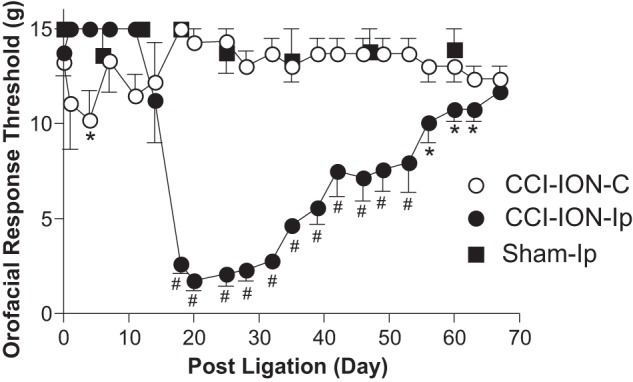
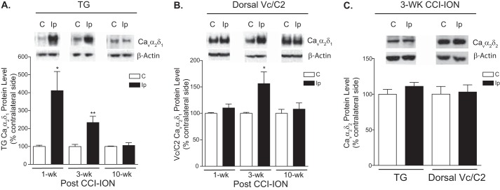
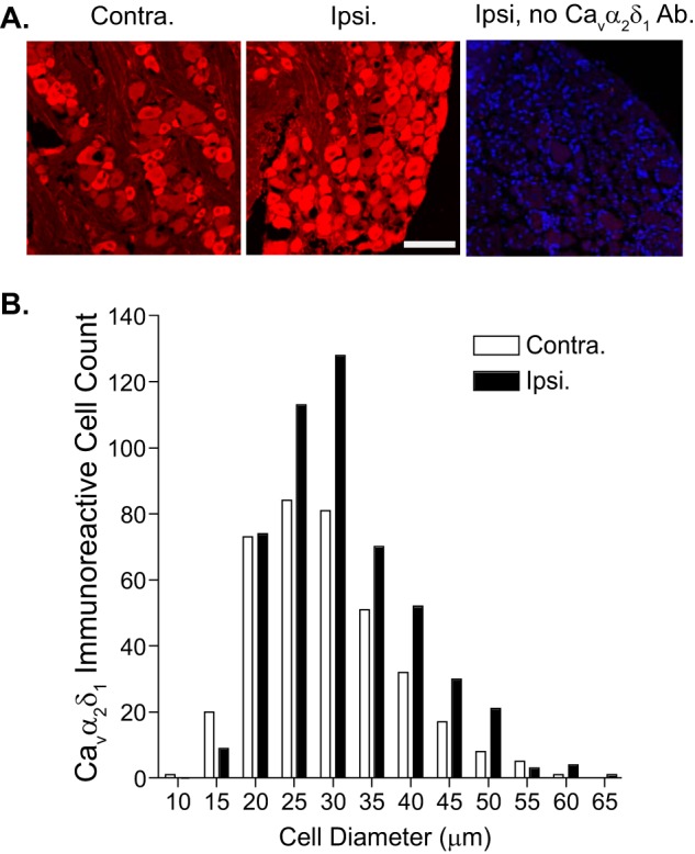
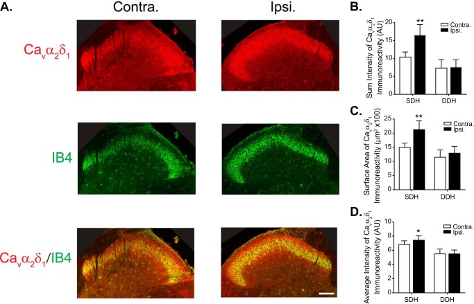
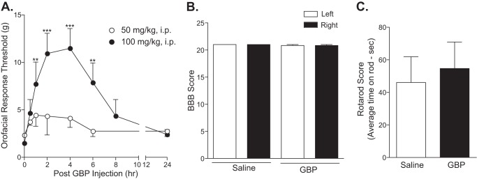
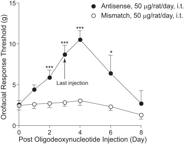
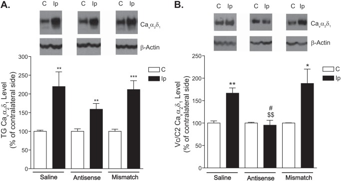
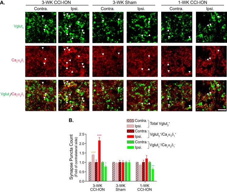
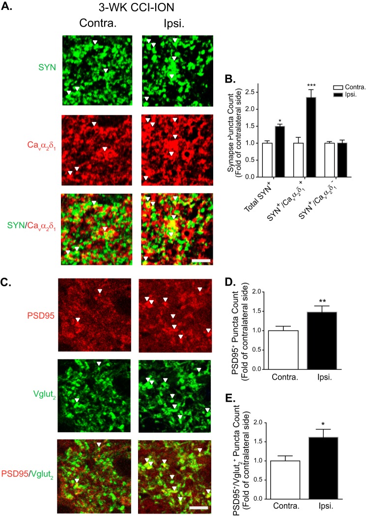
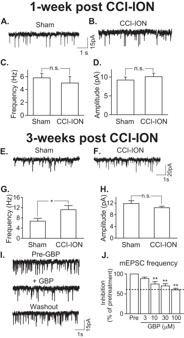
To investigate a potential mechanism underlying trigeminal nerve injury-induced orofacial hypersensitivity, we used a rat model of chronic constriction injury to the infraorbital nerve (CCI-ION) to study whether CCI-ION caused calcium channel α2δ1 (Cavα2δ1) protein dysregulation in trigeminal ganglia and associated spinal subnucleus caudalis and C1/C2 cervical dorsal spinal cord (Vc/C2). Furthermore, we studied whether this neuroplasticity contributed to spinal neuron sensitization and neuropathic pain states. CCI-ION caused orofacial hypersensitivity that correlated with Cavα2δ1 up-regulation in trigeminal ganglion neurons and Vc/C2. Blocking Cavα2δ1 with gabapentin, a ligand for the Cavα2δ1 proteins, or Cavα2δ1 antisense oligodeoxynucleotides led to a reversal of orofacial hypersensitivity, supporting an important role of Cavα2δ1 in orofacial pain processing. Importantly, increased Cavα2δ1 in Vc/C2 superficial dorsal horn was associated with increased excitatory synaptogenesis and increased frequency, but not the amplitude, of miniature excitatory postsynaptic currents in dorsal horn neurons that could be blocked by gabapentin. Thus, CCI-ION-induced Cavα2δ1 up-regulation may contribute to orofacial neuropathic pain states through abnormal excitatory synapse formation and enhanced presynaptic excitatory neurotransmitter release in Vc/C2.

Keywords: Gene Regulation; Molecular Pharmacology; Neuroscience; Pain; Synaptic Plasticity.


Figures

FIGURE 1.
FIGURE 1.
CCI-ION led to development of orofacial tactile allodynia in adult male rats. Orofacial sensitivity to von Frey filament stimulation was tested in the whisker pad area of rats before and at the designated times after CCI-ION or sham operations. Data presented are the means ± S.E. (error bars) from five to seven rats in each group. C, contralateral to injury; Ip, ipsilateral to injury. *, p < 0.05; #, p < 0.001 compared with preinjury level by two-way analysis of variance with Bonferroni post-tests.
FIGURE 2.
FIGURE 2.
CCI-ION led to increased expression of Cavα2δ1 proteins in TG and Vc/C2. Western blots were used to examine Cavα2δ1 protein levels in TG (A) and dorsal Vc/C2 (B) samples at the designated time points post-CCI-ION as well as Cavα2δ2 protein levels in these samples 3 weeks (wk) post-CCI-ION (C). Representative Western blot data from TG or dorsal Vc/C2 are shown on top of each bar graph summarizing respective Western blot data. For normalizing sample loading, ratios of Cavα2δ over β-actin band densities were taken within each sample group before comparisons were made between the injury side and non-injury side. Data presented are the means ± S.E. (error bars) from four to six rats. *, p < 0.05; **, p < 0.01 compared with the non-injury side by Student's t test. C, contralateral to injury; Ip, ipsilateral to injury.
FIGURE 3.
FIGURE 3.
Localization of CCI-ION-induced Cavα2δ1 immunoreactivity in TG. Immunostaining was performed in TG samples collected from 3-week CCI-ION rats with orofacial hypersensitivity in the injury side. A, representative Cavα2δ1 immunoreactive profiles (red) in injury (Ipsi.) and non-injury (Contra.) sides of TG. The right panel shows control staining with omission of the primary antibody (Ab.) in a section from the injury side. Blue, DAPI staining of nuclei. Scale bar, 100 μm for all image panels. B, summarized data quantifying the numbers of Cavα2δ1-immunoreactive profiles of different sizes of neurons from three rats.
FIGURE 4.
FIGURE 4.
Localization of CCI-ION-induced Cavα2δ1 immunoreactivity in Vc/C2. Immunostaining was performed in Vc/C2 samples collected from 3-week CCI-ION rats with orofacial hypersensitivity in the injury side. A, representative Cavα2δ1 immunoreactivity (red) in injury (Ipsi.) and non-injury sides (Contra.) of Vc/C2. IB4 reactivity specific to α-d-galactose-terminal glycoconjugates expressed on the cell surface of a subpopulation of non-peptidergic neuronal terminals (33) (green) is shown for anatomical localization of superficial dorsal horn. Scale bar, 140 μm for all image panels. B, summarized data of total intensity of Cavα2δ1 immunoreactivity in superficial (SDH) and deep (DDH) dorsal horn of Vc/C2 samples (AU, artificial units). C, summarized data of the surface area of Cavα2δ1 immunoreactivity in superficial and deep dorsal horn of Vc/C2 samples. D, summarized data of the average intensity of Cavα2δ1 immunoreactivity in superficial and deep dorsal horn of Vc/C2 samples. Data presented are the means ± S.E. (error bars) from nine images in each side from three rats. *, p < 0.05; **, p < 0.01 compared with non-injury side by Student's t test. Contra., contralateral to injury; Ipsi., ipsilateral to injury.
FIGURE 5.
FIGURE 5.
Injury-induced orofacial allodynia could be blocked by gabapentin. Rats with established orofacial allodynia after 3-week CCI-ION were given a bolus intraperitoneal (i.p.) injection of gabapentin with the doses indicated. Behavioral testing was performed in the injury side before the injection and at the indicated time points after the injection (A). Data presented are the means ± S.E. (error bars) from three to four rats. **, p < 0.01; ***, p < 0.001 compared with the pretreatment level by two-way analysis of variance with Bonferroni post-tests. To test whether gabapentin treatment impaired locomotor functions, Basso, Beattie, Bresnahan Locomotor Rating Scale (BBB) (B) and rotarod (C) tests were performed before and 4 h after intraperitoneal gabapentin (100 mg/kg) injection, a time point correlated with maximal reversal of orofacial allodynia in gabapentin-treated CCI-ION rats. Data presented are the means ± S.E. (error bars) from six rats in each group. GBP, gabapentin.
FIGURE 6.
FIGURE 6.
Injury-induced orofacial allodynia could be blocked by intrathecal treatment with Cavα2δ1 antisense, but not mismatched, oligodeoxynucleotides. Rats with established orofacial allodynia after 3-week CCI-ION were given a daily intrathecal (i.t.) injection of Cavα2δ1 antisense (AS) or mismatched (MM) oligodeoxynucleotides (50 μg/rat/day) for 4 consecutive days. Behavioral tests were performed daily in the injury side before the injection and at the designated time points after the last injection. Data presented are the means ± S.E. (error bars) from 12–14 rats up to day 4 after treatment initiation and from three to six rats afterward. *, p < 0.05; ***, p < 0.001 compared with the pretreatment level by two-way analysis of variance with Bonferroni post-tests.
FIGURE 7.
FIGURE 7.
Cavα2δ1 protein levels in TG and Vc/C2 after treatment with antisense and mismatched oligodeoxynucleotides. Cavα2δ1 protein levels in TG and dorsal Vc/C2 samples collected 1 day after the last injection of the 4-day treatment were subjected to Western blot analyses. Representative Western blot data from TG (A) or Vc/C2 (B) are shown on top of each bar graph summarizing respective Western blot data. For normalizing sample loading, ratios of Cavα2δ1 over β-actin band densities were taken within each sample group before comparisons were made between the injury side and non-injury side. Data presented are the means ± S.E. (error bars) from four rats in the saline group and six to seven rats in the treatment groups. *, p < 0.05; **, p < 0.01; and ***, p < 0.001 compared with the non-injury side; $$, p < 0.01 compared with the injury side of the saline-treated group; #, p < 0.05 compared with the injury side of the mismatch group with Student's t test. C, contralateral to injury; Ip, ipsilateral to injury.
FIGURE 8.
FIGURE 8.
CCI-ION-induced Vc/C2 Cavα2δ1 immunoreactivity at excitatory presynaptic terminals correlated with orofacial allodynia development. Co-immunostaining of Cavα2δ1 with excitatory presynaptic marker Vglut2 was performed in Vc/C2 samples collected from CCI-ION rats 1 week (1-WK CCI-ION) or 3 weeks (3-WK CCI-ION) postinjury that correlated with the absence or presence of orofacial hypersensitivity, respectively. A, representative images showing Vglut2 (green) and/or Cavα2δ1 (red) immunoreactivity in superficial dorsal horn of Vc/C2. Arrowheads indicate representative positive immunoreactivities of Vglut2 or Cavα2δ1, respectively, and their co-localization (yellow). Scale bar, 5 μm for all image panels. B, summarized total Vglut2-immunoreactive puncta with (yellow) or without (green) co-localization with Cavα2δ1 immunoreactivity in superficial dorsal horn of Vc/C2 samples collected at the designated time post sham or CCI-ION. Data presented are the means ± S.E. (error bars) collected from 27 images in each side of three rats. *, p < 0.05; **, p < 0.01; ***, p < 0.001 compared with non-injury (Contra.) side by Student's t test. Contra., contralateral to injury; Ipsi., ipsilateral to injury.
FIGURE 9.
FIGURE 9.
CCI-ION-induced Vc/C2 Cavα2δ1 immunoreactivity correlated with excitatory synaptogenesis. Co-immunostaining of Cavα2δ1 with synaptic markers was performed in Vc/C2 samples collected from 3-week (3-WK) CCI-ION rats with orofacial hypersensitivity in the injury side. A, representative images showing SYN (green), a presynaptic marker, and/or Cavα2δ1 (red) immunoreactivity in Vc/C2 superficial dorsal horn. Arrowheads indicate representative positive immunoreactivities of SYN or Cavα2δ1, respectively, and their co-localization (yellow). Scale bar, 5 μm for all image panels. B, summarized total SYN-immunoreactive puncta and those with (yellow) or without (green) co-localization with Cavα2δ1 immunoreactivity in Vc/C2 superficial dorsal horn. Data presented are the means ± S.E. (error bars) collected from nine images in each side of three rats. *, p < 0.05; ***, p < 0.001 compared with non-injury (Contra.) side by Student's t test. C, representative images showing PSD95 (red) and/or Vglut2 (green) immunoreactivity in Vc/C2 superficial dorsal horn. Arrowheads indicate representative positive immunoreactivities of PSD95 or Vglut2, respectively, and their co-localization (yellow). Scale bar, 5 μm for all image panels. D, summarized total PSD95-immunoreactive puncta in Vc/C2 superficial dorsal horn. E, summarized PSD95+ and Vglut2+ immunoreactive puncta in Vc/C2 superficial dorsal horn. For both D and E, data presented are the means ± S.E. (error bars) from 18 images in each side of three rats. *, p < 0.05; **, p < 0.01 compared with non-injury (Contra.) side by Student's t test. Contra., contralateral to injury; Ipsi., ipsilateral to injury.
FIGURE 10.
FIGURE 10.
CCI-ION enhanced presynaptic excitatory neurotransmitter release that could be blocked by gabapentin and correlated with orofacial allodynia. A–D, mEPSCs in superficial dorsal horn neurons of Vc/C2 spinal cord slices 1-week post sham or post-CCI-ION. A and B, representative traces of mEPSCs from neurons in the injury side of sham (A) and CCI-ION (B) rats. C and D, summary of mEPSC frequency (C) and amplitude (D) in superficial dorsal horn neurons from the injury side of sham and CCI-ION rats (means ± S.E. (error bars) from 17 neurons of three sham rats and 19 neurons of three CCI-ION rats). n.s., not significant by Student's t test. E–H, mEPSCs in superficial dorsal horn neurons of Vc/C2 spinal cord slices 3-weeks post sham or post-CCI-ION. E and F, representative traces of mEPSCs from neurons in the injury side of sham (E) and CCI-ION (F) rats. G and H, summary of mEPSC frequency (G) and amplitude (H) in superficial dorsal horn neurons from the injury side of sham and CCI-ION rats (means ± S.E. (error bars) from 28 neurons each of four sham rats and four CCI-ION rats, respectively). n.s., not significant; *, p < 0.05 compared with sham control by Student's t test. I, representative mEPSC traces before (upper panel), during (middle panel) 10-min 100 μm gabapentin (GBP) perfusion, and after 15-min washout (lower panel). J, gabapentin inhibition of enhanced mEPSC frequency in Vc/C2 superficial dorsal horn neurons of the injury side from 3-week CCI-ION rats (means ± S.E. (error bars) from three to four neurons in each group). The dotted line represents the baseline level of mEPSC frequency in superficial dorsal horn neurons from sham control rats. **, p < 0.01 compared with the pretreatment level by Student's t test.

Similar articles

Cited by

References

    1. Hardt J., Jacobsen C., Goldberg J., Nickel R., Buchwald D. (2008) Prevalence of chronic pain in a representative sample in the United States. Pain Med. 9, 803–812 - PubMed
    1. Clark G. T. (2006) Persistent orodental pain, atypical odontalgia, and phantom tooth pain: when are they neuropathic disorders? J. Calif. Dent. Assoc. 34, 599–609 - PubMed
    1. Vickers E. R., Cousins M. J. (2000) Neuropathic orofacial pain. Part 2—diagnostic procedures, treatment guidelines and case reports. Aust. Endod. J. 26, 53–63 - PubMed
    1. Gee N. S., Brown J. P., Dissanayake V. U., Offord J., Thurlow R., Woodruff G. N. (1996) The novel anticonvulsant drug, gabapentin (Neurontin), binds to the α2δ subunit of a calcium channel. J. Biol. Chem. 271, 5768–5776 - PubMed
    1. Marais E., Klugbauer N., Hofmann F. (2001) Calcium channel α2δ subunits—structure and gabapentin binding. Mol. Pharmacol. 59, 1243–1248 - PubMed

Publication types