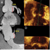
Imaging to select and guide transcatheter aortic valve implantation
- PMID: 24459198
- PMCID: PMC4416118
- DOI: 10.1093/eurheartj/eht569
Imaging to select and guide transcatheter aortic valve implantation
Abstract
Transcatheter aortic valve implantation (TAVI) is indicated for patients with severe aortic stenosis and high or prohibitive surgical risk. Patients' selection requires clinical and anatomical selection criteria, being the later determined by multimodality imaging evaluation. Echocardiography, multislice computed tomography (MSCT), angiography, and cardiovascular magnetic resonance (CMR) are the methods available to determine the anatomical suitability for the procedure. Imaging assists in the selection of bioprosthesis type, prosthetic sizing and in the decision of the best vascular access. In this review, we present our critical appraisal on the use of imaging to best patients' selection and procedure guidance in TAVI.
Keywords: Aortic stenosis; Imaging; Transcatheter aortic valve implantation.
Published on behalf of the European Society of Cardiology. All rights reserved. © The Author 2014. For permissions please email: journals.permissions@oup.com.
Figures


Similar articles
-
Patient selection for transcatheter aortic valve implantation: patient risk profile and anatomical selection criteria.Arch Cardiovasc Dis. 2012 Mar;105(3):165-73. doi: 10.1016/j.acvd.2012.02.007. Epub 2012 Mar 15. Arch Cardiovasc Dis. 2012. PMID: 22520800 Review.
-
Imaging for planning of transcatheter aortic valve implantation.Herz. 2017 Sep;42(6):554-563. doi: 10.1007/s00059-017-4587-9. Herz. 2017. PMID: 28608132 Review. English.
-
Minimalistic Approach for Transcatheter Aortic Valve Implantation (TAVI): Open Vascular Vs. Fully Percutaneous Approach.Pril (Makedon Akad Nauk Umet Odd Med Nauki). 2019 Oct 1;40(2):5-14. doi: 10.2478/prilozi-2019-0009. Pril (Makedon Akad Nauk Umet Odd Med Nauki). 2019. PMID: 31605589
-
Role of Echocardiography Before Transcatheter Aortic Valve Implantation (TAVI).Curr Cardiol Rep. 2016 Apr;18(4):38. doi: 10.1007/s11886-016-0715-z. Curr Cardiol Rep. 2016. PMID: 26960423 Free PMC article. Review.
-
Vascular Imaging Before Transcatheter Aortic Valve Replacement (TAVR): Why and How?Curr Cardiol Rep. 2016 Feb;18(2):14. doi: 10.1007/s11886-015-0694-5. Curr Cardiol Rep. 2016. PMID: 26768740 Review.
Cited by
-
Vascular approaches for transcatheter aortic valve implantation.J Thorac Dis. 2017 May;9(Suppl 6):S478-S487. doi: 10.21037/jtd.2017.05.73. J Thorac Dis. 2017. PMID: 28616344 Free PMC article. Review.
-
Practical update on imaging and transcatheter aortic valve implantation.World J Cardiol. 2015 Apr 26;7(4):178-86. doi: 10.4330/wjc.v7.i4.178. World J Cardiol. 2015. PMID: 25914787 Free PMC article.
-
Use of Internal Endoconduit for Unfavorable Iliac Artery Anatomy in Patients Undergoing Transcatheter Aortic Valve Replacement - A Single Center Experience.Acta Cardiol Sin. 2018 Jan;34(1):37-48. doi: 10.6515/ACS.201801_34(1).20170911A. Acta Cardiol Sin. 2018. PMID: 29375223 Free PMC article.
-
[Imaging in structural heart disease : Impact on interventional therapy].Herz. 2016 Nov;41(7):639-652. doi: 10.1007/s00059-016-4481-x. Herz. 2016. PMID: 27646067 Review. German.
-
Immersogeometric fluid-structure interaction modeling and simulation of transcatheter aortic valve replacement.Comput Methods Appl Mech Eng. 2019 Dec 1;357:112556. doi: 10.1016/j.cma.2019.07.025. Epub 2019 Aug 14. Comput Methods Appl Mech Eng. 2019. PMID: 32831419 Free PMC article.
References
-
- Vahanian A, Alfieri O, Al-Attar N, Antunes M, Bax J, Cormier B, Cribier A, De Jaegere P, Fournial G, Kappetein AP, Kovac J, Ludgate S, Maisano F, Moat N, Mohr F, Nataf P, Pierard L, Pomar JL, Schofer J, Tornos P, Tuzcu M, van Hout B, Von Segesser LK, Walther T. Transcatheter valve implantation for patients with aortic stenosis: a position statement from the European Association of Cardio-Thoracic Surgery (EACTS) and the European Society of Cardiology (ESC), in collaboration with the European Association of Percutaneous Cardiovascular Interventions (EAPCI) Eur Heart J. 2008;29:1463–1470. - PubMed
-
- Rodes-Cabau J, Webb JG, Cheung A, Ye J, Dumont E, Feindel CM, Osten M, Natarajan MK, Velianou JL, Martucci G, DeVarennes B, Chisholm R, Peterson MD, Lichtenstein SV, Nietlispach F, Doyle D, DeLarochelliere R, Teoh K, Chu V, Dancea A, Lachapelle K, Cheema A, Latter D, Horlick E. Transcatheter aortic valve implantation for the treatment of severe symptomatic aortic stenosis in patients at very high or prohibitive surgical risk: acute and late outcomes of the multicenter Canadian experience. J Am Coll Cardiol. 2010;55:1080–1090. - PubMed
-
- Leon MB, Smith CR, Mack M, Miller DC, Moses JW, Svensson LG, Tuzcu EM, Webb JG, Fontana GP, Makkar RR, Brown DL, Block PC, Guyton RA, Pichard AD, Bavaria JE, Herrmann HC, Douglas PS, Petersen JL, Akin JJ, Anderson WN, Wang D, Pocock S. Transcatheter aortic-valve implantation for aortic stenosis in patients who cannot undergo surgery. N Engl J Med. 2010;363:1597–1607. - PubMed
-
- Smith CR, Leon MB, Mack MJ, Miller DC, Moses JW, Svensson LG, Tuzcu EM, Webb JG, Fontana GP, Makkar RR, Williams M, Dewey T, Kapadia S, Babaliaros V, Thourani VH, Corso P, Pichard AD, Bavaria JE, Herrmann HC, Akin JJ, Anderson WN, Wang D, Pocock SJ. Transcatheter versus surgical aortic-valve replacement in high-risk patients. N Engl J Med. 2011;364:2187–2198. - PubMed
-
- Lange R, Schreiber C, Gotz W, Hettich I, Will A, Libera P, Laborde JC, Bauernschmitt R. First successful transapical aortic valve implantation with the Corevalve Revalving system: a case report. Heart Surg Forum. 2007;10:E478–E479. - PubMed
Publication types
MeSH terms
Grants and funding
LinkOut - more resources
Full Text Sources
Other Literature Sources
Medical

