. 2014 Apr;349(1):56-65.
doi: 10.1124/jpet.113.210708. Epub 2014 Jan 23.

BK channel-mediated relaxation of urinary bladder smooth muscle: a novel paradigm for phosphodiesterase type 4 regulation of bladder function

Affiliations

BK channel-mediated relaxation of urinary bladder smooth muscle: a novel paradigm for phosphodiesterase type 4 regulation of bladder function

Wenkuan Xin et al. J Pharmacol Exp Ther. 2014 Apr.

Abstract

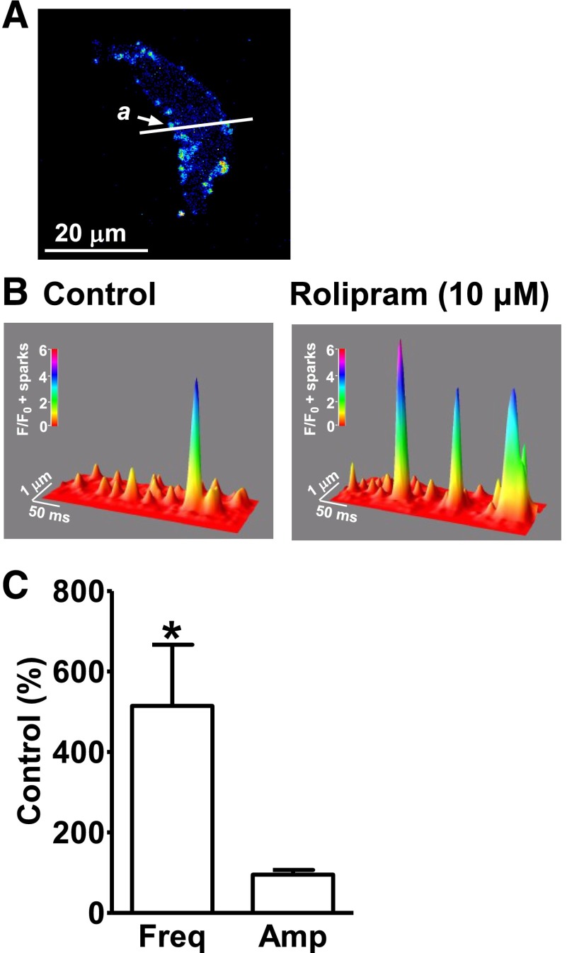
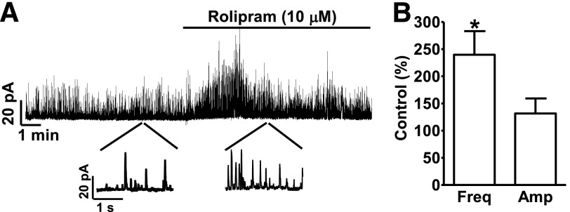
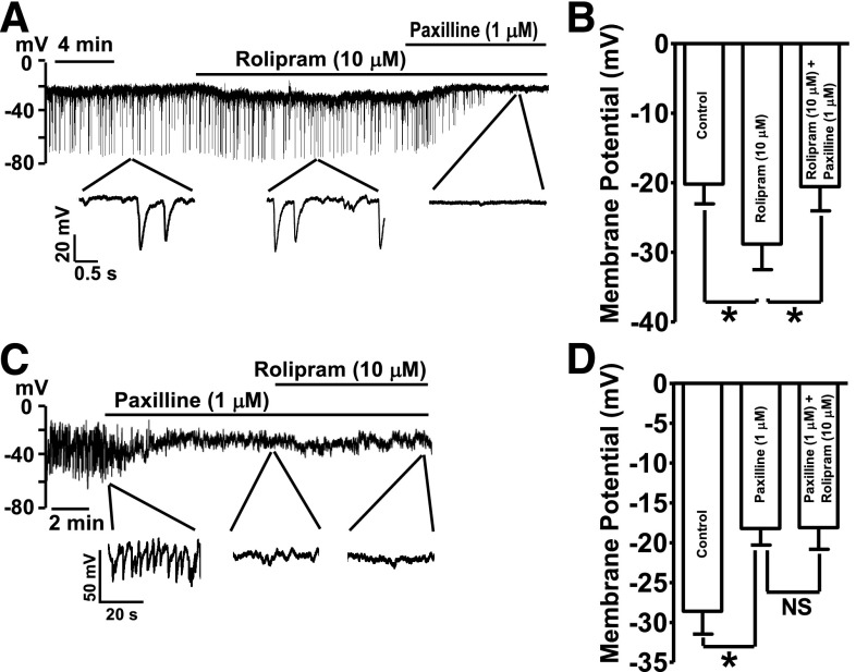
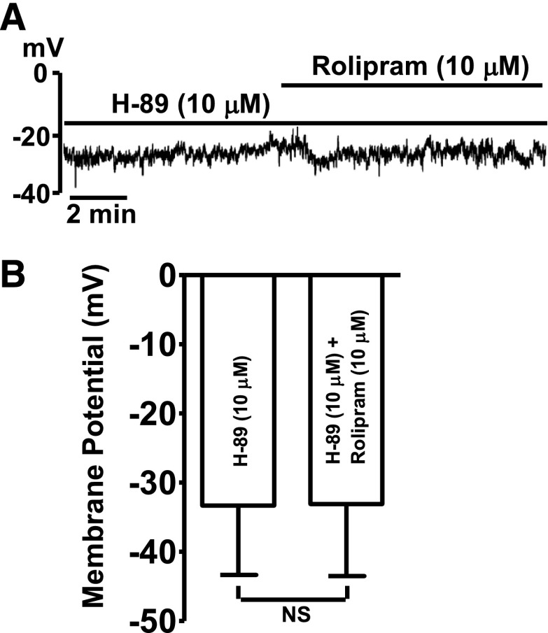
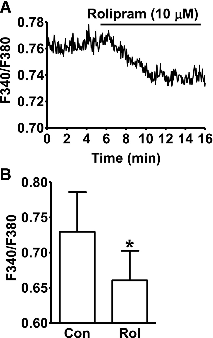
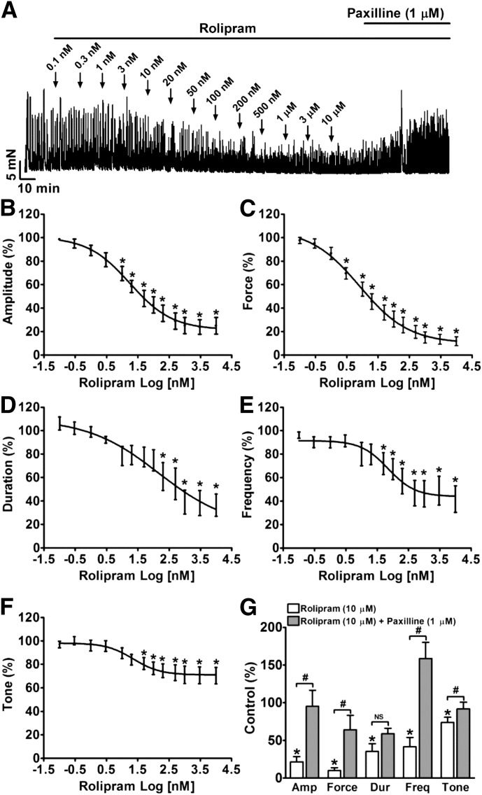
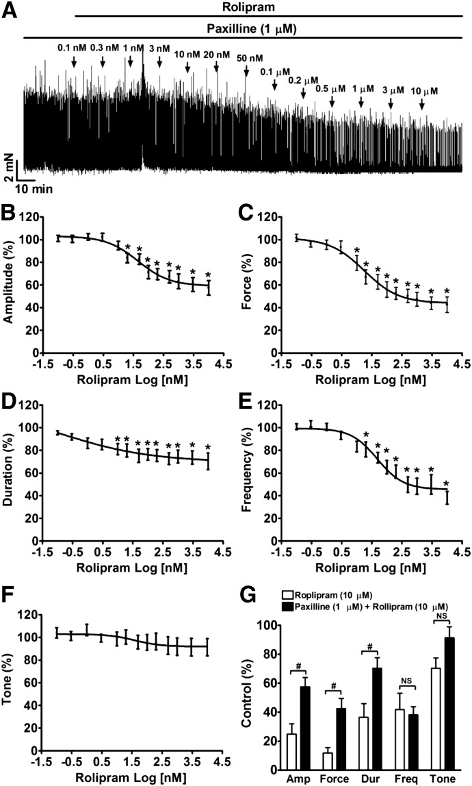
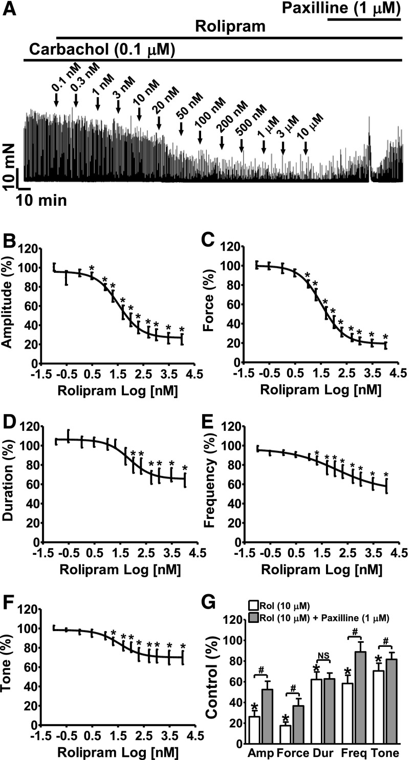
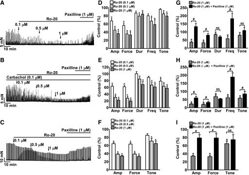
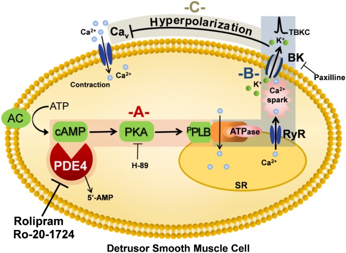
Elevation of intracellular cAMP and activation of protein kinase A (PKA) lead to activation of large conductance voltage- and Ca(2+)-activated K(+) (BK) channels, thus attenuation of detrusor smooth muscle (DSM) contractility. In this study, we investigated the mechanism by which pharmacological inhibition of cAMP-specific phosphodiesterase 4 (PDE4) with rolipram or Ro-20-1724 (C(15)H(22)N(2)O(3)) suppresses guinea pig DSM excitability and contractility. We used high-speed line-scanning confocal microscopy, ratiometric fluorescence Ca(2+) imaging, and perforated whole-cell patch-clamp techniques on freshly isolated DSM cells, along with isometric tension recordings of DSM isolated strips. Rolipram caused an increase in the frequency of Ca(2+) sparks and the spontaneous transient BK currents (TBKCs), hyperpolarized the cell membrane potential (MP), and decreased the intracellular Ca(2+) levels. Blocking BK channels with paxilline reversed the hyperpolarizing effect of rolipram and depolarized the MP back to the control levels. In the presence of H-89 [N-[2-[[3-(4-bromophenyl)-2-propenyl]amino]ethyl]-5-isoquinolinesulfonamide dihydrochloride], a PKA inhibitor, rolipram did not cause MP hyperpolarization. Rolipram or Ro-20-1724 reduced DSM spontaneous and carbachol-induced phasic contraction amplitude, muscle force, duration, and frequency, and electrical field stimulation-induced contraction amplitude, muscle force, and tone. Paxilline recovered DSM contractility, which was suppressed by pretreatment with PDE4 inhibitors. Rolipram had reduced inhibitory effects on DSM contractility in DSM strips pretreated with paxilline. This study revealed a novel cellular mechanism whereby pharmacological inhibition of PDE4 leads to suppression of guinea pig DSM contractility by increasing the frequency of Ca(2+) sparks and the functionally coupled TBKCs, consequently hyperpolarizing DSM cell MP. Collectively, this decreases the global intracellular Ca(2+) levels and DSM contractility in a BK channel-dependent manner.


Figures

Fig. 1.
Fig. 1.
Selective phosphodiesterase 4 (PDE4) inhibition with rolipram increases Ca2+ spark frequency in freshly isolated detrusor smooth muscle (DSM) cells. (A) An image of a freshly isolated DSM cell loaded with fluo-4-AM. The white line passing the active site a is the laser beam scanning pathway (1-pixel width). (B) A three-dimensional view of the recordings illustrating the relative fluorescence intensity profiles of the Ca2+ sparks. The color scale indicates the relative fluorescence intensity F/F0. (C) Summary data illustrating that rolipram (10 µM) increased the Ca2+ spark frequency without a significant change of Ca2+ spark amplitude (n = 15, N = 9; *P < 0.05). Amp, amplitude; Freq, frequency.
Fig. 2.
Fig. 2.
Selective PDE4 inhibition with rolipram increases spontaneous transient BK current (TBKC) frequency in freshly isolated DSM cells. (A) An original recording illustrating that rolipram (10 µM) increased the frequency of TBKCs in a single DSM cell. A portion of the recording is shown on an expanded time scale before and after rolipram application. (B) Summary data illustrating that rolipram (10 µM) increased the TBKCs’ frequency without significant change in TBKCs’ amplitude (n = 7, N = 6; *P < 0.05).
Fig. 3.
Fig. 3.
Selective PDE4 inhibition with rolipram hyperpolarizes DSM cell resting membrane potential (MP) in a BK channel-dependent manner. (A) An original current-clamp recording illustrating that rolipram (10 µM) hyperpolarized cell MP. The subsequent addition of the BK channel blocker, paxilline (1 µM), inhibited the spontaneous transient hyperpolarizations and depolarized the MP. (B) Summary data illustrating that rolipram (10 µM) hyperpolarized the MP. The subsequent addition of 1 µM paxilline reversed the rolipram-induced hyperpolarization and further depolarized the cell MP back to the control level (n = 8, N = 6; *P < 0.05). (C) An original current-clamp recording illustrating that paxilline (1 µM) abolished the spontaneous transient hyperpolarizations. Rolipram (10 µM) did not exhibit a hyperpolarizing effect on the MP in the presence of 1 µM paxilline. (D) Summary data illustrating that rolipram (10 µM) did not have any effect on the MP in DSM cells pretreated with 1 µM paxilline (n = 7, N = 5; P > 0.05; NS, nonsignificant).
Fig. 4.
Fig. 4.
Inhibition of PKA with H-89 abolishes the hyperpolarizing effect of rolipram in DSM cells. (A) An original current-clamp recording illustrating that, in the presence of H-89 (10 µM), rolipram (10 µM) did not have any effect on the MP. (B) Summary data illustrating that rolipram (10 µM) did not have any effect on the average MP when PKA was inhibited (n = 6, N = 4; P > 0.05; NS, nonsignificant).
Fig. 5.
Fig. 5.
Selective PDE4 inhibition with rolipram reduces the global intracellular Ca2+ levels in freshly isolated DSM cells. (A) An original recording illustrating that rolipram (10 µM) decreased the global intracellular Ca2+ level in a single DSM cell shown as a ratio of fura-2 fluorescence emission intensities at 510 nm with excitation at 340 and 380 nm. (B) Summary data indicating that rolipram (10 µM) significantly decreased the average intracellular Ca2+ levels (n = 8, N = 5; *P < 0.05).
Fig. 6.
Fig. 6.
Selective PDE4 inhibition with rolipram significantly reduces DSM spontaneous phasic contractions in a concentration-dependent manner. (A) A representative recording of a DSM strip depicts the concentration-dependent inhibitory effects of rolipram (0.1 nM–10 μM) on spontaneous phasic contractions. Post-treatment of the DSM strip with 1 μM paxilline in the presence of rolipram (10 μM) recovered DSM contractility. (B–F) Cumulative concentration-response curves illustrating the inhibitory effects of rolipram (0.1 nM–10 μM) on the amplitude (B), muscle force integral (C), duration (D), frequency (E), and muscle tone (F) of DSM spontaneous contractions (n = 12, N = 8; *P < 0.05 versus control). (G) Summary data illustrating that addition of 1 μM paxilline following application of 10 µM rolipram significantly recovered the phasic contraction amplitude, muscle force integral, and frequency of DSM-isolated strips (n = 11, N = 7; #P < 0.05 paxilline versus 10 μM rolipram; P > 0.05; NS, nonsignificant). TTX (1 μM) was present throughout the experiments. Amp, amplitude; Dur, duration; Freq, frequency.
Fig. 7.
Fig. 7.
Blocking BK channels with paxilline significantly attenuates rolipram-induced relaxation in DSM-isolated strips. (A) A representative recording of a DSM strip depicts the inhibitory effects of rolipram (0.1 nM–10 µM) on 1 μM paxilline-induced DSM spontaneous contractions. (B–F) Summary data illustrating the effect of rolipram on the amplitude (B), muscle force integral (C), duration (D), frequency (E), and tone (F) of paxilline-induced contractions of DSM strips pretreated with paxilline (n = 10, N = 4; *P < 0.05, versus control). (G) Summary data illustrating that pretreatment of DSM strips with 1 μM paxilline significantly reduced the maximal relaxation effect of 10 µM rolipram on the contraction amplitude, muscle force integral, frequency, and tone in DSM strips. #P < 0.05, paxilline pretreatment (n = 10, N = 4) versus control (without paxilline) (n = 12, N = 8). TTX (1 μM) was present throughout the experiments. Amp, amplitude; Dur, duration; Freq, frequency.
Fig. 8.
Fig. 8.
Selective PDE4 inhibition with rolipram significantly reduces carbachol-induced contractions of DSM strips in a concentration-dependent manner. (A) A representative recording of a DSM strip depicts the concentration-dependent inhibitory effects of rolipram (0.1 nM–10 µM) on 0.1 µM carbachol-induced contractions. Post-treatment of the DSM strip with paxilline (1 μM) reversed the inhibitory effect of rolipram. (B–F) Cumulative concentration-response curves illustrating the effect of rolipram on the amplitude (B), muscle force integral (C), duration (D), frequency (E), and tone (F) of carbachol-induced contractions in DSM strips (n = 13, N = 7; *P < 0.05 versus control). (G) Summary data illustrating that addition of 1 μM paxilline following application of 10 µM rolipram significantly recovered contraction amplitude, muscle force integral, frequency, and tone in DSM-isolated strips (n = 12, N = 6; #P < 0.05, paxilline versus 10 μM rolipram; P > 0.05; NS, nonsignificant). TTX (1 μM) was present throughout the experiments. Amp, amplitude; Dur, duration; Freq, frequency; Rol, rolipram.
Fig. 9.
Fig. 9.
Selective PDE4 inhibition with rolipram significantly reduces 20 Hz EFS-induced contractions of DSM-isolated strips in a concentration-dependent manner. (A) A representative recording of a DSM strip showing the concentration-dependent inhibitory effects of rolipram (0.1 nM-10 µM) on EFS-induced contractions. Post-treatment of the DSM strip with paxilline (1 μM) recovered the EFS-induced contractions. (B–D) Cumulative concentration-response curves for the inhibitory effects of rolipram on DSM contraction amplitude (B), muscle force integral (C), and muscle tone (D) (n = 10, N = 6; *P < 0.05, versus control). (E) Summary data illustrating that addition of 1 μM paxilline following application of 10 µM rolipram significantly recovered the EFS-induced contraction amplitude and muscle force in DSM-isolated strips (n = 8, N = 5; #P < 0.05 paxilline versus 10 μM rolipram; P > 0.05; NS, nonsignificant). Amp, amplitude; pax, paxilline; Rol, rolipram.
Fig. 10.
Fig. 10.
Selective PDE4 inhibition with Ro-20-1724 reduces the spontaneous, carbachol-, and EFS-induced contractions of DSM-isolated strips in a concentration-dependent manner. (A–C) Original recordings of DSM strips showing the concentration-dependent inhibitory effects of Ro-20-1724 (0.1 µM–1 µM) on spontaneous phasic, carbachol-induced, and EFS-induced contractions. (D–F) Summary data showing the concentration-dependent inhibitory effect of Ro-20-1724 on the spontaneous phasic (n = 7, N = 6), carbachol-induced (n = 12, N = 5), and EFS-induced (n = 10, N = 5) contractions in DSM-isolated strips (*P < 0.05 versus control). (G–I) Summary data illustrating that addition of 1 μM paxilline following application of 1 µM Ro-20-1724 significantly recovered the spontaneous phasic (n = 7, N = 6), carbachol-induced (n = 12, N = 5), and EFS-induced (n = 9, N = 5) contractions (*P < 0.05 versus control; #P < 0.05 paxilline versus 1 μM Ro-20-1724; P > 0.05; NS, nonsignificant). TTX (1 μM) was present throughout the experiments except the EFS-induced contractions. Ro-20 refers to Ro-20-1724. Amp, amplitude; Dur, duration; Freq, frequency.
Fig. 11.
Fig. 11.
Proposed novel signaling pathway by which PDE4 regulates BK channel activity in DSM cells. This diagram illustrates that the pharmacological inhibition of PDE4 leads to activation of PKA (A), increase in Ca2+ spark and TBKC activities (B), and hyperpolarization of DSM cell membrane and a consequent decrease in global Ca2+ levels (C). AC, adenylyl cyclase; BK, large conductance voltage- and Ca2+-activated K+ channel; CaV, L-type voltage-gated Ca2+ channel; pPLB, phosphorylated phospholamban; RyR, ryanodine receptor; SR, sarcoplasmic reticulum; TBKC, transient BK current.

Similar articles

Cited by

References

    1. Beca S, Helli PB, Simpson JA, Zhao D, Farman GP, Jones PP, Tian X, Wilson LS, Ahmad F, Chen SR, et al. (2011) Phosphodiesterase 4D regulates baseline sarcoplasmic reticulum Ca2+ release and cardiac contractility, independently of L-type Ca2+ current. Circ Res 109:1024–1030 - PMC - PubMed
    1. Bender AT, Beavo JA. (2006) Cyclic nucleotide phosphodiesterases: molecular regulation to clinical use. Pharmacol Rev 58:488–520 - PubMed
    1. Blackman BE, Horner K, Heidmann J, Wang D, Richter W, Rich TC, Conti M. (2011) PDE4D and PDE4B function in distinct subcellular compartments in mouse embryonic fibroblasts. J Biol Chem 286:12590–12601 - PMC - PubMed
    1. Brading AF. (2006) Spontaneous activity of lower urinary tract smooth muscles: correlation between ion channels and tissue function. J Physiol 570:13–22 - PMC - PubMed
    1. Brown SM, Bentcheva-Petkova LM, Liu L, Hristov KL, Chen M, Kellett WF, Meredith AL, Aldrich RW, Nelson MT, Petkov GV. (2008) β-adrenergic relaxation of mouse urinary bladder smooth muscle in the absence of large-conductance Ca2+-activated K+ channel. Am J Physiol Renal Physiol 295:F1149–F1157 - PMC - PubMed

Publication types

MeSH terms

Substances