. 2014 Jun 15;23(12):3157-65.
doi: 10.1093/hmg/ddu026. Epub 2014 Jan 23.

Thiol peroxidases ameliorate LRRK2 mutant-induced mitochondrial and dopaminergic neuronal degeneration in Drosophila

Affiliations

Thiol peroxidases ameliorate LRRK2 mutant-induced mitochondrial and dopaminergic neuronal degeneration in Drosophila

Dario C Angeles et al. Hum Mol Genet. .

Abstract

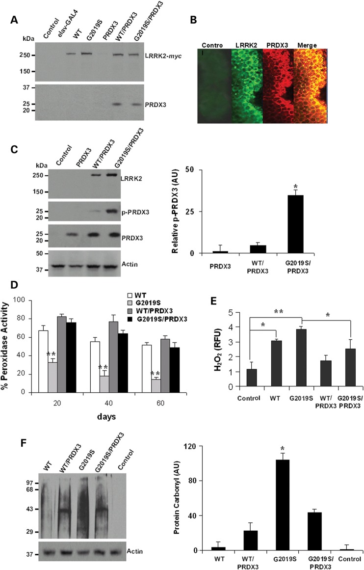
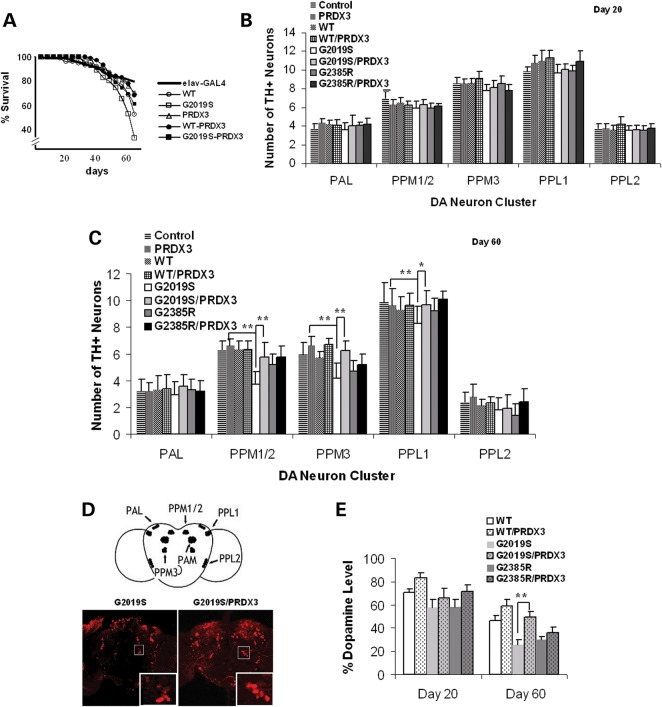
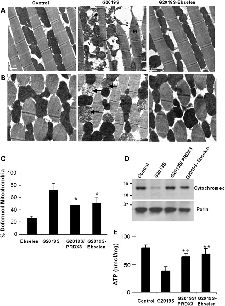
Mutations in leucine-rich repeat kinase 2 (LRRK2) are common causes of familial Parkinson's disease (PD). LRRK2 has been shown to bind peroxiredoxin-3 (PRDX3), the most important scavenger of hydrogen peroxide in the mitochondria, in vitro. Here, we examined the interactions of LRRK2 and PRDX3 in Drosophila models by crossing transgenic LRRK2 and PRDX3 flies. As proof of principle experiments, we subsequently challenged LRRK2 and LRRK2/PRDX3 flies with a peroxidase mimic, Ebselen. We demonstrated that co-expression of PRDX3 with the LRRK2 kinase mutant G2019S in bigenic Drosophila ameliorated the G2019S mutant-induced reduction in peroxidase capacity, loss of dopaminergic neurons, shortened lifespan and mitochondrial defects of flight muscles in monogenic flies expressing the G2019S alone. Challenges with Ebselen recapitulated similar rescue of these phenotypic features in mutant-expressing Drosophila. The peroxidase mimic preserved neuronal and mitochondrial and neuronal integrity and improved mobility and survival in mutant-expressing Drosophila. Taken together, our study provides the first in vivo evidence to suggest that phosphoinhibition of endogenous peroxidases could be a mechanism in LRRK2-induced oxidant-mediated neurotoxicity. Our therapeutic experiments also highlight the potential of thiol peroxidases as neuroprotective agents in PD patients carrying LRRK2 mutations.


Figures

Figure 1.
Figure 1.
LRRK2 kinase mutant inhibits activity of PRDX3 in transgenic Drosophila. (A) Immunoblot of head lysates from 20 d.p.e. flies that were subjected to immunoprecipitation using myc-specific antibody. WT refers to the LRRK2-WT line, G2019S is the LRRK2-G2019S line and the control is UAS-LRRK2 line that was not crossed with the driver. (B) Images showing colocalization of LRRK2 and PRDX3 signals in brain whole mounts. The control was probed with anti-LRRK2 as primary antibody. (C) Representative immunoblot showing p-PRDX3 detected by phospho-specific antibody. Chart showing relative p-PRDX3 levels over total PRDX3. *Significant increase from PRDX3 values at P < 0.01 by Student's t-test. AU, arbitrary unit. (D) Chart showing peroxidase activities as percent mean fluorescence ± SEM of values in PRDX3 lysate. Data were derived from three independent crosses that were background corrected and normalized to PRDX3 lysate values. **Significant decrease from PRDX3 values at P < 0.01 by Student's t-test. (E) Level of H2O2 as relative fulorescence unit ± SEM (n = 4) normalized to PRDX3 values. *,**Significant difference at P < 0.05 and P < 0.01 by one-way ANOVA with Bonferroni corrections. (F) Level of oxidized protein in LRRK2-variant-expressing flies relative to loading control. *Significant increase from all values at P < 0.01 by one-way ANOVA. Genotypes: elav-GAL4/+, elav-GAL4-hLRRK2 variants, elav-GAL4-PRDX3, elav-GAL4-hLRRK2; elav-GAL4-PRDX3.
Figure 2.
Figure 2.
PRDX3 ameliorate mutant-induced DA neuronal degeneration. (A) Survival curves as percent living flies (n = 3, cohort of 50). Intergenotypic differences were analyzed by log-rank test, significant at P < 0.05. (B) Chart showing mean DA neuronal counts per cluster in the brain at 20 d.p.e. (n = 3; cohort of 20). (C) DA neuronal counts at 60 d.p.e. (n = 3; cohort of 20). *,**Significant difference at P < 0.05 and P < 0.01 by one-way ANOVA with Bonferroni corrections. The elav-GAL4 driver line was used as control. (D) Representative images of brain whole mount at 60 d.p.e. showing the PPM3 cluster (boxed) labeled with anti-TH antibody (red signal). Scale bar: 100 μm. (E) Dopamine level in fly brains as percent of mean absorbance ± SEM (n = 4) normalized to control. **Significant increase from counts in G2019S values at P < 0.01 by Student's t-test. PPL, protocerebral posterior lateral; PPM, protocerebral posterior medial; PAL, protocerebral anterior lateral. Genotypes: ddc-GAL4/+, ddc-GAL4-hLRRK2, ddc-GAL4-hLRRK2; ddc-GAL4-PRDX3.
Figure 3
Figure 3
PRDX3 and Ebselen ameliorate mutant-induced motor dysfunction. (A) Age-dependent climbing scores as percent of mean ± SEM (n = 3, cohort of 50) normalized to control. **Significant increase from G2019S values at P < 0.01 by one-way ANOVA with Bonferroni corrections. (B) Climbing scores as in (A). (C) DA neuronal counts by cluster (n = 3, cohort of 20). *,**Significant difference at P < 0.05 and P < 0.01 by one-way ANOVA with Bonferroni corrections.
Figure 4
Figure 4
PRDX3 and Ebselen ameliorate mutant-induced degeneration of muscle and mitochondria. (A) Transmission electron micrograph of longitudinal thoracic muscle sections showing the effects of Ebselen on kinase mutant-induced disruption of myofibrils with diffused Z-lines (Z) and M-bands (M). Swollen and vacuolated mitochondria (V) are also shown. (B) Transverse sections showing reduction in kinase mutant-induced fragmentation of cristae (arrows). Scale bars: 1 μm. (C) Chart showing percent of deformed mitochondria ± SEM normalized to driver control. Data were derived from the relative number of deformed over healthy mitochondria in 20 fields of five sections for each genotypic line. *Significant decrease from kinase mutant values at P < 0.05 by Student's t-test. (D) Immunoblot analysis of extracts for the level of cytochrome c in the mitochondria treated and untreated LRRK2-G2019S and driver control. (E) Comparison of ATP levels in the mitochondrial extracts showing mean ATP ± SEM (n = 4, cohort of 20 thoraces), corrected for blank and calculated from standard curve plot. **Significant increase from kinase mutant values at P < 0.01 by one-way ANOVA with Bonferroni corrections. Genotypes: 24B-GAL4/+, 24B-GAL4-hLRRK2, 24B-GAL4-hLRRK2; 24B-GAL4-PRDX3.

Similar articles

Cited by

References

    1. Zimprich A., Biskup S., Leitner P., Lichtner P., Farrer M., Lincoln S., Kachergus J., Hulihan M., Uitti R.J., Calne D.B., et al. Mutations in LRRK2 cause autosomal-dominant parkinsonism with pleomorphic pathology. Neuron. 2004;44:601–607. - PubMed
    1. Hernandez D.G., Paisán-Ruíz C., McInerney-Leo A., Jain S., Meyer-Lindenberg A., Evans E.W., Berman K.F., Johnson J., Auburger G., Schäffer A.A., et al. Clinical and positron emission tomography of Parkinson's disease caused by LRRK2. Ann. Neurol. 2005;57:453–456. - PubMed
    1. Paisan-Ruiz C., Jain S., Evans E.W., Gilks W.P., Simon J., van der Brug M., Lopez de Munain A., Aparicio S., Gil A.M., Khan N., et al. Cloning of the gene containing mutations that cause PARK8-linked Parkinson's disease. Neuron. 2004;44:595–600. - PubMed
    1. Kachergus J., Mata I.F., Hulihan M., Taylor J.P., Lincoln S., Aasly J., Gibson J.M., Ross O.A., Lynch T., Wiley J., et al. Identification of a novel LRRK2 mutation linked to autosomal dominant parkinsonism: evidence of a common founder across European populations. Am. J. Hum. Genet. 2005;76:672–680. - PMC - PubMed
    1. Di Fonzo A., Tassorelli C., De Mari M., Chien H.F., Ferreira J., Rohe C.F., Riboldazzi G., Antonini A., Albani G., Mauro A., et al. Comprehensive analysis of the LRRK2 gene in sixty families with Parkinson's disease. Eur. J. Hum. Genet. 2006;14:322–331. - PubMed

Publication types

MeSH terms