

Dysregulation of ENaC in Animal Models of Nephrotic Syndrome and Liver Cirrhosis
- PMID: 24459482
- PMCID: PMC3894541
- DOI: 10.5049/EBP.2006.4.1.23
Dysregulation of ENaC in Animal Models of Nephrotic Syndrome and Liver Cirrhosis
Abstract
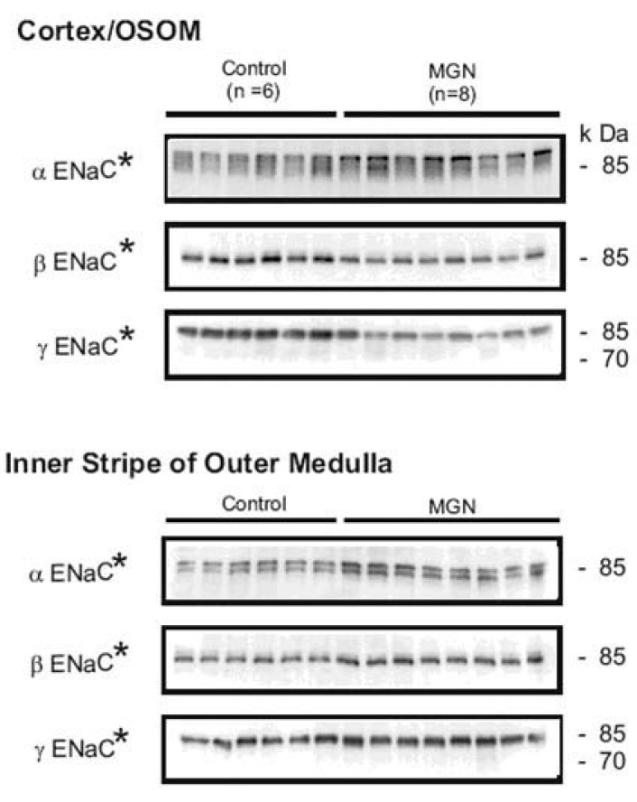
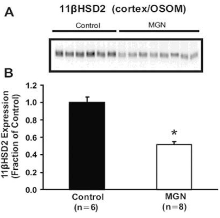
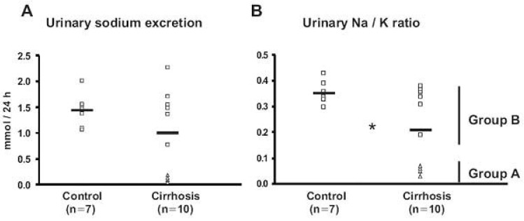
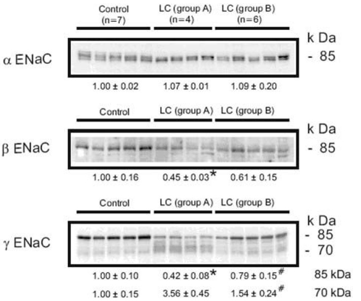
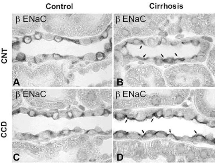
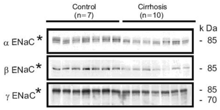
Nephrotic syndrome and liver cirrhosis are common clinical manifestations, and are associated with avid sodium retention leading to the development of edema and ascites. However, the mechanism for the sodium retention is still incompletely understood and the molecular basis remains undefined. We examined the changes of sodium (co)transporters and epithelial sodium channels (ENaCs) in the kidneys of experimental nephrotic syndrome and liver cirrhosis. The results demonstrated that puromycin- or HgCl2-induced nephrotic syndrome was associated with 1) sodium retention, decreased urinary sodium excretion, development of ascites, and increased plasma aldosterone level; 2) increased apical targeting of ENaC subunits in connecting tubule and collecting duct segments; and 3) decreased protein abundance of type 2 11β-hydroxysteroid dehydrogenase (11βHSD2). Experimental liver cirrhosis was induced in rats by CCl4 treatment or common bile duct ligation. An increased apical targeting of alpha-, beta-, and gamma-ENaC subunits in connecting tubule, and cortical and medullary collecting duct segments in sodium retaining phase of liver cirhosis but not in escape phase of sodium retention. Immunolabeling intensity of 11βHSD2 in the connecting tubule and cortical collecting duct was significantly reduced in sodium retaining phase of liver cirrhosis, and this was confirmed by immunoblotting. These observations therefore strongly support the view that the renal sodium retention associated with nephrotic syndrome and liver cirrhosis is caused by increased sodium reabsorption in the aldosterone sensitive distal nephron including the connecting tubule and collecting duct, and increased apical targeting of ENaC subunits plays a role in the development of sodium retention in nephrotic syndrome and liver cirrhosis.
Keywords: Aldosterone sensitive distal nephron; Edema; Epithelial sodium channel; Sodium retention.
Figures
Similar articles
-
Increased apical targeting of renal epithelial sodium channel subunits and decreased expression of type 2 11beta-hydroxysteroid dehydrogenase in rats with CCl4-induced decompensated liver cirrhosis.J Am Soc Nephrol. 2005 Nov;16(11):3196-210. doi: 10.1681/ASN.2004080721. Epub 2005 Sep 28. J Am Soc Nephrol. 2005. PMID: 16192424
-
Increased apical targeting of renal ENaC subunits and decreased expression of 11betaHSD2 in HgCl2-induced nephrotic syndrome in rats.Am J Physiol Renal Physiol. 2006 Mar;290(3):F674-87. doi: 10.1152/ajprenal.00084.2005. Epub 2005 Sep 27. Am J Physiol Renal Physiol. 2006. PMID: 16189294
-
Biphasic changes of epithelial sodium channel abundance and trafficking in common bile duct ligation-induced liver cirrhosis.Kidney Int. 2006 Jan;69(1):89-98. doi: 10.1038/sj.ki.5000018. Kidney Int. 2006. PMID: 16374428
-
Pathogenesis of oedema in nephrotic syndrome: role of epithelial sodium channel.Nephrology (Carlton). 2007 Dec;12 Suppl 3:S8-10. doi: 10.1111/j.1440-1797.2007.00874.x. Nephrology (Carlton). 2007. PMID: 17995529 Review.
-
Regulation of renal sodium and water excretion in the nephrotic syndrome and cirrhosis of the liver.Dan Med Bull. 1997 Apr;44(2):191-207. Dan Med Bull. 1997. PMID: 9151012 Review.
Cited by
-
Role of the cytochrome P-450/ epoxyeicosatrienoic acids pathway in the pathogenesis of renal dysfunction in cirrhosis.Nephrol Dial Transplant. 2018 Aug 1;33(8):1333-1343. doi: 10.1093/ndt/gfx354. Nephrol Dial Transplant. 2018. PMID: 29361048 Free PMC article.
-
ENaC activation by proteases.Acta Physiol (Oxf). 2022 May;235(1):e13811. doi: 10.1111/apha.13811. Epub 2022 Mar 21. Acta Physiol (Oxf). 2022. PMID: 35276025 Free PMC article. Review.
References
-
- Hager H, Kwon TH, Vinnikova AK, Masilamani S, Brooks HL, Frokiaer J, Knepper MA, Nielsen S. Immunocytochemical and immunoelectron microscopic localization of alpha-, beta-, and gamma-ENaC in rat kidney. Am J Physiol. 2001;280:F1093–F1106. - PubMed
-
- Kim SW, Wang W, Kwon TH, Knepper MA, Frokiaer J, Nielsen S. Increased expression of ENaC subunits and increased apical targeting of AQP2 in the kidneys of spontaneously hypertensive rats. Am J Physiol Renal Physiol. 2005;289:F957–F968. - PubMed
-
- Kim SW, Gresz Veronika, Rojek Alexandra, Wang Weidong, Verkman A.S., Frøkiær Jørgen, Nielsen Søren. Decreased expression of AQP2 and AQP4 water channels and Na,K-ATPase in kidney collecting duct in AQP3 null mice. Biol Cell. 2005;97:765–778. - PubMed
-
- Awayda MS, Tousson A, Benos DJ. Regulation of a cloned epithelial Na+ channel by its β- and γ-subunits. Am J Physiol. 1997;273:C1889–C1899. - PubMed
-
- Barker PM, Nguyen MS, Gatzy JT, Grubb B, Norman H, Hummler E, Rossier B, Boucher RC, Koller B. Role of gammaENaC subunit in lung liquid clearance and electrolyte balance in newborn mice. Insights into perinatal adaptation and pseudohypoaldosteronism. J Clin Invest. 1998;102:1634–1640. - PMC - PubMed
Publication types
LinkOut - more resources
Full Text Sources