

Case Reports
doi: 10.4250/jcu.2013.21.4.200.
Epub 2013 Dec 27.
Early complication of mustard procedure after late repair
Affiliations
- PMID: 24459571
- PMCID: PMC3894375
- DOI: 10.4250/jcu.2013.21.4.200
Item in Clipboard
Case Reports
Early complication of mustard procedure after late repair
J Cardiovasc Ultrasound.
2013 Dec.
No abstract available
Keywords: Baffle stenosis; Echocardiogram; Magnetic resonance; Mustard procedure; Transposition of great arteries.
Figures
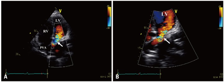
Transthoracic echocardiogram apical 4-chamber view (A) with zoom mode (B) demonstrating the connections of the systemic venous circulation with significant color turbulence suggestive of baffle stenosis at the veno-atrial junction (arrow). LV: left ventricle, PVA: pulmonary venous atrium, RV: right ventricle.
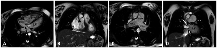
A: Cardiovascular magnetic resonance 4-chamber steady-state free precession (SSFP) image showing the trabeculated dilated systemic RV and patent pulmonary venous baffle (arrow). B: Cardiovascular magnetic resonance coronal SSFP image showing the typical discordant ventriculararterial relationship in complete transposition of the great arteries with the AO arising from the RV and MPA arising from the LV. C: Cardiovascular magnetic resonance axial SSFP image demonstrating severely dilated main PA and both branches. D: Cardiovascular magnetic resonance coronal SSFP image revealing significant stenosis of both superior vena cava (long arrow) and inferior vena cava (short arrow) limbs of the Mustard baffle at the veno-atrial junction. LV: left ventricle, PVA: pulmonary venous atrium, RV: right ventricle, AO: aorta, PA: pulmonary artery, LPA: left pulmonary artery, MPA: main pulmonary artery, RPA: right pulmonary artery.
Similar articles
-
Resolution of Hypoxia and Ascites With Percutaneous Intervention of Mustard Baffle Obstruction and Leak.JACC Case Rep. 2020 Jun 17;2(7):1079-1083. doi: 10.1016/j.jaccas.2020.05.004. eCollection 2020 Jun 17. JACC Case Rep. 2020. PMID: 34317419 Free PMC article.
-
The Mustard procedure for correction of simple transposition of the great arteries before 1 month of age.J Thorac Cardiovasc Surg. 1992 Nov;104(5):1218-24. J Thorac Cardiovasc Surg. 1992. PMID: 1434698
-
Factors influencing choice of procedure in transposition of the great arteries: a decision analysis approach.J Am Coll Cardiol. 1990 Aug;16(2):471-5. doi: 10.1016/0735-1097(90)90605-o. J Am Coll Cardiol. 1990. PMID: 2197318
-
Corrected Transposition: Anatomic Repair Using the Hemi-Mustard Atrial Baffle and Bidirectional Superior Cavopulmonary Connection.Semin Thorac Cardiovasc Surg Pediatr Card Surg Annu. 2019;22:51-56. doi: 10.1053/j.pcsu.2019.02.002. Semin Thorac Cardiovasc Surg Pediatr Card Surg Annu. 2019. PMID: 31027564 Review.
-
Pregnancy outcome following Mustard procedure for transposition of the great arteries: a report of five cases and review of the literature.Obstet Gynecol. 1994 Apr;83(4):512-6. doi: 10.1097/00006250-199404000-00005. Obstet Gynecol. 1994. PMID: 8134059 Review.
References
-
- Cohen MD, Johnson T, Ramrakhiani S. MRI of surgical repair of transposition of the great vessels. AJR Am J Roentgenol. 2010;194:250–260. - PubMed
-
- Kouchoukos NT, Blackstone EH, Doty DB, Hanley FL, Karp RB. Kirklin/Barratt-Boyes cardiac surgery. 3rd ed. Philadelphia: Churchill Livingstone; 2003. pp. 1439–1507.
Publication types
LinkOut - more resources
Full Text Sources
Other Literature Sources
