. 2013 Aug;1(2):99-111.
doi: 10.1158/2326-6066.CIR-13-0047.

Transnuclear TRP1-specific CD8 T cells with high or low affinity TCRs show equivalent antitumor activity

Affiliations

Transnuclear TRP1-specific CD8 T cells with high or low affinity TCRs show equivalent antitumor activity

Stephanie K Dougan et al. Cancer Immunol Res. 2013 Aug.

Abstract

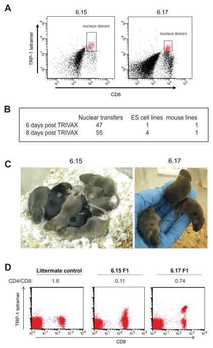
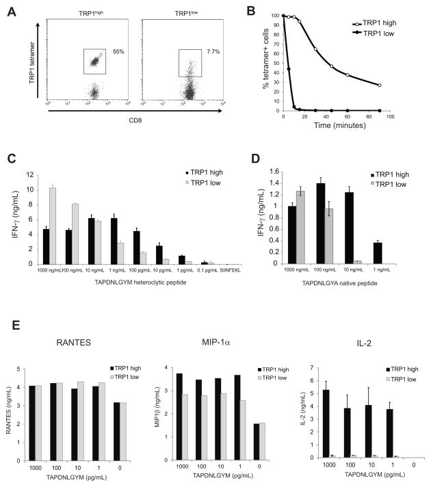
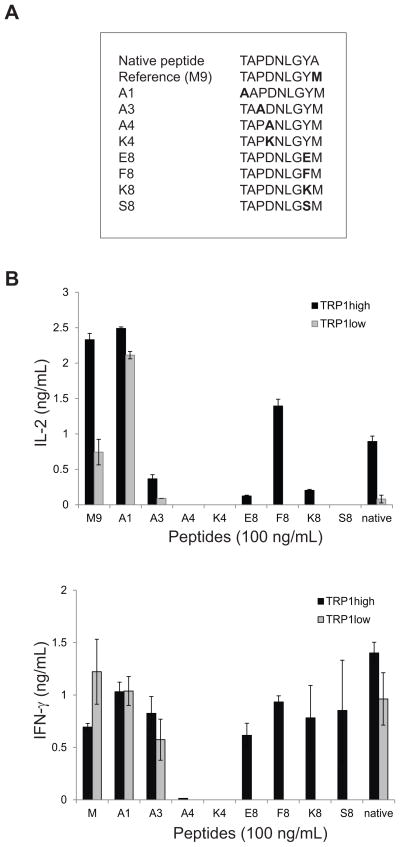
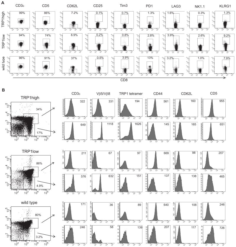
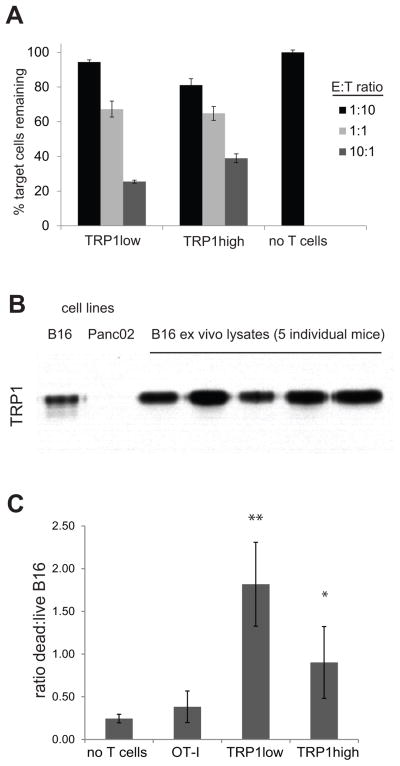
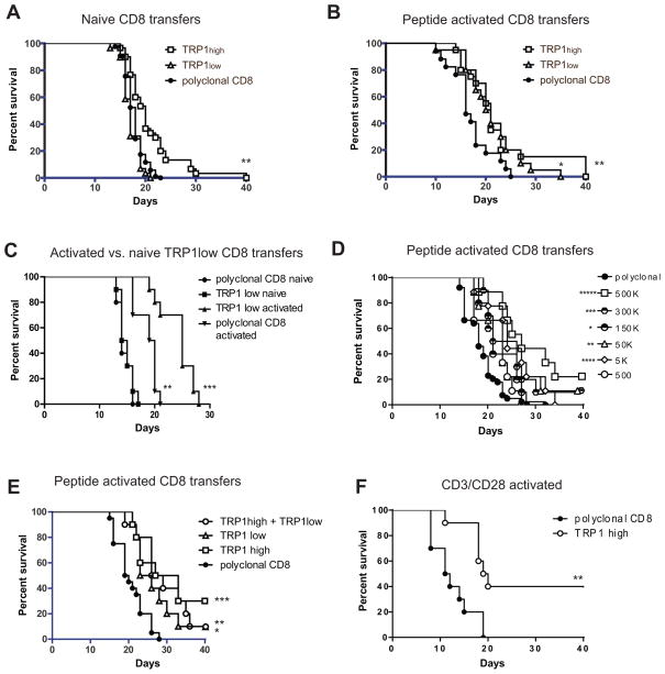
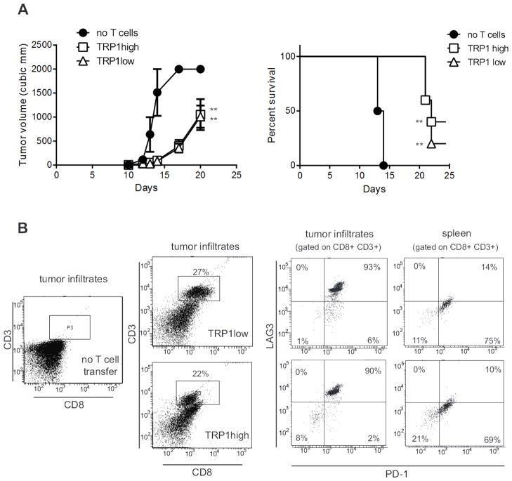
We have generated, via somatic cell nuclear transfer, two independent lines of transnuclear (TN) mice, using as nuclear donors CD8 T cells, sorted by tetramer staining, that recognize the endogenous melanoma antigen TRP1. These two lines of nominally identical specificity differ greatly in their affinity for antigen (TRP1(high) or TRP1(low)) as inferred from tetramer dissociation and peptide responsiveness. Ex vivo-activated CD8 T cells from either TRP1(high) or TRP1(low) mice show cytolytic activity in 3D tissue culture and in vivo, and slow the progression of subcutaneous B16 melanoma. Although naïve TRP1(low) CD8 T cells do not affect tumor growth, upon activation these cells function indistinguishably from TRP1(high) cells in vivo, limiting tumor cell growth and increasing mouse survival. The anti-tumor effect of both TRP1(high) and TRP1(low) CD8 T cells is enhanced in RAG-deficient hosts. However, tumor outgrowth eventually occurs, likely due to T cell exhaustion. The TRP1 TN mice are an excellent model for examining the functional attributes of T cells conferred by TCR affinity, and they may serve as a platform for screening immunomodulatory cancer therapies.

Keywords: B16; T cell receptor; melanoma; somatic cell nuclear transfer; tyrosinase related protein 1.


Conflict of interest statement

Conflict of interest: SO is an employee of Janssen Pharmaceuticals. The authors have no other conflicts of interest to disclose.

Figures

Figure 1
Figure 1
TRP1 TN mice were derived from TRP1-specific CD8 T cells. A) C57BL/6x129 F1 agouti male mice were immunized with TRIVAX (palmitoylated TAPDNLGYM 10μg/mouse, anti-CD40 30μg/mouse and poly-IC 15 μg/mouse) intravenously. Splenocytes were harvested 6 or 8 days post-immunization and stained with anti-CD8 and TRP1 tetramer. Gated cells were sorted by FACS and used as nucleus donors for SCNT. B) Success rate of nuclear transfer. C) ES cell lines 6.15 and 6.17 were injected into wild type blastocysts to generate chimeric mice as shown. Germline transmission was achieved by crossing chimeras to C57BL/6 females. D) Peripheral blood mononuclear cells were harvested from germline transmitted 6.15 or 6.17 mice and stained with TRP1 tetramer, anti-CD4 and anti-CD8. The calculated CD4:CD8 ratio is shown below each plot. Note the lower intensity tetramer staining in the 6.15 mouse, representative of more than 100 mice analyzed.
Figure 2
Figure 2
TRP1 TN lines differ in TCR affinity. A) Splenocytes from TRP1low (6.15) and TRP1high (6.17) TN mice were stained with TRP1 tetramer to saturation, washed, and incubated at room temperature for various times prior to fixation and analysis by flow cytometry. Time 0 is shown. B) Percent tetramer+ cells as defined by the gates shown in A were calculated for each time point and normalized to Time 0. Representative of 3 independent experiments. C) CD8 T cells isolated from TRP1low or TRP1high mice were cocultured with BMDCs pulsed with TAPDNLGYM at the indicated concentrations. IFN-γ was measured by ELISA of 48 hr culture supernatants. Representative of 5 independent experiments. D) CD8 T cells were cocultured as in (C) using BMDCs pulsed with the native TRP1 peptide (TAPDNLGYA). Representative of 3 independent experiments. E) CD8 T cells were cultured as in (C). RANTES and MIP-1α were analyzed by cytokine bead array using 24 hr culture supernatants; IL-2 was measured by ELISA of the same supernatants. Representative of 3 independent experiments.
Figure 3
Figure 3
Fine specificity mapping of TRP TN TCR specificities. A) An altered peptide library (as shown) was generated by substitution of non MHC contact residues. B) CD8 T cells isolated from TRP1high or TRP1low mice were cocultured with BMDCs pulsed with the indicated peptides at 100 ng/mL. IL-2 was measured by ELISA of 24hr culture supernatants. IFN-γ was measured by ELISA of 72hr culture supernatants. Representative of 2 independent experiments. Error bars are SD of triplicate samples.
Figure 4
Figure 4
Surface profiling and thymic development of TRP1 TN CD8 cells. A) Pooled spleen and lymph node cells from 6 week old male TRP1high, TRP1low and C57BL/6 mice were stained with the indicated antibodies. Plots shown are gated on CD8+ cells. B) Thymocytes from 6 week old male TRP1high, TRP1low and C57BL/6 mice were stained with the indicated antibodies. Histogram plots are gated on CD4+CD8+ double positive cells (top rows) or on CD8+ single positive cells (bottom rows). Numbers indicate median fluorescent intensities of histograms. TRP1low cells show negligible tetramer staining. To confirm genotype, naïve CD8 T cells from the same mice were stimulated with APCs and TRP1 A1 peptide in tissue culture, and activation was assessed by CD69 expression. %CD69+: TRP1high = 81% (no peptide =0.5%); TRP1low = 35% (no peptide = 0.3%).
Figure 5
Figure 5
TRP1 TN CD8 T cells are tumoricidal in vitro. A) CD8+ T cells were harvested from the indicated mice and cultured for 8 days with anti-CD3/CD28 beads and IL-2 (20 U/mL). Activated T cells were washed, counted, and aliquoted into a 96 well round bottom plate at 50,000 T cells per well. B cells were harvested from a wildtype or MHCII-GFP mouse. GFP+ B cells were incubated with 100uM TRP1 A1 peptide for 2 hours, washed twice, counted and mixed with non-GFP cells at a 1:1 ratio. This B cell mixture was then added to the plated T cells at the indicated ratios where E = effector T cells and T = total pooled B cells. After 48 hours of coculture, the cells were stained with anti-CD19 and the viability dye 7-AAD. B) Lysates were prepared from cultured B16 or Panc02 cells, and from B16 tumors >1cm in diameter resected from tumor-bearing mice. Immunoblotting shows TRP1 protein expression. C) CD8 T cells were cultured for 18 hours with BMDCs and their cognate peptide to induce activated. Peptide activated CD8 T cells were counted, washed, and seeded with B16 cells in Matrigel at 10:1 ratio. 3D cultures were incubated for 4 days; cells were reisolated and stained with anti-CD8 and the viability dye 7-AAD. B16 cells were gated by FSC/SSC profile of CD8− cells. The ratio of 7AAD+:7AAD− cells is shown. Representative of 2 independent experiments. Error bars are SD. * p=0.026, ** p=0.005 vs. OT-I.
Figure 6
Figure 6
TRP1 CD8 T cells delay tumor growth in vivo. A) Naïve CD8 T cells were harvested from TRP1high, TRP1low or wild type mice and transferred at 3 million cells per mouse into female C57BL/6 recipients. On the same day, 50,000 B16 tumor cells were inoculated subcutaneously. Tumor volume was measured over time, and mice were euthanized when tumor size reached 2 cm3. n=30 mice per group, pooled from 3 independent experiments. **p=0.0001 vs. polyclonal CD8. B) CD8 T cells were harvested from TRP1 TN mice and cultured with BMDCs pulsed with TRP1 heteroclytic peptide for 18 hours. CD8 cells were harvested, washed, counted and transferred to recipient C57BL/6 mice at 3 million cells per mouse one day after inoculation with 50,000 B16 cells. n=30 mice per group, pooled from 3 independent experiments. *p=0.0119, **p=0.0161 vs. polyclonal CD8. C) CD8 T cells were harvested from TRP1low or C57BL/6 donor mice and transferred into recipients either directly (naïve) or after 18 hours of culture with peptide-pulsed BMDCs (activated). Recipient mice were inoculated with B16 cells on the same day as naïve cell transfer, or one day prior to activated cell transfer. **p=0.001 vs polyclonal CD8 naïve, ***p=0.0005 vs, polyclonal CD8 activated. D) CD8 T cells were harvested from TRP1 TN mice and cultured with BMDCs pulsed with TRP1 heteroclytic peptide for 18 hours. CD8 cells were harvested, washed, counted and transferred to recipient C57BL/6 mice at the indicated number of cells per mouse one day after inoculation with 50,000 B16 cells. n=10 mice per group. *p=0.0361, **p=0.012, ***p=.0045, ****p=0.0011, *****p<0.0001 vs. polyclonal CD8. E) CD8 T cells were harvested from TRP1 TN mice and cultured with BMDCs pulsed with TRP1 heteroclytic peptide for 18 hours. CD8 cells were harvested, washed, counted and transferred to recipient C57BL/6 mice at 300,000 cells per mouse one day after inoculation with 50,000 B16 cells. n=10 mice per group. *p=0.0047, **p=0.0008, *** p=0.0004 vs. polyclonal CD8. F) CD8 T cells were harvested from C57BL/6 mice or TRP1high mice and cultured for 5 days with anti-CD3/CD28 coated beads. CD8 cells were harvested, washed, counted and transferred into C57BL/6 recipient mice at 300,000 cells per mouse one day after inoculation with 50,000 B16 cells. **p=0.0021.
Figure 7
Figure 7
TRP1 TN cells migrating to the tumor show an exhausted phenotype. A) RAG2−/− mice were inoculated with 100,000 B16 cells one day prior to transfer of naïve CD8+ T cells from TRP1high or TRP1low TN mice. Tumor volume was measured over time. **p=0.001. Representative of 3 independent experiments. B) End stage tumors from mice in A) were harvested, digested with collagenase and tumor infiltrating lymphocytes were analyzed by flow cytometry after staining with the indicated antibodies. n=5 mice per group.

Similar articles

Cited by

References

    1. Galon J, Costes A, Sanchez-Cabo F, Kirilovsky A, Mlecnik B, Lagorce-Pages C, et al. Type, density, and location of immune cells within human colorectal tumors predict clinical outcome. Science. 2006;313(5795):1960–4. - PubMed
    1. Hodi FS, Butler M, Oble DA, Seiden MV, Haluska FG, Kruse A, et al. Immunologic and clinical effects of antibody blockade of cytotoxic T lymphocyte-associated antigen 4 in previously vaccinated cancer patients. Proc Natl Acad Sci U S A. 2008;105(8):3005–10. - PMC - PubMed
    1. Dougan M, Dranoff G. Immune therapy for cancer. Annu Rev Immunol. 2009;27:83–117. - PubMed
    1. Hodi FS, O’Day SJ, McDermott DF, Weber RW, Sosman JA, Haanen JB, et al. Improved survival with ipilimumab in patients with metastatic melanoma. N Engl J Med. 2010;363(8):711–23. - PMC - PubMed
    1. Korman AJ, Peggs KS, Allison JP. Checkpoint blockade in cancer immunotherapy. Adv Immunol. 2006;90:297–339. - PMC - PubMed

Publication types