. 2014 May 1;31(9):857-71.
doi: 10.1089/neu.2013.3162.

Progesterone reduces secondary damage, preserves white matter, and improves locomotor outcome after spinal cord contusion

Affiliations

Progesterone reduces secondary damage, preserves white matter, and improves locomotor outcome after spinal cord contusion

Daniel Garcia-Ovejero et al. J Neurotrauma. .

Abstract

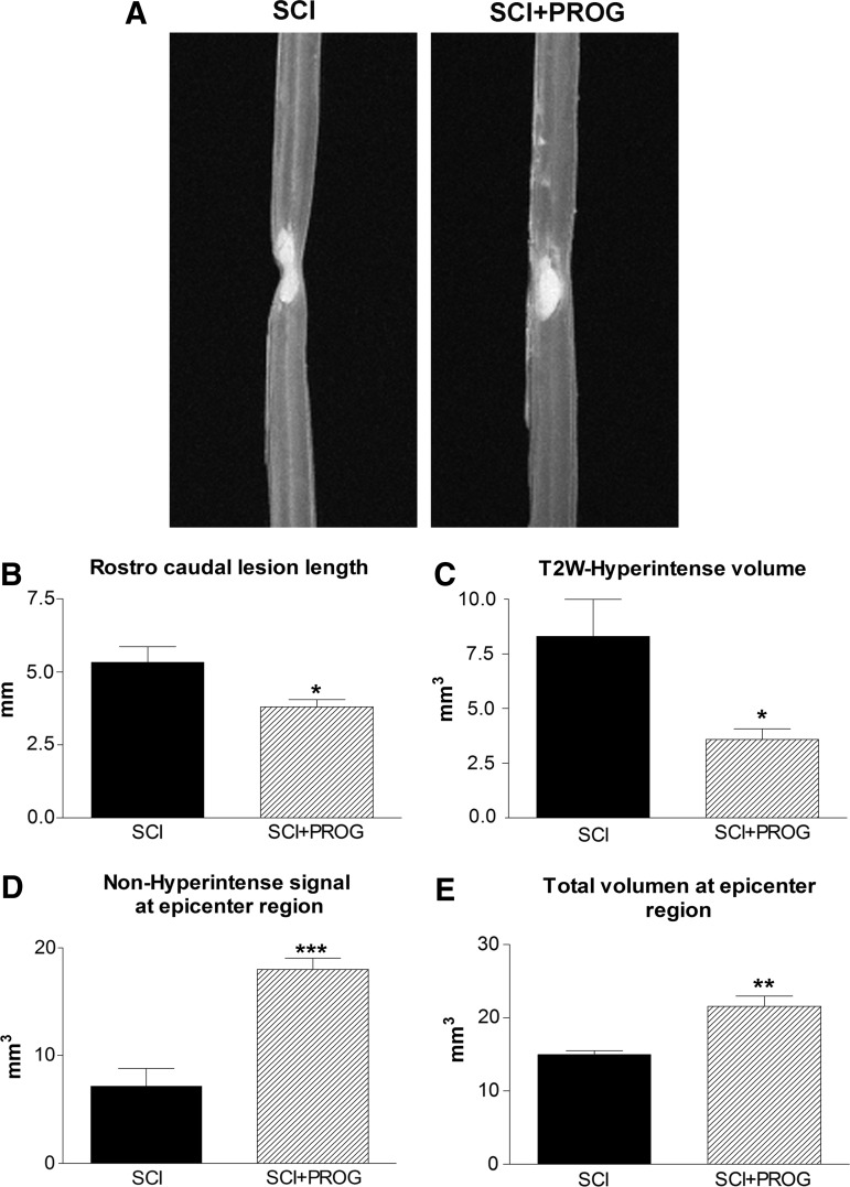
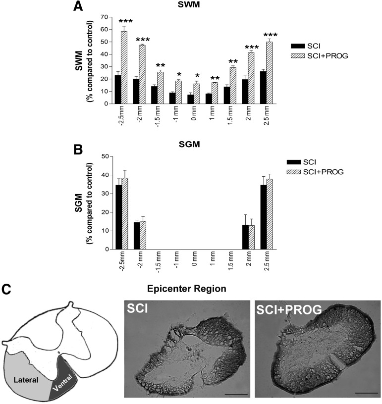
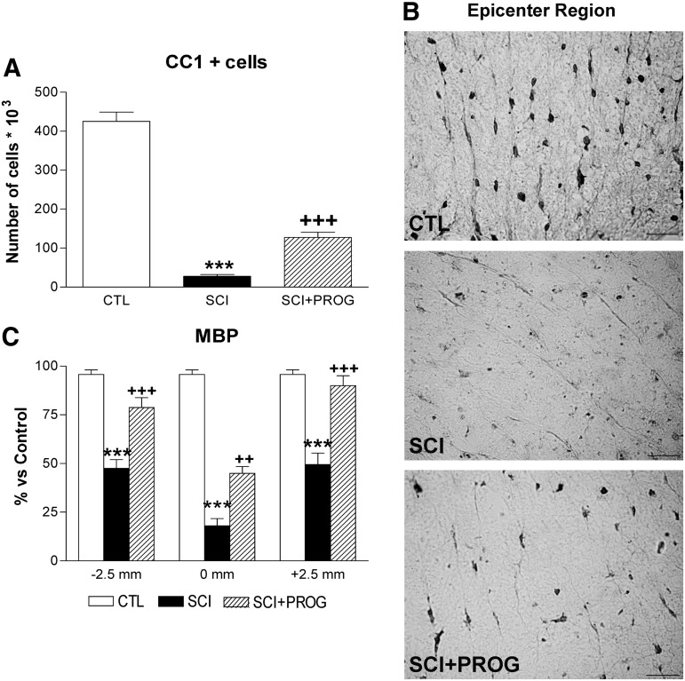
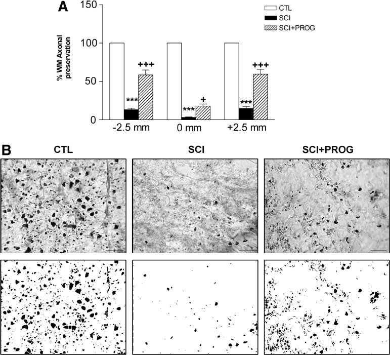
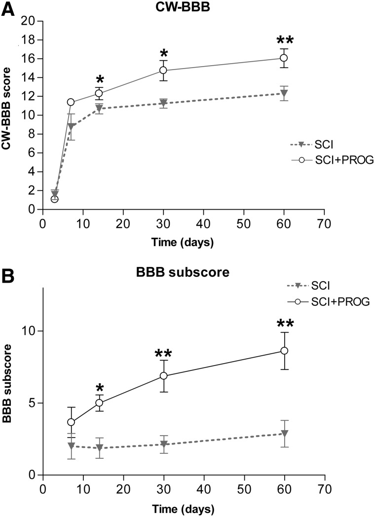
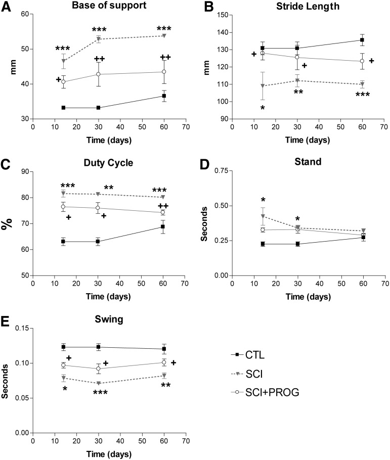
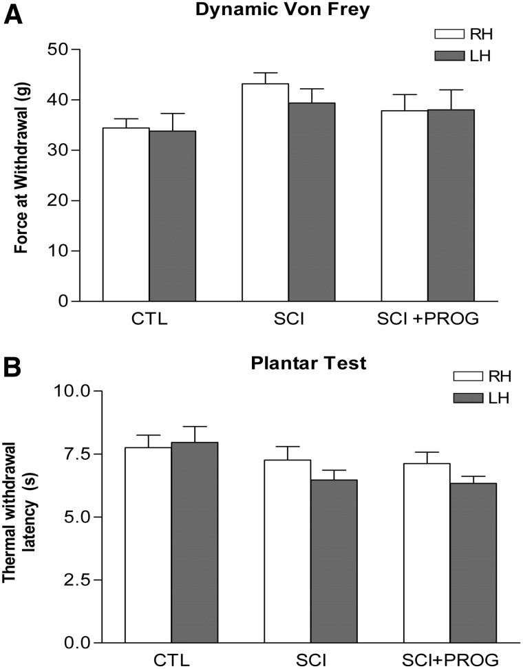
Progesterone is an anti-inflammatory and promyelinating agent after spinal cord injury, but its effectiveness on functional recovery is still controversial. In the current study, we tested the effects of chronic progesterone administration on tissue preservation and functional recovery in a clinically relevant model of spinal cord lesion (thoracic contusion). Using magnetic resonance imaging, we observed that progesterone reduced both volume and rostrocaudal extension of the lesion at 60 days post-injury. In addition, progesterone increased the number of total mature oligodendrocytes, myelin basic protein immunoreactivity, and the number of axonal profiles at the epicenter of the lesion. Further, progesterone treatment significantly improved motor outcome as assessed using the Basso-Bresnahan-Beattie scale for locomotion and CatWalk gait analysis. These data suggest that progesterone could be considered a promising therapeutical candidate for spinal cord injury.


Figures

<b>FIG. 1.</b>
FIG. 1.
Representative T2-weighted three-dimensional magnetic resonance imaging (T2W-3D MRI) showing smaller hyperintense region in spinal cord injury plus progesterone (SCI+PROG) rats (A). Effects of PROG treatment on reducing lesion extension (B), cyst formation (C), preservation of non-hyperintense tissue (D), and total spinal cord volume (E) at the epicenter region after 60 days of injury. All values are expressed as mean±standard error of the mean, *p<0.05 vs. SCI, **p<0.01 vs. SCI, ***p<0.001 vs. SCI, t test.
<b>FIG. 2.</b>
FIG. 2.
Effect of progesterone (PROG) on white and grey matter damage measured at 60 days after lesion. An increase in spred white matter (SWM) preservation was observed in spinal cord injury (SCI)+PROG rats at all distances measured from the epicenter (A). PROG did not modify the volume of spared grey matter (SGM) along the segment (B). Micrographs showing coronal section of spinal cord stained with Luxol Fast Blue from the epicenter region. Spinal cord contusion produced a cavitation at the epicenter, leaving a greater sparse region of intact white matter in the ventral-lateral margins (lateral and ventral funiculus) in SCI+PROG group (C). All values are expressed as mean±standard error of the mean. For A and B: ***p<0.001 vs. SCI, **p<0.01 vs. SCI, *p<0.05 vs. SCI, two-way analysis of variance of repeated measures followed by Bonferroni post-test. For C: scale bar=200 μm.
<b>FIG. 3.</b>
FIG. 3.
Progesterone (PROG) increased the oligodendrocytes number and reduced myelin damage 60 days after lesion. Spinal cord injury (SCI) animals showed a reduction in the total number of CC1+cells throughout the white matter compared with control (CTL) rats, while the in SCI+PROG group, the reduction was lower (A). Micrographs showing CC1 immunohistochemistry in ventrolateral white matter at the epicenter region in the different groups of animals (B). Myelin basic protein (MBP) immunoreactivity increased in SCI+PROG rats compared with SCI rats at 2.5 mm rostral, 2.5 mm caudal, and the midpoint−0mm- from the epicenter (C). All values are expressed as mean±standard error of the mean. For A: ***p<0.001 vs. CTL, +++ p<0.001 vs. SCI, one-way analysis of variance (ANOVA) followed by Newman Keuls post-test. For C: ***p<0.001 vs. CTL, ++ p<0.01 vs. SCI, +++ p<0.001 vs. SCI, two-way ANOVA of repeated measures followed by Bonferroni post-test. For B: scale bar=20 μm.
<b>FIG. 4.</b>
FIG. 4.
Progesterone (PROG) improved axonal preservation 60 days after injury at 2.5 mm rostral, 2.5 mm caudal, and at the midpoint−0mm- of the epicenter (A). Micrographs show neurofilament (NF-H) immohistochemistry in ventrolateral white matter at the epicenter region in the different groups of animals. Spinal cord injury (SCI) group showed less axonal profiles than control (CTL), while in SCI+PROG rats, the number was recovered (B upper panel). Examples of processed images used for quantification are shown (B lower panel). All values are expressed as mean±standard error of the mean. For A: ***p<0.001 vs. CTL, +++ p<0.001 vs. SCI, two-way analysis of variance of repeated measures followed by Bonferroni post-test. For B: scale bar=20 μm.
<b>FIG. 5.</b>
FIG. 5.
Integration of the CatWalk(CW)-based coordination into the Basso-Bresnahan-Beattie (BBB) locomotor scale results in the CatWalk-based BBB, which showed that spinal cord injury plus progesterone (SCI+PROG) animals regained better locomotor function when compared with SCI rats (A). BBB subscore also showed marked differences between SCI and SCI+PROG rats (B). All values are expressed as mean±standard error of the mean, *p<0.05 vs. SCI, **p<0.01 vs. SCI, ***p<0.001 vs. SCI, nonparametric Mann-Whitney U test.
<b>FIG. 6.</b>
FIG. 6.
CatWalk gait parameters analysis showed that progesterone (PROG) improved overground locomotion. Base of support (A), Stride length (B), Duty cycle (C), Stand phase (D), and Swing phase (E). All values are expressed as mean±standard error of the mean, *p<0.05 vs. contgrol (CTL), **p<0.01 vs. CTL, ***p<0.001 vs. CTL, + p<0.05 vs. spinal cord injury (SCI), ++ p<0.01 vs. SCI, two-way analysis of variance of repeated measures followed by Bonferroni post-test.
<b>FIG. 7.</b>
FIG. 7.
PROG treatment improved interlimb coordination after injury. Spinal cord contusion decreased RI in SCI rats compared to CTL animals, but in a lesser extent in SCI+PROG (A). PD variability was increased in SCI rats compared to CTL in all the studied pairs of paw. However, in SCI+PROG rats, PD variability was lower compared to SCI in diagonal pair (B), ipsilateral pair (C) a pelvic girdle (D). All values are expressed as mean±SEM, ***p<0.001 vs CTL, +p<0.05 vs SCI. A two-way ANOVA of repeated measures followed by Bonferroni post-text.
<b>FIG. 8.</b>
FIG. 8.
Neither PROG nor spinal cord injury induced mechanical or thermal allodynia after 60 days of spinal cord contusion. Dynamic Von Frey (A). Plantar test (B). All values are expressed as mean ±SEM and no statistical differences were found by two-way ANOVA. RH, right hindlimb; LH, left hindlimb.

References

    1. Ambrozaitis K.V., Kontautas E., Spakauskas B., and Vaitkaitis D. (2006). [Pathophysiology of acute spinal cord injury.] (Lith) Medicina (Kaunas) 42, 255–261 - PubMed
    1. Beattie M.S., Farooqui A.A., and Bresnahan J.C. (2000). Review of current evidence for apoptosis after spinal cord injury. J. Neurotrauma 17, 915–925 - PubMed
    1. Borgens R.B., and Liu-Snyder P. (2012). Understanding secondary injury. Q. Rev. Biol. 87, 89–127 - PubMed
    1. Kwon B.K., Okon E., Hillyer J., Mann C., Baptiste D., Weaver L.C., Fehlings M.G., and Tetzlaff W. (2011). A systematic review of non-invasive pharmacologic neuroprotective treatments for acute spinal cord injury. J. Neurotrauma 28, 1545–1588 - PMC - PubMed
    1. Lammertse D.P. (2013). Clinical trials in spinal cord injury: lessons learned on the path to translation. The 2011 International Spinal Cord Society Sir Ludwig Guttmann Lecture. Spinal Cord51, 2–9 - PubMed

Publication types

LinkOut - more resources