Comparative Study
. 2014 Mar;35(10):3281-97.
doi: 10.1016/j.biomaterials.2013.12.103. Epub 2014 Jan 22.

Comparative decellularization and recellularization of normal versus emphysematous human lungs

Affiliations
Comparative Study

Comparative decellularization and recellularization of normal versus emphysematous human lungs

Darcy E Wagner et al. Biomaterials. 2014 Mar.

Abstract

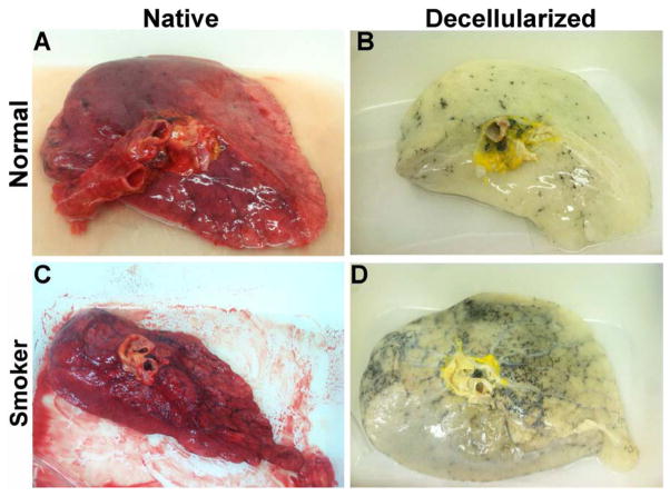
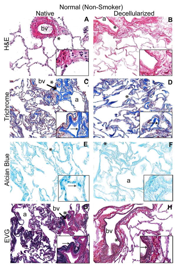
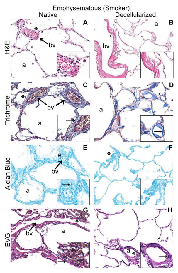
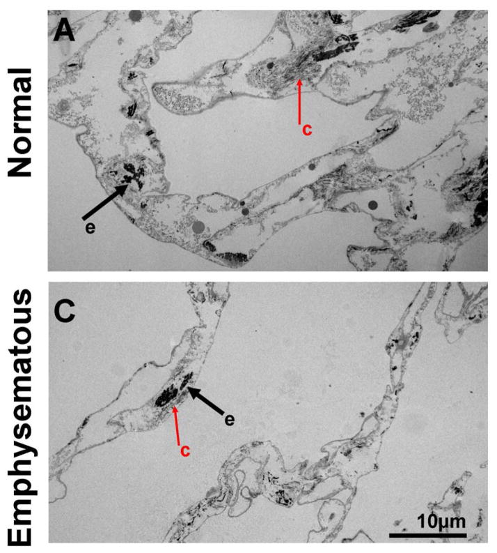
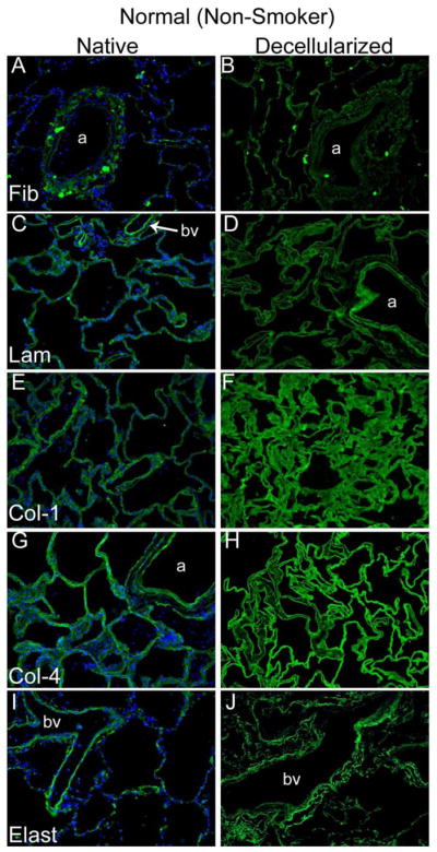
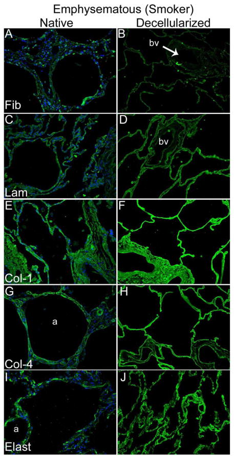
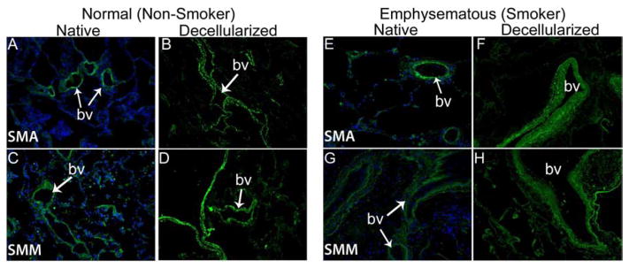
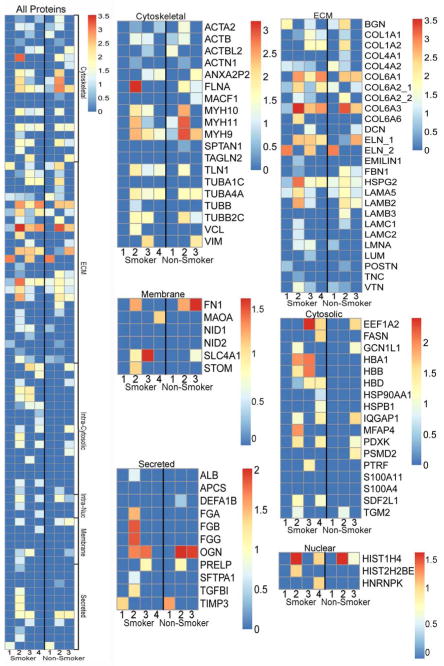
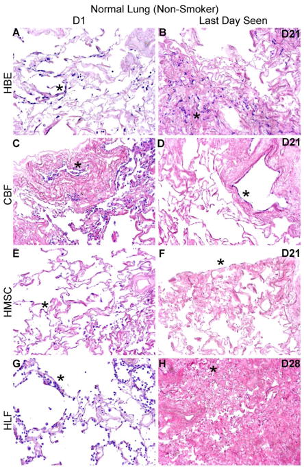
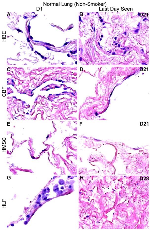
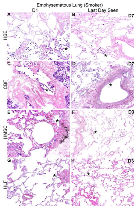
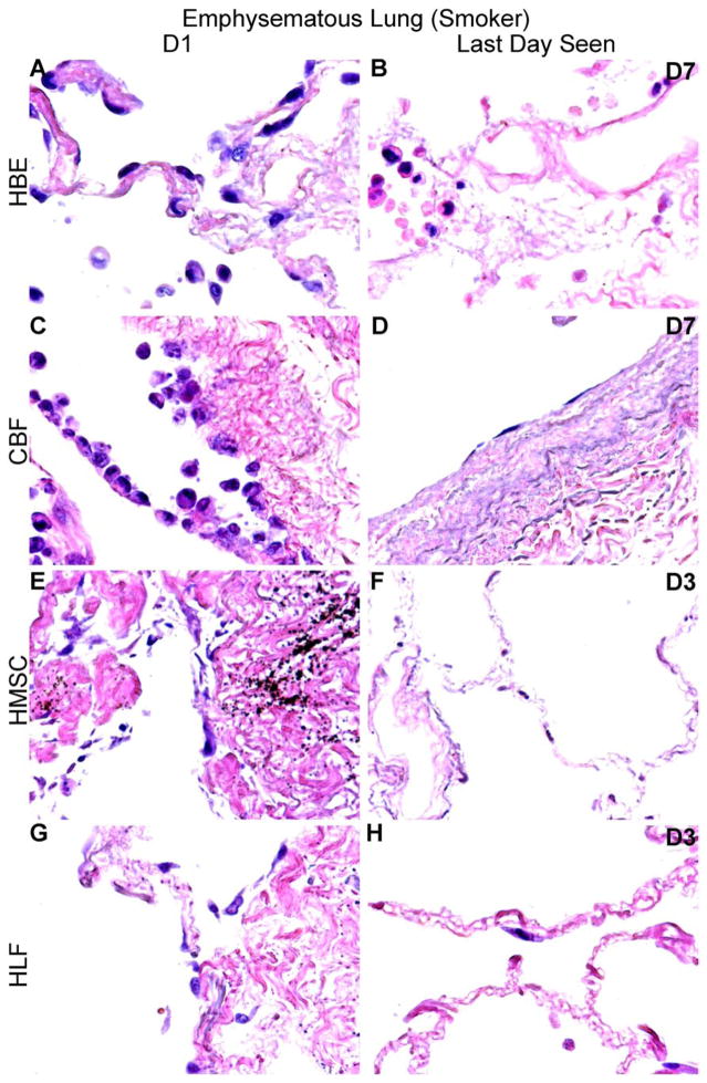
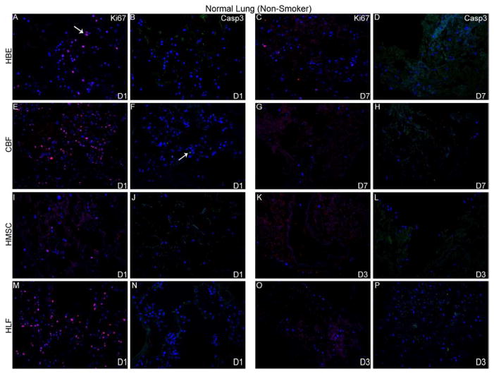
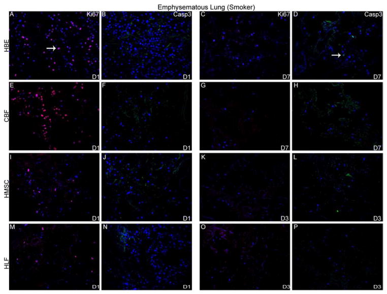
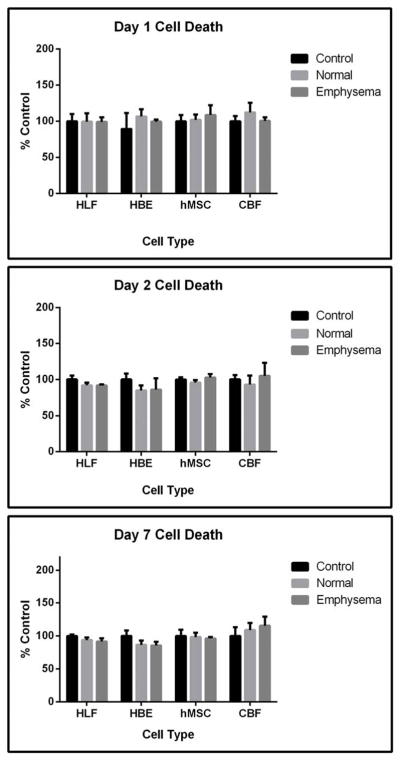
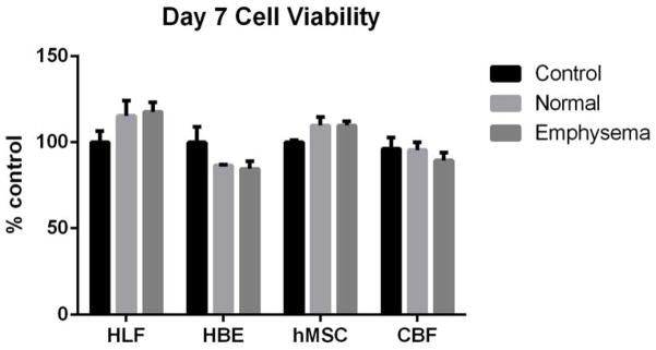
Acellular whole human lung scaffolds represent a unique opportunity for ex vivo tissue engineering. However, it remains unclear whether lungs from individuals with chronic lung diseases such as chronic obstructive pulmonary disease (COPD) can be appropriately decellularized and recellularized. To assess this, cadaveric human lungs from normal (non-smoking) patients and from patients with COPD (smoking history) were decellularized and found by histochemical and immunohistochemical staining, electron microscopy, and mass spectrometry to retain characteristic histological architecture and extracellular matrix components (ECM) reflecting either normal or COPD, particularly emphysematous, origin. Inoculation of human bronchial epithelial cells, endothelial progenitor cells, bone marrow-derived mesenchymal stem cells, and lung fibroblasts via airway or vascular routes into small, excised segments of the decellularized lungs demonstrated that normal lung scaffolds robustly supported initial engraftment and growth of each cell type for up to one month. In contrast, despite initial binding, all cell types inoculated into decellularized emphysematous lungs did not survive beyond one week. However, cell attachment and proliferation on solubilized ECM homogenates of decellularized normal and emphysematous lungs coated onto tissue culture plates was comparable and not impaired, suggesting that the 3-dimensional decellularized emphysematous scaffolds may lack the necessary ECM architecture to support sustained cell growth.

Keywords: Acellular matrix; Emphysema; Endothelial cell; Epithelial cell; Extracellular matrix (ECM); Human lung fibroblast.


Figures

Figure 1
Figure 1. Decellularized normal and emphysematous human lungs retain characteristic gross and histologic appearances
1A) Representative images of naïve and decellularized normal and emphysematous human lungs. Anthracotic pigment (black, dense deposits) is grossly retained following decellularization (panels B,D). 1B,C) Representative photomicrographs demonstrate maintenance of characteristic histologic and collagen content but loss of elastin and glycosaminoglycans following decellularization of both normal (1B) and emphysematous human lungs (1C) a = airways, bv = blood vessels. Arrows highlight individual blood vessels. Original magnifications 100X. Inserts indicate higher power images (200X) of areas indicated by *. 1D) Electron micrographs demonstrate retention of characteristic normal and emphysematous alveolar septa following decellularization with alveolar wall thinning observed in emphysematous samples. Collagen (labeled c) and elastin (labeled e) are indicated with black and red arrows, respectively. All scale bars are 10μm.
Figure 2
Figure 2. Decellularization preserves major ECM proteins in normal and emphysematous human lungs
Representative photomicrographs comparing native normal human lung and freshly decellularized human lung (2A) and native and freshly decellularized emphysematous lung (2B) are depicted. Smooth muscle actin and smooth muscle myosin similarly are retained following decellularization of both normal and emphysematous human lungs (2C). Nuclear DAPI staining is depicted in blue, and the stain(s) of interest are depicted in green in each respective panel. Lam = laminin, Col-1 = type I collagen, Col-4 = type 4 collagen, Elast = elastin, Fib = fibronectin. SMA = smooth muscle actin, SMM = smooth. a = airways, bv = blood vessels. Original magnifications 200x.
Figure 3
Figure 3. Decellularized normal and emphysematous human lungs retain both ECM and non-ECM proteins
Positively identified proteins (ie. those with unique peptide hits greater than or equal to two) were manually grouped according to their subcellular location. Heat maps for all proteins detected, grouped by category, as well as individually protein categories with identified proteins listed are shown. All unique peptide hit values were log normal transformed to more readily show the relative abundance of each protein. Individual heat maps are shown for scaffolds produced from lungs obtained from obtained from 4 normal (non-smoking) and 3 emphysematous (smoking) lungs. The key to protein identification is depicted in Supplemental Table 1.
Figure 4
Figure 4. HBE, CBF, hMSC, and HLF cells engraft and proliferate in decellularized normal human lungs for up to 28 days, but fail to thrive in emphysematous lungs beyond 3–7 days
Representative H and E photomicrographs depict the characteristic recellularization patterns one day post-inoculation of each cell type and the last day viable cells were observed. Engrafted cells in decellularized normal human lungs predominantly acquired characteristic adherent, flattened phenotypes, while those in emphysematous lungs do not appear to similarly engraft. All cell types were viable up to 21 days post-inoculation in acellular normal tissue, except for the HLFs which were viable until day 28 in normal acellular tissue. hMSCs and HLFs remained viable in acellular emphysematous lungs for three days whereas the HBEs and CBFs remained viable for seven days. Normal lungs: N = 3 for HBEs, N = 13 for CBFs, N = 3 for hMSCs, and N = 6 for HLFs. Emphysematous lungs: N = 7 for HBEs, N = 10 for CBFs, N = 5 for hMSCs, and N = 3 for HLFs. Asterisk indicates enlarged region shown in lower power images. Original magnification 100x (low power images) and 400x (high power images)
Figure 5
Figure 5. HBE, CBF, hMSC, and HLFs all demonstrate initial proliferation post-inoculation in decellularized normal and emphysematous human lungs by positive Ki67 staining
Representative photomicrographs of Ki67 (red) or caspase-3 (green) staining are shown at 1 day post-inoculation and at the last viable time point cells were observed in acellular normal (Panel A) or emphysematous lung (Panel B). DAPI nuclear staining is depicted in blue. White arrow indicates positive staining. Original magnification 200x.
Figure 6
Figure 6. No significant differences in cell death or viability are detected when cells are cultured wells coated with ECM from normal or emphysematous decellularized human lungs
Cells grown on ECM derived from normal or emphysematous human lungs show no detectable differences in cell death between day 1 (6A) and day 7 (6B). Quantitatively, there were no differences in cell death at days 1, 2, and 7 as assessed by an LDH assay (6C) or in cell viability on day 7 as assessed by MTS assay (6D) indicating that the scaffolds are nontoxic and that the ECM components support cellular proliferation for 7 days. Data are presented as mean+/− standard deviation as percentage compared to control cells grown on standard tissue culture plastic (HBE, HLF, hMSC) or collagen I-coated tissue culture plastic (CBF). No statistically significant differences were found. N=4 for each cell grown on control wells, N=3 for each cell type grown on wells coated with either solubilized ECM lyophilates of normal or emphysematous decellularized human lungs. Data shown is representative of one of two similar experiments.

References

    1. Orens JB, Garrity ER., Jr General overview of lung transplantation and review of organ allocation. Proc Am Thorac Soc. 2009;6:13–9. - PubMed
    1. Panoskaltsis-Mortari A, Weiss DJ. Breathing new life into lung transplantation therapy. Mol Ther. 2010;18:1581–3. - PMC - PubMed
    1. Price AP, England KA, Matson AM, Blazar BR, Panoskaltsis-Mortari A. Development of a decellularized lung bioreactor system for bioengineering the lung: the matrix reloaded. Tissue Eng Part A. 2010;16:2581–91. - PMC - PubMed
    1. Cortiella J, Niles J, Cantu A, Brettler A, Pham A, Vargas G, et al. Influence of acellular natural lung matrix on murine embryonic stem cell differentiation and tissue formation. Tissue Eng Part A. 2010;16:2565–80. - PubMed
    1. Petersen TH, Calle EA, Zhao L, Lee EJ, Gui L, Raredon MB, et al. Tissue-engineered lungs for in vivo implantation. Science. 2010;329:538–41. - PMC - PubMed

Publication types

Substances