. 2013 Sep;1(7):534-42.
doi: 10.1016/S2213-2600(13)70138-3. Epub 2013 Jul 25.

Tropism and innate host responses of a novel avian influenza A H7N9 virus: an analysis of ex-vivo and in-vitro cultures of the human respiratory tract

Affiliations

Tropism and innate host responses of a novel avian influenza A H7N9 virus: an analysis of ex-vivo and in-vitro cultures of the human respiratory tract

Michael C W Chan et al. Lancet Respir Med. 2013 Sep.

Abstract

Background: Since March, 2013, an avian-origin influenza A H7N9 virus has caused severe pneumonia in China. The aim of this study was to investigate the pathogenesis of this new virus in human beings.

Methods: We obtained ex-vivo cultures of the human bronchus, lung, nasopharynx, and tonsil and in-vitro cultures of primary human alveolar epithelial cells and peripheral blood monocyte-derived macrophages. We compared virus tropism and induction of proinflammatory cytokine responses of two human influenza A H7N9 virus isolates, A/Shanghai/1/2013 and A/Shanghai/2/2013; a highly pathogenic avian influenza H5N1 virus; the highly pathogenic avian influenza H7N7 virus that infected human beings in the Netherlands in 2003; the 2009 pandemic influenza H1N1 virus, and a low pathogenic duck H7N9 virus that was genetically different to the human disease causing A H7N9 viruses.

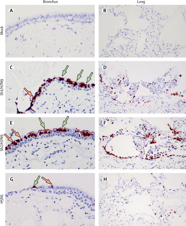
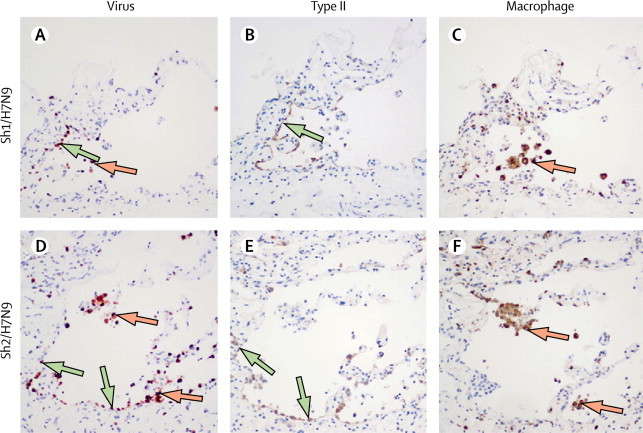
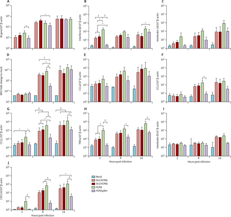
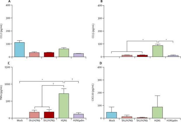
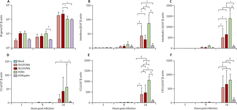
Findings: Both human H7N9 viruses replicated efficiently in human bronchus and lung ex-vivo cultures, whereas duck/H7N9 virus failed to replicate in either. Both human A H7N9 viruses infected both ciliated and non-ciliated human bronchial epithelial cells and replicated to higher titres than did H5N1 (p<0.0001 to 0.0046) and A/Shanghai/1/2013 replicated to higher titres than did H7N7 (p=0.0002-0.01). Both human A H7N9 viruses predominantly infected type II alveolar epithelial cells and alveolar macrophages in the human lung and replicated to higher titres than did H5N1 (p<0.0001 to 0.0078); A/Shanghai/1/2013 replicated to higher titres than did H1N1 (p=0.0052-0.05) and H7N7 (p=0.0031-0.0151). Human H7N9 viruses were less potent inducers of proinflammatory cytokines compared with H5N1 virus.

Interpretation: Collectively, the results suggest that the novel H7N9 viruses are better adapted to infect and replicate in the human conducting and lower airways than are other avian influenza viruses, including H5N1, and pose an important pandemic threat.

Funding: Area of Excellence Scheme of the University Grants Committee (AoE/M-12/96), Hong Kong Special Administrative Region.


Figures

Figure 1
Figure 1
Viral replication kinetics of influenza A H7N9 virus in ex-vivo cultures of bronchus (A) and lung (B), infected with 10 TCID50/mL of influenza viruses at 37°C Barcharts show the mean virus titre pooled from at least three independent experiments. The horizontal dotted line denotes the limit of detection in the TCID50 assay.; error bars show SEM. Tables show statistical significance between virus titres at each timepoint. Red shows that the reference virus titre (listed at the top of the column) is significantly higher than the comparator (viruses listed on the left) and black shows that it is significantly lower. TCID50=tissue culture infective dose. ns=non-significant. *p<0·0001.
Figure 2
Figure 2
Tissue tropism of influenza A H7N9 virus in ex-vivo cultures of bronchus and lung Formalin-fixed paraffin-embedded sections of bronchus (A, C, E, G) and lung (B, D, F, H) after 24 h infection with mock (A, B), Sh1/H7N9 (C, D), Sh2/H7N9 (E, F), and H5N1 (G, H) viruses. Sections were stained with a monoclonal antibody against the influenza nucleoprotein with positive cells identified as a red-brown colour. Ciliated and non-ciliated cells are identified with green and orange arrows, respectively.
Figure 3
Figure 3
Cellular localisation of influenza antigen in human lung tissues infected with influenza A H7N9 virus Serial sections of ex-vivo cultures of human lungs infected with Sh1/H7N9 (A–C) and Sh2/H7N9 viruses (D–F). Sections were stained with monoclonal antibodies to influenza nucleoprotein (A, D), surfactant protein D (B, E), and CD68 (C, F). Green arrows show type II pneumocytes and orange arrows show alveolar macrophages.
Figure 4
Figure 4
Cytokine and chemokine mRNA expression profile in peripheral blood monocyte-derived macrophages infected with mock, Sh1/H7N9, Sh2/H7N9, H5N1, and H1N1pdm viruses Expression of the influenza matrix (M) gene (A), interferon β (B), interleukin 29 (C), MX1 (D), CCL4 (E), CCL2 (F), CCL5 (G), tumour necrosis factor α (TNFα; H), interleukin 8 (I), and CXCL10 (J) at 3, 8, and 24 h after infection. Graphs show mean mRNA copies expressed per 10 β-actin copies from three independent experiments; error bars show SEM. MX1=myxovirus resistance 1. CCL=chemokine (C-C motif) ligand. CXCL10=chemokine (C-X-C motif) ligand 10. *p<0·05. † p<0·01. ‡p<0·005.
Figure 5
Figure 5
Cytokine and chemokine protein expression in cell culture supernatants of peripheral blood monocyte-derived macrophages infected with mock, Sh1/H7N9, Sh2/H7N9, H5N1, and H1N1pdm viruses Expression of CCL2 (A), CCL5 (B) tumour necrosis factor α (TNFα; C), and CXCL10 (D) at 24 h post infection by ELISA (R&D Systems, Minneapolis, MN, USA). Graphs show mean protein from three independent experiments; error bars show SEM. CCL=chemokine (C-C motif) ligand. CXCL10=chemokine (C-X-C motif) ligand 10. *p<0·005. † p<0·01.
Figure 6
Figure 6
Cytokine and chemokine mRNA expression profile in human pneumocytes infected with mock, Sh1/H7N9, Sh2/H7N9, H5N1, and H1N1pdm viruses Expression of influenza matrix (M) gene (A), interferon β (B), interleukin 29 (C), CCL4 (D), CCL5 (E), and CXCL10 (F) at 1, 6, and 24 h after infection. Graphs show mean mRNA copies expressed per 10 β-actin copies from three independent experiments; error bars show SEM. CCL=chemokine (C-C motif) ligand. CXCL10=chemokine (C-X-C motif) ligand 10. *p<0·05. † p<0·01. ‡p<0·005.
Figure 7
Figure 7
Cytokine and chemokine protein expression in cell culture supernatants of pneumocytes infected with mock, Sh1/H7N9, Sh2/H7N9, H5N1, and H1N1pdm viruses Expression of CCL5 (A) and CXCL10 (B) at 24 h post infection by ELISA (R&D Systems, Minneapolis, MN, USA). Graphs show mean protein from three independent experiments; error bars show SEM. CCL=chemokine (C-C motif) ligand. CXCL10=chemokine (C-X-C motif) ligand 10. *p<0·05. † p<0·01.

Comment in

References

    1. Gao R, Cao B, Hu Y. Human infection with a novel avian-origin influenza A (H7N9) virus. N Engl J Med. 2013;368:1888–1897. - PubMed
    1. WHO Human infection with avian influenza A(H7N9) virus—update. http://www.who.int/csr/don/2013_07_04/en/index.html (accessed July 7, 2013).
    1. Li Q, Zhou L, Zhou M. Preliminary report: epidemiology of the avian influenza A (H7N9) outbreak in China. N Engl J Med. 2013 doi: 10.1056/NEJMoa1304617. published online April 24. - DOI - PubMed
    1. Kageyama T, Fujisaki S, Takashita E. Genetic analysis of novel avian A(H7N9) influenza viruses isolated from patients in China, February to April 2013. Euro Surveill. 2013;18:20453. - PMC - PubMed
    1. Chen Y, Liang W, Yang S. Human infections with the emerging avian influenza A H7N9 virus from wet market poultry: clinical analysis and characterisation of viral genome. Lancet. 2013;381:1916–1925. - PMC - PubMed

Publication types