Review
. 2014 Mar;37(3):146-58.
doi: 10.1016/j.tins.2013.12.002. Epub 2014 Jan 21.

Opening paths to novel analgesics: the role of potassium channels in chronic pain

Affiliations
Review

Opening paths to novel analgesics: the role of potassium channels in chronic pain

Christoforos Tsantoulas et al. Trends Neurosci. 2014 Mar.

Abstract

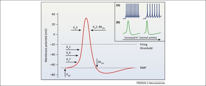
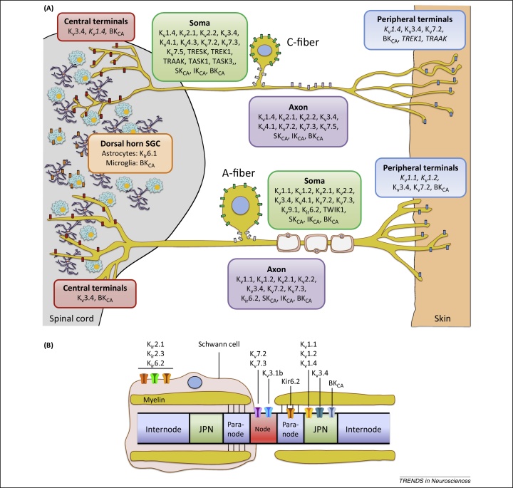
Chronic pain is associated with abnormal excitability of the somatosensory system and remains poorly treated in the clinic. Potassium (K⁺) channels are crucial determinants of neuronal activity throughout the nervous system. Opening of these channels facilitates a hyperpolarizing K⁺ efflux across the plasma membrane that counteracts inward ion conductance and therefore limits neuronal excitability. Accumulating research has highlighted a prominent involvement of K⁺ channels in nociceptive processing, particularly in determining peripheral hyperexcitability. We review salient findings from expression, pharmacological, and genetic studies that have untangled a hitherto undervalued contribution of K⁺ channels in maladaptive pain signaling. These emerging data provide a framework to explain enigmatic pain syndromes and to design novel pharmacological treatments for these debilitating states.

Keywords: dorsal root ganglia; pain; pharmacotherapy; potassium channel.


Figures

Figure 1
Figure 1
Potassium channel activation during action potential (AP) firing in sensory neurons. A depiction of the sequential engagement of different K+ channels during neuronal activity, and typical effects of K+ channel opening on AP waveform and frequency (inset). The resting membrane potential (RMP) is primarily stabilized by two-pore K+ (K2P) channels and Kv7 background conductance, whereas KATP channels may also contribute in large neurons . Basal excitability is also influenced by the opening of low-threshold Kv1 and Kv4 channels which filter out small depolarizations and therefore control the number of triggered APs. Kv4 channels are normally inactivated at RMP and require prior hyperpolarization (achieved during AP generation) to remove this steady-state inactivation. Once activated, however, Kv4 and other A-type channels may modulate firing threshold as well as repetitive spiking rate owing to their very fast kinetics [inset (A)] . Following suprathreshold stimulation and initiation of an AP, high-threshold Kv3 channels open to limit AP duration and ensure quick recovery of voltage-gated Na+ channels from inactivation [inset (B)]. Kv2 channels are also high-threshold but with much slower activation and inactivation kinetics; they mainly contribute to the repolarizing/after-hyperpolarizing phases and are hence important for regulating interspike interval and conduction fidelity during sustained stimulation [inset (B)] . Upon neuronal activity, Ca2+-activated K+ channels are engaged during repolarization (BKCA) and after-hyperpolarization (SKCA) to provide feedback inhibition at nerve terminals by restricting AP duration and thus neurotransmitter release [(inset (B)]. It is emphasized that this schematic is a simplified representation of most prominent K+ channel contributions to AP firing, based on in vitro assessment of recombinant counterparts. In vivo, however, the oligomeric composition, association with auxiliary proteins, post-translational modifications, and regulation by intracellular messengers can yield divergent biophysical properties. K+ channel opening will also have concurrent effects on the function of other ion channels, for instance by affecting their inactivation. For this reason the combined effect on firing behavior in a physiological context is often hard to predict. Abbreviations: BKCA, big conductance and SKCA, small conductance Ca2+-activated K+ channels.
Figure 2
Figure 2
Expression and function of K+ channels in sensory neurons. (A) Subcellular localization of K+ channel subunits in unmyelinated (top) and myelinated (bottom) murine dorsal root ganglia (DRG) neurons. The panoply of K+ channels endows sensory neurons with a sophisticated machinery for the regulation of neuronal excitability. The depiction illustrated here is not absolute but rather reflects most prominent expression patterns in pain-relevant subpopulations, as reported in the literature. In addition, it is noted that K+ channel distribution patterns can vary tremendously between species, and validation against human data is currently very limited , . In the pain pathway, the TWIK-related (TR) channels TREK1 and TRAAK (and possibly TRESK) located at C-fiber terminals can counteract the activation of inward-conducting ion channels by pressure, heat or cold, whereas steady Kv7 currents also stabilize RMP and regulate action potential (AP) threshold. In myelinated neurons, low-threshold Kv1.1/Kv1.2 heterotetramers appear to modulate acute and neuropathic pain modalities , , , whereas Kv1.4 and Kv4 members may exert similar roles in small nociceptors , . In addition, recent evidence suggest that Kv1.1/Kv1.2 may also function as mechanoreceptors in some C-fibers (not shown) . Transmission of signals generated at the periphery is reliant on numerous axonal K+ channels, which influence the fidelity of AP conduction and therefore the fiber following frequency. Although normal sensory transduction is independent of spiking in the DRG soma, this can become a site of spontaneous firing in neuropathic conditions. In these scenarios, the activity of somal K+ channels may become an important regulator of excitability by influencing somal AP generation as well as propagation past the DRG T-junction. Potential candidates here are channels that preferentially localize at the soma or axon initial segments (in grey), such as Kv4.3 in mechanosensitive C-fibers or Kv2/Kv9.1 in A-fibers , . At the central terminals, Ca2+-activated channels BKCA fine-tune activity and regulate neurotransmitter release in the spinal cord in response to calcium influx during AP firing. The high-threshold Kv3.4 limits AP duration and thus may play a key role in synaptic transmission, whereas Kv1.2 may also regulate presynaptic terminal excitability . Finally, pain processing can be influenced by K+ channels expressed by glial satellite cells (GSC) in the dorsal horn. Astrocyte-expressed Kir6.1 (and perhaps Kir3.1 [86]) buffers the extracellular K+ to maintain equilibrium potential during neuronal firing , and BKCA conduction is involved in microgliosis following injury . In addition, satellite cell-expressed Kir4.1 is involved in facial pain processing in the trigeminal ganglion (not shown). Subunits denoted in italics represent localizations that are indirectly implied by pharmacological profiling in DRG neurons, or by extrapolating on known localization in other neuronal types. For example, the Kv1.1 and Kv1.2 subunits are typically detected in dendrites and terminals of CNS neurons , , whereas TREK1 and TRAAK are axonally trafficked in sciatic nerves and are present at synaptic sites in cerebellar cultures . (B) K+ channel composition of a myelinated DRG axon, illustrating nodes, paranodes, juxtaparanodes (JPN), internode segments, and a myelinating Schwann cell. The Kv7.2 and Kv7.3 subunits (together with a splice variant of Kv3.1) are found in the nodes, and may therefore more prominently affect saltatory conduction under physiological conditions. Following axonal injury and demyelination, however, other channels such as the juxtaparanodal Kv1 subunits may become exposed, leading to reduced conduction velocity and negative symptoms including sensory loss. In other cases, reduced axonal K+ channel function due to disrupted node organization (e.g., autoantibodies against Kv complex proteins) may induce peripheral hyperexcitability. Schwann cells also express inward rectifiers that regulate the node microenvironment during neuronal activity.

References

    1. van Hecke O. Chronic pain epidemiology and its clinical relevance. Br. J. Anaesth. 2013;111:13–18. - PubMed
    1. Labianca R. Adverse effects associated with non-opioid and opioid treatment in patients with chronic pain. Clin. Drug Investig. 2012;32(Suppl. 1):53–63. - PubMed
    1. D’Mello R., Dickenson A.H. Spinal cord mechanisms of pain. Br. J. Anaesth. 2008;101:8–16. - PubMed
    1. Kajander K.C. Spontaneous discharge originates in the dorsal root ganglion at the onset of a painful peripheral neuropathy in the rat. Neurosci. Lett. 1992;138:225–228. - PubMed
    1. Kajander K.C., Bennett G.J. Onset of a painful peripheral neuropathy in rat: a partial and differential deafferentation and spontaneous discharge in Abeta and Adelta primary afferent neurons. J. Neurophysiol. 1992;68:734–744. - PubMed

Publication types