. 2014 Feb 19;81(4):873-887.
doi: 10.1016/j.neuron.2013.12.011. Epub 2014 Jan 23.

Central terminal sensitization of TRPV1 by descending serotonergic facilitation modulates chronic pain

Affiliations

Central terminal sensitization of TRPV1 by descending serotonergic facilitation modulates chronic pain

Yu Shin Kim et al. Neuron. .

Abstract

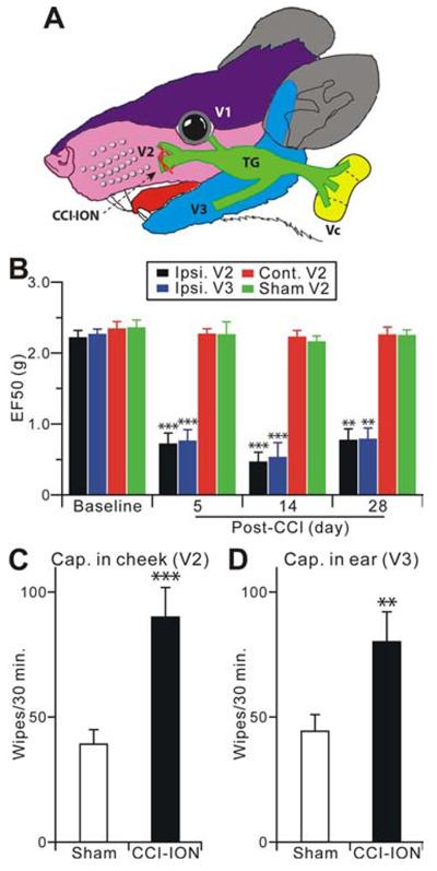
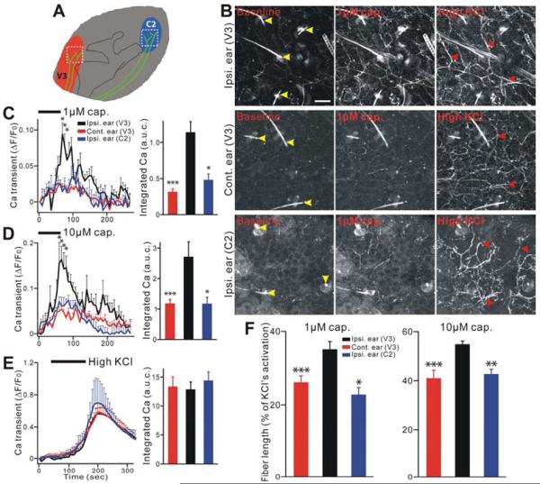
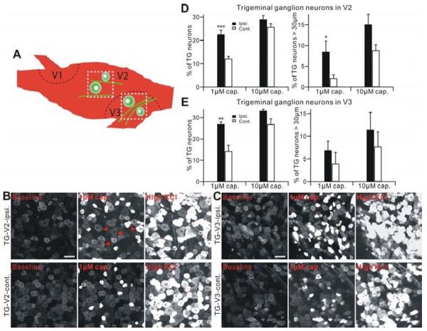
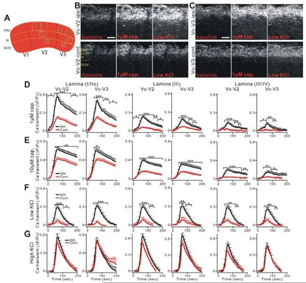
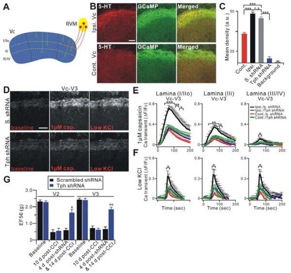
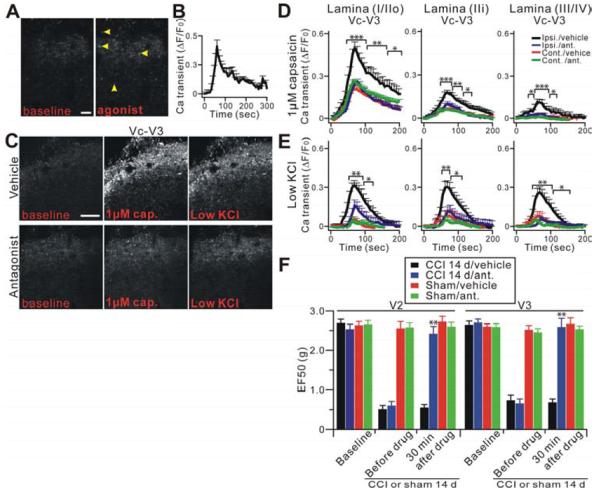
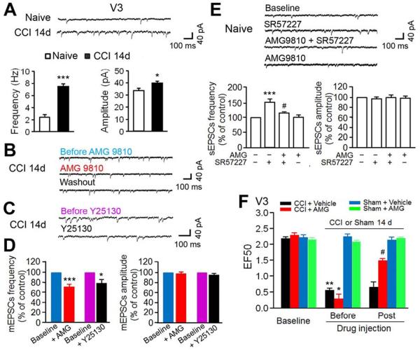
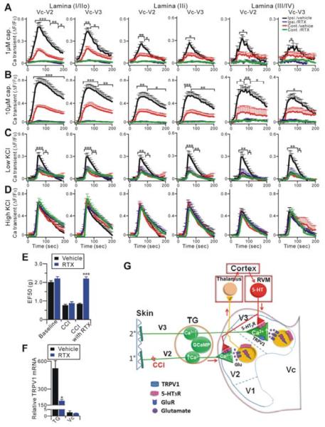
The peripheral terminals of primary nociceptive neurons play an essential role in pain detection mediated by membrane receptors like TRPV1, a molecular sensor of heat and capsaicin. However, the contribution of central terminal TRPV1 in the dorsal horn to chronic pain has not been investigated directly. Combining primary sensory neuron-specific GCaMP3 imaging with a trigeminal neuropathic pain model, we detected robust neuronal hyperactivity in injured and uninjured nerves in the skin, soma in trigeminal ganglion, and central terminals in the spinal trigeminal nucleus. Extensive TRPV1 hyperactivity was observed in central terminals innervating all dorsal horn laminae. The central terminal TRPV1 sensitization was maintained by descending serotonergic (5-HT) input from the brainstem. Central blockade of TRPV1 or 5-HT/5-HT3A receptors attenuated central terminal sensitization, excitatory primary afferent inputs, and mechanical hyperalgesia in the territories of injured and uninjured nerves. Our results reveal central mechanisms facilitating central terminal sensitization underlying chronic pain.


Figures

Figure 1
Figure 1
Trigeminal CCI-ION in Pirt-GCaMP3 mice shows long-lasting hyperalgesia from territories innervated by injured and uninjured nerves. (A) The CCI-ION and the anatomy in orofacial and trigeminal system are shown. (B) Mechanical hyperalgesia was tested at the ipsilateral V2 whisker pad (injured), the V3 lower jaw (uninjured), and contralateral V2 regions at 5, 14 and 28 days after CCI-ION (n=8) or sham (n=6) in mice. The data are expressed as an EF50 value and are presented as mean ± s.e.m. (C,D) Cheek injection of capsaicin evokes nocifensive facial wiping by the forepaw. The number of face wipes were measured within 30 minutes of capsaicin (0.5 μg) injection into the cheek (V2, injured) and the anterior part of ear skin (V3, uninjured; n=10 for each group). Data are presented as mean ± s.e.m. **p<0.01; ***p<0.001 vs. baseline (B), or sham (C,D).
Figure 2
Figure 2
Ear skin nerve fibers in the secondary hyperalgesia region show peripheral hypersensitivity after CCI-ION in Pirt-GCaMP3 mice. (A) Schematic diagram of a mouse ear. Red or blue color indicates where V3 nerve fibers (green) or C2 nerve fibers are innervated into ear skin, respectively. White square dash lines indicate where the image was taken. (B) Representative GCaMP3 imaging of ear skin explants. Ear skin after CCI-ION in Pirt-GCaMP3 mice were activated by capsaicin (1 μM) or high KCl (100 mM). Upper rows show ipsilateral (on the CCI-ION side) V3 region. Middle rows show contralateral (uninjured side) V3 region. Lower rows show ipsilateral C2 region. Yellow arrow heads indicate hairs and hair follicles which are autofluoresecent. Red arrow heads indicate activated fibers and endings. Ipsi., ipsilateral; cont., contralateral. Scale bar: 50 μm. (CE) In left panels time course of the amplitude of the Ca2+ transient that was evoked by capsaicin (C,1 μM; D, 10 μM) or KCl (E, 100 mM) application in ear skin. In right panels Ca2+ transient amounts with area under curve (a.u.c.) that was evoked by capsaicin (C,1 μM; D, 10 μM) or KCl (E, 100 mM) in ear skin. All population data for Ca2+ transient are expressed as the percentage of baseline Ca2+ transient (ΔF/F0) and are presented as mean ± s.e.m. Black bars indicate when stimuli were applied. (F) Length of ear skin fibers and endings after activation by capsaicin (1 μM and 10 μM), which was normalized to the length activated by KCl. Significant increases were found in ipsilateral ear (V3) as compared to contralateral ear (V3) or ipsilateral ear (C2). *p<0.05; **p<0.01; ***p<0.001.
Figure 3
Figure 3
TG neurons show neuronal hyperactivity after CCI-ION in Pirt-GCaMP3 mice. (A) Schematic diagram of a mouse TG. Black dash lines indicate the border of three large divisions (V1, V2, V3) in TG (green cells). White square dash lines indicate where the image was taken. (B and C) Representative GCaMP3 imaging of TG explants. TG neurons of V2 (B) or V3 (C) division after CCI-ION in Pirt-GCaMP3 mice were activated by capsaicin (1 μM) or KCl (100 mM). Upper panels show ipsilateral V2 (B) or V3 (C) division of TG. Lower panels show contralateral V2 (B) or V3 (C) division of TG. Scale bar: 50 μm for both. Arrow heads in red indicate TG neurons which are over 30 μm diameter and activated by capsaicin. (D and E) Population data for V2 shown in (D) or for V3 shown in (E) are expressed as the percentage of KCl sensitive TG neurons and are presented as mean ± s.e.m. ipsi., ipsilateral; cont., contralateral. *p<0.05; **p<0.01; ***p<0.001.
Figure 4
Figure 4
Central nerve fibers and terminals of Vc in TG system show strong central terminal hypersensitivity after CCI-ION in Pirt-GCaMP3 mice. (A) Schematic diagram of a mouse Vc. Black dash lines indicate the border of three large divisions (V1, V2, V3) in Vc. White square dash lines indicate where the region of the nerve fibers and terminals (green) was selected and analyzed to measure Ca2+ transient at different lamina (yellow dash lines). (B and C) Representative GCaMP3 imaging of Vc slices. Central fibers and terminals in V2 (B) or V3 (C) division of Vc after CCI-ION in Pirt-GCaMP3 mice were activated by capsaicin (1 μM) or 20 mM KCl. Upper panels show ipsilateral V2 (B) or V3 (C) division of Vc. Lower panels show contralateral V2 (B) or V3 (C) division of Vc. Scale bar: 50 μm. (D–F) Time course of the amplitude of the Ca2+ transient that was evoked by capsaicin (1 μM, 10 μM) or KCl (20 mM) application at different lamina (lamina I/IIo; IIi; III/IV) of V2 and V3 of Vc. Capsaicin was bath applied from 0 to 60 sec during the time course. (G) Ca2+ transients induced by KCl (100 mM) show no significant differences in ipsilateral and contralateral V2 and V3 of Vc after CCI-ION. The Ca2+ transient (ΔF/F0) was normalized to the value imaged in baseline. Ipsi., ipsilateral; cont., contralateral. *p<0.05; **p<0.01; ***p<0.001.
Figure 5
Figure 5
Descending 5-HT from RVM is upregulated after CCI-ION and nerve injury induced central terminal sensitization in Vc is blocked by depletion of descending 5-HT in the RVM. (A) Schematic diagram of descending RVM innervations into Vc. RVM (yellow) nerve fibers (red) innervate into dorsal lamina layers of Vc (blue). (B) Vc slices from mice 14 days after CCI-ION were doubly stained with anti-GFP (for GCaMP3; green) and anti-5-HT (red) antibodies. Scale bar: 50 μm. (C) Quantification of 5-HT levels in (B) and Figure S4B are expressed as the mean density. 5-HT level in ipsilateral Vc (Ipsi.; n=13, 48.40±1.35 arbitrary unit (a.u.)) after nerve injured is significantly higher than contralateral side (cont.; n=8, 34.33±1.55). Local gene transfer of Tph-2 shRNA into the RVM attenuated 5-HT in Vc (n=21, 17.18±1.59) compared with that by scrambled shRNA treatment (S.; n=10, 45.26±1.27) and background (n=21, 14.71±0.87). (D) Representative GCaMP3 imaging of Vc slices. Fourteen days after CCI-ION, central terminals in V3 of ipsilateral Vc slices, from mice, whose RVM was treated 3 days earlier with either Tph-2 (lower panels) or scrambled shRNA (upper panels), were activated by capsaicin (1 μM) or low KCl (20 mM). Scale bar: 50 μm. (E and F) Time course of the amplitude of the Ca2+ transient that was evoked by capsaicin (1 μM) or low KCl application at V3 in Vc. Capsaicin was bath applied from 0 to 60 sec during the time course. Ipsi./s. shRNA, ipsilateral scrambled shRNA (n=15); cont./s. shRNA, contralateral scrambled shRNA (n=15); ipsi./Tph shRNA, ipsilateral Tph-2 shRNA (n=17); cont./Tph shRNA, contralateral Tph-2 shRNA (n=17). (G) The effects of molecular depletion of descending 5-HT on mechanical hyperalgesia induced by CCI-ION at the V2 (injured) or V3 (uninjured) region. Behavioral measures were done before shRNAi (at 10 d after CCI-ION) and at 4 day after shRNAi (14 day after CCI-ION) with intra-RVM gene transfer of Tph-2 shRNA (n=8) or scrambled shRNA (n=7) *p<0.05; **p<0.01; ***p<0.001.
Figure 6
Figure 6
Functional 5-HT3 receptors are present on central terminals of primary sensory neurons and mediate the sensitization of central terminals. (A) Representative Pirt-GCaMP3 imaging of Vc slices treated with 5-HT3AR agonist by bath application. Many central terminals in Vc after nerve injury were activated by the agonist (yellow arrowheads; n=9). Scale bar: 20 μm. (B) Time course of the amplitude of the Ca2+ transient that was evoked by 5-HT3AR agonist from (A). (C) Central terminals in V3 of ipsilateral Vc slices after nerve injury were bath pretreated with either the 5-HT3AR antagonist (lower panels) or vehicle (upper panels) for 30 min and then were activated by capsaicin (1 μM) or low KCl. Scale bar: 50 μm. (D and E) Strong hypersensitivity of central nerve fibers and terminals in Vc after nerve injury is significantly blocked by the antagonist. Time course of the amplitude of the Ca2+ transient that was evoked by capsaicin (1 μM) or low KCl application at V3 in Vc. Ipsi., ipsilateral (n=15); cont., contralateral (n=14); ipsi./ant., ipsilateral antagonist (n=12); cont./ant., contralateral antagonist (n=8). (F) Mechanical hyperalgesia was tested in V2 (injured) or V3 (uninjured) region at 14 days before and 30 min after intra-Vc 5-HT3AR antagonist (n=8) or vehicle (n=6) injection in nerve injury or sham mice (n=8, n=6 for antagonist or vehicle, respectively). *p<0.05; **p<0.01; ***p<0.001.
Figure 7
Figure 7
Central terminal TRPV1 and 5-HT3AR facilitate synaptic transmission to V3 lamina II neurons of the ipsilateral Vc after CCI-ION. (A) Upper: representative traces showed mEPSCs in one neuron of V3 division of the Vc from naïve or CCI-treated mice at 14d. Bottom: summarized mEPSCs frequency (left) and amplitude (right) from V3 neurons in the Vc of different groups (n=5–6 neurons per group). *p<0.05; ***p<0.001 versus naïve. (B) Original traces show mEPSCs during baseline control, application of selective TRPV1 antagonist AMG9810 (10 μM, 3 min) into Vc slices, and washout in one V3 neuron of the Vc from CCI 14 day-treated mice (right). (C) Original traces showed mEPSCs during baseline control and application of 5-HT3AR antagonist Y25130 (5 μM, 3 min) in one V3 lamina II neuron of Vc from CCI 14d-treated mice. (D) Normalized to the pre-drug treatment baseline mEPSCs, pooled data show significant reduction of frequencies (left) of mEPSCs in V3 neurons with blockade of Vc TRPV1 (n=9) or 5-HT3AR function (n=6) 14 days after CCI. However, there was no change of the amplitudes of mEPSCs (right). *p<0.05, ***p<0.001 vs. before drugs. (E) Upper: Typical traces showed sEPSCs (in the absence of TTX) during control, application of 5-HT3AR agonist SR57227 (10 μM, 3 min), pretreatment of AMG9810 plus application of SR57227, and application of AMG9810 alone in one V3 neuron of the Vc slices from naïve mice. Bottom: Normalized to the baseline sEPSCs, pooled data demonstrated that 10 μM SR57227-induced increase of the frequency (the left) but not the amplitude (the right) of sEPSCs in 7 neurons from the Vc slices of naïve mice. Pretreatment of AMG9810 attenuated SR57227-induced increase of the frequency of sEPSCs without effects on baseline control. ***p<0.001, vs. baseline control; #p<0.05 vs. SR57227 alone. (F) Effects of intra-Vc injection of AMG9810 (5 pmol/0.5μl) on CCI-induced mechanical hyperalgesia 14 days after nerve injury (n=6–8 mice per groups). **p<0.01, vs. baseline; #, p<0.05 vs. before drug treatment.
Figure 8
Figure 8
Selective silencing of TRPV1 in the central terminals of primary sensory neurons blocks neuronal hypersensitivity of the central terminals and neuropathic pain. (A–D) Vc slices from CCI-ION in Pirt-GCaMP3 mice which were i.p. injected with either RTX (n=17) or vehicle (n=14), were imaged in response to capsaicin and KCl. The time course of the amplitude of the Ca2+ transient that was evoked by capsaicin (1 μM, 10 μM) or KCl (20 mM) application at different lamina (lamina I/IIo; lamina IIi; lamina III/IV) of V2 and V3 of Vc is shown. Capsaicin was bath applied from 0 to 60 sec during the time course. Capsaicin-evoked responses were completely gone in the Vc from RTX treated mice (A, B). (D) Ca2+ transients induced by high KCl (100 mM) show no significant differences in ipsilateral and contralateral V2 and V3 of Vc after CCI-ION in Pirt-GCaMP3 mice. The Ca2+ transient (ΔF/F0) was normalized to the value imaged in baseline. Ipsi., ipsilateral; cont., contralateral. (E) Effects of i.p. injection of RTX on CCI-induced mechanical hyperalgesia 14 days after nerve injury. (F) Quantitative real time RT-PCR was performed on total RNAs isolated from TG and Vc from RTX or vehicle-injected CCI-ION mice using TRPV1 specific primers (n=4 per group). TRPV1 levels were normalized by β-actin in each sample. *p<0.05; **p<0.01; ***p<0.001. (G) Schematic illustration of the proposed mechanism. CCI-ION-induced hyperactivity of primary nociceptive fibers in TG V2 subdivision neurons and nociceptive signal input leads to dorsal horn V2 neuron activation in the Vc; neuronal activation then ascends to the thalamus and the cerebral cortex. Pain modulatory signals then descend to the RVM. Enhanced 5-HT release from the RVM leads to enhanced activation of 5-HT3ARs and subsenquent TRPV1 on the central terminals of primary afferents in the V2 and V3 subdivision of the ipsilateral Vc resulting in central terminal sensitization of injury and uninjured nerve fibers and a spread of behavioral hypersensitivity to uninjured nearby facial skin.

Similar articles

Cited by

References

    1. Aita M, Byers MR, Chavkin C, Xu M. Trigeminal injury causes kappa opioid-dependent allodynic, glial and immune cell responses in mice. Molecular pain. 2010;6:8. - PMC - PubMed
    1. Basbaum AI, Bautista DM, Scherrer G, Julius D. Cellular and molecular mechanisms of pain. Cell. 2009;139:267–284. - PMC - PubMed
    1. Bhave G, Zhu W, Wang H, Brasier DJ, Oxford GS, Gereau R.W.t. cAMP-dependent protein kinase regulates desensitization of the capsaicin receptor (VR1) by direct phosphorylation. Neuron. 2002;35:721–731. - PubMed
    1. Caterina MJ, Leffler A, Malmberg AB, Martin WJ, Trafton J, Petersen-Zeitz KR, Koltzenburg M, Basbaum AI, Julius D. Impaired nociception and pain sensation in mice lacking the capsaicin receptor. Science. 2000;288:306–313. - PubMed
    1. Caterina MJ, Schumacher MA, Tominaga M, Rosen TA, Levine JD, Julius D. The capsaicin receptor: a heat-activated ion channel in the pain pathway. Nature. 1997;389:816–824. - PubMed

Publication types