Review
. 2014 Jan 22;81(2):229-48.
doi: 10.1016/j.neuron.2013.12.034.

Reactive gliosis and the multicellular response to CNS damage and disease

Affiliations
Review

Reactive gliosis and the multicellular response to CNS damage and disease

Joshua E Burda et al. Neuron. .

Abstract

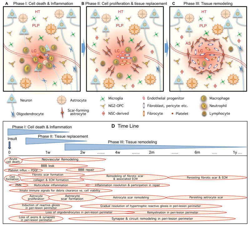
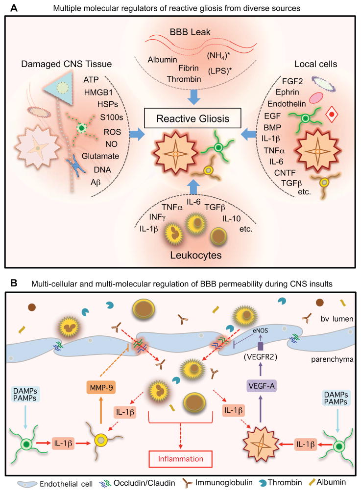
The CNS is prone to heterogeneous insults of diverse etiologies that elicit multifaceted responses. Acute and focal injuries trigger wound repair with tissue replacement. Diffuse and chronic diseases provoke gradually escalating tissue changes. The responses to CNS insults involve complex interactions among cells of numerous lineages and functions, including CNS intrinsic neural cells, CNS intrinsic nonneural cells, and CNS extrinsic cells that enter from the circulation. The contributions of diverse nonneuronal cell types to outcome after acute injury, or to the progression of chronic disease, are of increasing interest as the push toward understanding and ameliorating CNS afflictions accelerates. In some cases, considerable information is available, in others, comparatively little, as examined and reviewed here.


Figures

Figure 1
Figure 1. Phases and time course of multicellular responses to acute focal CNS damage
During periods of cell death (A), cell replacement (B) and tissue remodeling (C), numerous overlapping events occur (D) that involve interactions among CNS intrinsic neural cells, CNS intrinsic non-neural cells and cells infiltrating from the circulation.
Figure 2
Figure 2. Lesions exhibit tissue compartments and signaling gradients
(A, B) Focal damage gives rise to lesions with distinct tissue compartments in the form of non-neural lesion cores (LC), newly-proliferated compact astrocyte scars (AS) and peri-lesion perimeters (PLP) that exhibit diminishing gradients of reactive gliosis that transition gradually to healthy tissue (HT). AS abruptly demarcate the transition between non-neural LC and PLP with viable neural cells. Size of the lesion core and the location of AS and this transition is regulated by opposing molecular signals. Gradients of molecular signals emanating from LC impact on cells in PLP.
Figure 3
Figure 3. Diffuse tissue damage leads to extended regions of reactive gliosis
(A) Small chronic insults can lead to diffuse tissue damage with formation of dispersed small lesion cores (LC), astrocyte scars (AS) and peri-lesion perimeters (PLP) that can overlap and coalesce into regions of contiguous reactive gliosis that can extend over large areas. (B) Reactive astrocytes and reactive microglia in PLP can impact on synapses and neuronal functions in various ways.
Figure 4
Figure 4. Multi-molecular regulation and graded responses to damage
(A) Responses to CNS damage are regulated and influenced by diverse categories of molecular signals derived from many types of CNS intrinsic and extrinsic cells. (B) Low levels of ATP released by cells during mild CNS insults can act through (i) P2X receptors to induce rapid chemotaxis of microglial processes, and (ii) P2Y receptors on local astrocytes to evoke calcium signaling and connexin-dependent ATP release, which can increase extracellular ATP levels and thereby amplify this “danger” signal. (C) Following severe injury, signaling molecules from “altered self” and “healthy self” vie to balance debris clearance and preservation of viable tissue. Severely damaged or dead cells release a number of hydrophobic intracellular molecules (left), or “alarmins”, which express damage associated molecular patterns (DAMPs). These “danger” signals alert CNS innate immune cells to tissue injury and promote clearance of debris via activation of pattern recognition receptors on phagocytic immune cells (center). Viable CNS intrinsic cells at lesion borders (right) express membrane-bound and soluble neuroimmune regulatory molecules to prevent attack and phagocytosis by immune cells.
Figure 5
Figure 5. Multi-cellular and multi-molecular regulation of reactive gliosis and BBB
(A) Reactive gliosis can be induced, regulated and influenced by a wide variety of molecular signals that can derive from many types of CNS intrinsic and extrinsic sources. (B) BBB permeability can be regulated by specific molecular signaling cascades involving interactions among different cell types that converge on endothelial tight junctions (Argaw et al., 2012; Argaw et al., 2009; Seo et al., 2013).
Figure 6
Figure 6. Lesions as targets for therapeutic strategies
Non-neural lesion cores (LC) represent targets for biomaterial depots (A) or grafts of exogenous neural stem/progenitor cells (B), or mesenchymal stem cells (C) as potential therapeutic strategies. Molecules delivered by biomaterial depots (A) or certain cell grafts (C) have potential to support and attract axon growth (arrow a), modulate cells in peri-lesion perimeter (PLP) (arrow b), modulate cells in LC (arrow c) modulate or astrocyte scar (AS) (arrow c). New neurons generated by stem/progenitor cell grafts can receive and send neural connects in PLP and beyond, and may be able to form new, functional relay circuits (B).

Similar articles

Cited by

References

    1. Abbott NJ, Ronnback L, Hansson E. Astrocyte-endothelial interactions at the blood-brain barrier. Nat Rev Neurosci. 2006;7:41–53. - PubMed
    1. Abematsu M, Tsujimura K, Yamano M, Saito M, Kohno K, Kohyama J, Namihira M, Komiya S, Nakashima K. Neurons derived from transplanted neural stem cells restore disrupted neuronal circuitry in a mouse model of spinal cord injury. J Clin Invest. 2010;120:3255–3266. - PMC - PubMed
    1. Abeyama K, Stern DM, Ito Y, Kawahara K, Yoshimoto Y, Tanaka M, Uchimura T, Ida N, Yamazaki Y, Yamada S, et al. The N-terminal domain of thrombomodulin sequesters high-mobility group-B1 protein, a novel antiinflammatory mechanism. J Clin Invest. 2005;115:1267–1274. - PMC - PubMed
    1. Aldrich A, Kielian T. Central nervous system fibrosis is associated with fibrocyte-like infiltrates. Am J Pathol. 2011;179:2952–2962. - PMC - PubMed
    1. Argaw AT, Asp L, Zhang J, Navrazhina K, Pham T, Mariani JN, Mahase S, Dutta DJ, Seto J, Kramer EG, et al. Astrocyte-derived VEGF-A drives blood-brain barrier disruption in CNS inflammatory disease. J Clin Invest. 2012;122:2454–2468. - PMC - PubMed

Publication types