. 2014 Jan 22;81(2):333-48.
doi: 10.1016/j.neuron.2013.12.009.

Neuronal matrix metalloproteinase-9 is a determinant of selective neurodegeneration

Affiliations

Neuronal matrix metalloproteinase-9 is a determinant of selective neurodegeneration

Artem Kaplan et al. Neuron. .

Abstract

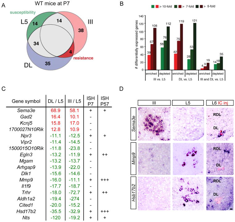
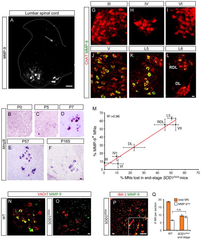
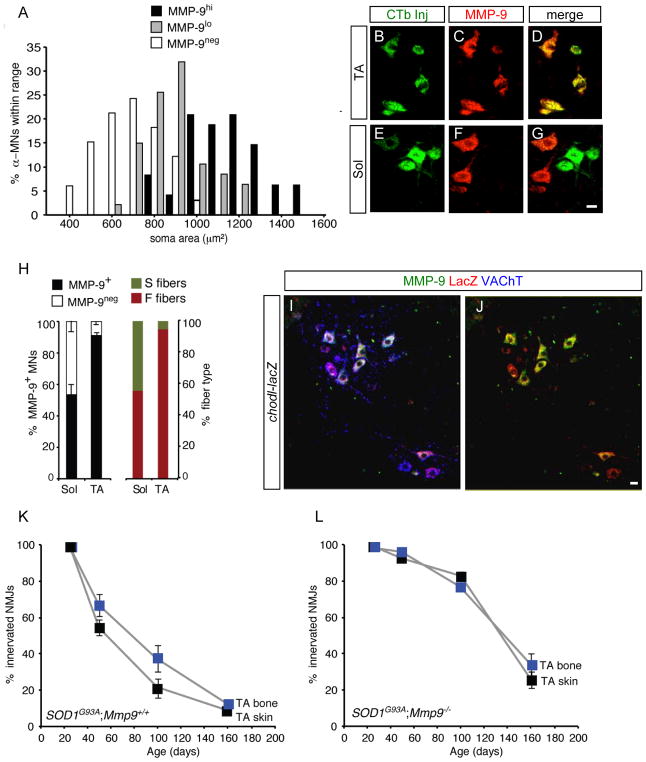
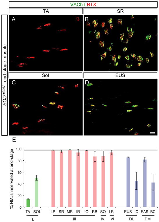
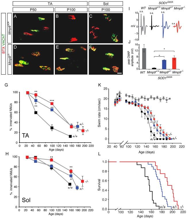
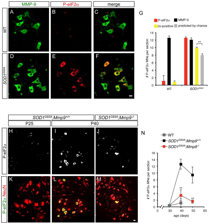
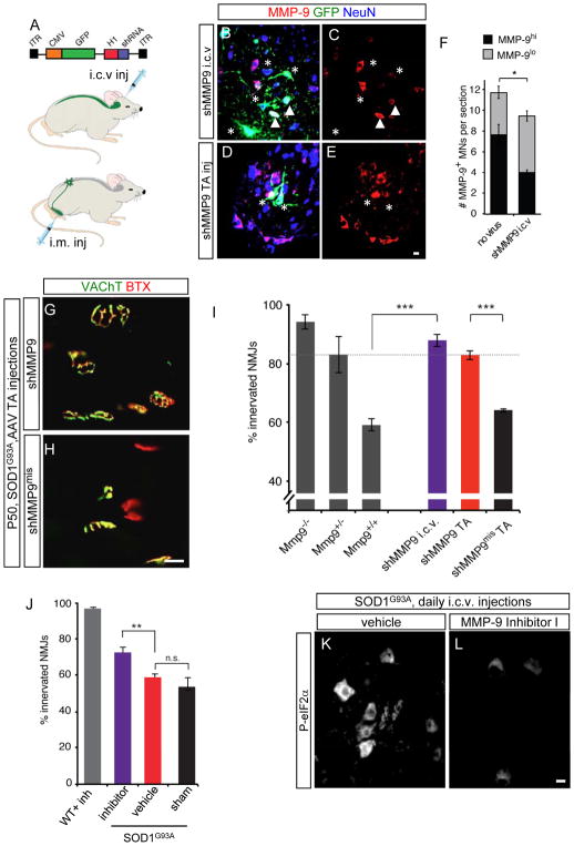
Selective neuronal loss is the hallmark of neurodegenerative diseases. In patients with amyotrophic lateral sclerosis (ALS), most motor neurons die but those innervating extraocular, pelvic sphincter, and slow limb muscles exhibit selective resistance. We identified 18 genes that show >10-fold differential expression between resistant and vulnerable motor neurons. One of these, matrix metalloproteinase-9 (MMP-9), is expressed only by fast motor neurons, which are selectively vulnerable. In ALS model mice expressing mutant superoxide dismutase (SOD1), reduction of MMP-9 function using gene ablation, viral gene therapy, or pharmacological inhibition significantly delayed muscle denervation. In the presence of mutant SOD1, MMP-9 expressed by fast motor neurons themselves enhances activation of ER stress and is sufficient to trigger axonal die-back. These findings define MMP-9 as a candidate therapeutic target for ALS. The molecular basis of neuronal diversity thus provides significant insights into mechanisms of selective vulnerability to neurodegeneration.


Figures

Figure 1
Figure 1. Identification of candidate ALS resistance and susceptibility genes by gene profiling of laser-captured motor neurons
(A) Gene expression profiles of mouse lumbar motor neurons (L5), oculomotor nucleus (III, pink) and Onuf’s nucleus (DL, blue) overlap but also reveal significant differences between motor neuron populations. Figures indicate number of genes expressed at >10-fold higher levels in each subset (P<0.05). ALS resistance genes are more likely to be found among genes enriched in both III and DL (red), while susceptibility genes (dark green) are predicted to be enriched in L5. (B) Numbers of genes showing significant differences (P<0.05 by one-way ANOVA) among the three populations at different fold ratios. (C) Identities of 18 genes enriched (red) or depleted (green) in the two resistant populations showing fold changes for 3 replicates. + signs in the P7 ISH column indicate levels of in situ hybridization (ISH) in motor neurons, - signs indicate no detectable expression. For P57, +++ differential expression maintained at comparable levels to P7, ++ more restricted expression, and + low levels. (D) Sample validation of microarray data by in situ hybridization for indicated genes in P7 midbrain and lumbar spinal cord. At L6, DL (Onuf’s) nucleus motor neurons were identified by injection of CTB (red) into the IC muscle at P5. Resistant motor neurons do not express Mmp9 or Hsd17b2. Scale bar, 20 μm. See also Figure S1 and Table S1.
Figure 2
Figure 2. Matrix metalloproteinase-9 (MMP-9) is a prospective marker for motor neurons lost at end-stage in ALS model mice
(A) In P25 SOD1G93A mouse lumbar spinal cord, intense MMP-9 immunostaining restricted to motor neurons, though some dorsal neurons stain weakly (arrow). Scale bar 100 μm. (B–F) MMP-9 is first detected by ISH in non-transgenic mouse lumbar spinal cord postnatally at P5 (C), and remains at high levels until the latest time point studied, P165 (F). (G–L) Immunostaining for MMP-9 (green) on P30 spinal cord of ChAT-Cre; Rosa-TdTomato (MN, red) reporter mice shows many motor neurons expressing high levels of MMP-9 in the vulnerable trigeminal nucleus (J), L5 spinal cord (K) and RDL in L6 (L). In contrast, resistant oculomotor (G), trochlear (H), abducens (I), and DL (L) nuclei show no or very little MMP-9 expression. Scale bars, 20 μm. (M) Tight correlation between MMP-9 expression in wildtype motor pools and their vulnerability to ALS. Absolute values fit a linear regression y = 1.07x + 3.99, R2 >0.96. Values are mean ± s.e.m. (n=3). (N, O and Q) By end-stage at L5 in SOD1G93A mice, 50% of motor neurons are lost (Q). Of those surviving, all are MMP-9-negative (compare O with P40 non-transgenic control in N). Scale bars, 20 μm. (P) All MMP-9 expressed in spinal cord at end-stage co-localizes with microglial marker IBA-1. Scale bars, 20 μm. See also Figure S2.
Figure 3
Figure 3. MMP-9 is a marker for fast motor neurons
(A) α-MNs (ChAT+/NeuN+) expressing MMP-9 are larger, consistent with a fast identity. MMP-9hi and MMP-9lo potentially reflect FF and FR, respectively. Staining intensity was determined as in Figure S3. (B to G) Many motor neurons of the fast TA pool, identified by retrograde labeling with CTB488 (green), are MMP-9-positive (red) whereas Sol motor neurons tend to be negative. Scale bar, 20 μm. (H) MMP-9 staining in the TA and Sol motor pools (mean ± s.e.m., n=3) (left) compared to fiber type distribution in the corresponding muscles replotted from Hegedus et al (2007) (right). Values for fast fibers are very close to those for MMP-9+. (I and J) Immunostaining for VAChT (blue), MMP-9 (green), and β-galactosidase (red) on spinal cord of an adult Chodl-lacZ mouse. There is strong overlap between MMP-9 and the Chodl reporter. (K) In SOD1G93A;Mmp9+/+ mice, denervation of the superficial, skin-facing compartment (black) precedes that of the deep, bone-facing fibers (blue) by >20 days (P = 0.039). (L) The two subcompartments of the TA become denervated at an identical rate in SOD1G93A;Mmp9−/− mice (P = 0.560). See also Figure S3.
Figure 4
Figure 4. Extraocular and pelvic sphincter motor units show selective resistance in ALS model mice
(A–D) Overlap of VAChT-positive motor terminals (green) with acetylcholine receptors stained using α-bungarotoxin (BTX, red) as an indicator of innervated motor endplates in end-stage SOD1G93A mice. (A) The fast tibialis anterior (TA) muscle exhibits marked denervation, whereas the slow soleus (Sol) remains partially innervated (C). (B) The extraocular muscle superior rectus (SR) and (D) the external urethral sphincter (EUS) show nearly complete preservation of motor units. Scale bar, 20 μm. (E) Percentage of intact neuromuscular junctions in muscles of end-stage SOD1G93A mice (mean ± s.e.m from 3 animals). Control, non-transgenic values are indicated by the grey-shaded region. Muscles (top row): levator palpebrae (LP), medial rectus (MR), inferior rectus (IR), inferior oblique (IO), retractor bulbi (RB), superior oblique (SO), lateral rectus (LR), ischiocavernosus (IC), external anal sphincter (EAS), bulbocavernosus (BC). Motor neurons (bottom row): lumbar spinal cord (L), oculomotor nucleus (III), trochlear nucleus (IV), abducens nucleus (VI), dorsolateral and dorsomedial components of Onuf’s nucleus (DL and DM).
Figure 5
Figure 5. Genetic ablation of MMP-9 markedly delays muscle denervation and loss of motor function in SOD1G93A mice
(A to F) Mmp9 deletion in SOD1G93A delays loss of muscle innervation in both TA and Sol muscles. Scale bars, 20 μm. (G to L) Morphological and functional evaluation of four different genotypes, color-coded in the same way in all panels: SOD1G93A;Mmp9+/+ (black; +/+), SOD1G93A;Mmp9+/− (blue; +/−), SOD1G93A;Mmp9−/− (red; −/−), and non-transgenic (gray, WT). (G) Denervation of tibialis anterior muscle is delayed by ~80 days by removal of either one or both alleles of Mmp9. (H) Denervation of slow soleus muscle later than TA, but is further delayed by 50 days in the absence of MMP-9. For both G and H, values are mean ± s.e.m of one muscle from each of 3-4 animals per genotype (***p<0.001, **p< 0.01). (I and J) MMP-9 ablation significantly rescues muscle function. Evoked compound muscle action potentials (CMAPs) in the TA muscle after stimulation of the sciatic nerve; s.a., stimulus artifact. (J) CMAP measurements from groups of the indicated genotypes aged between P105–130. Means ± s.e.m. (n= 3–4 animals per genotype); * p<0.05. (K) Mmp9 deletion significantly delays motor impairment in a swimming task that reflects hindlimb function. (n=10, p= 0.005). (L) Lifespan is prolonged by 14% following deletion of one Mmp9 allele and 25% following deletion of both. Kaplan–Meier plot showing the cumulative probability of survival of indicated genotypes (n=12–19 animals per genotype; log-rank test= 49.2, p<0.001). See also Figure S4, and Movie S1.
Figure 6
Figure 6. MMP-9 is required for early activation of ER stress in fast SOD1G93A motor neurons
(A to F) Immunostaining for MMP-9 (green) and ER stress marker phospho-EIF2α (red) in P40 spinal cord of non-transgenic (WT) and SOD1G93A mice. P-EIF2α is restricted to mSOD1 mice (compare E with B) and co-localizes with MMP-9 (F). Scale bars, 20 μm. (G) MMP-9-expressing motor neurons preferentially activate ER stress. There are significantly more SOD1G93A motor neurons co-positive for P-EIF2α and MMP-9 (yellow) than would be predicted by chance (grey bar). (H to N) The normal abrupt appearance of P-EIF2α at P40 in SOD1G93A motor neurons is severely damped in the absence of MMP-9. Scale bars, 20 μm. **p<0.01 compared to P-EIF2α-positive cells in normal mSOD1 mice.
Figure 7
Figure 7. MMP-9 is sufficient to induce degeneration in fast motor neurons of ALS mice
(A) (Top) AAV6 expressing full-length mouse MMP-9 under CMV promoter. (Bottom) Injections into TA or soleus muscles or intraorbitally were performed at P4 (n=3–4 animals per genotype; control AAV6-GFP injection on contralateral side). (B) Immunostaining for MMP-9 (red) of the oculomotor nuclei (ChAT+, green) of a P150 SOD1G93A mouse following intraorbital (EOM) injection of AAV6-MMP9. (C, D) High levels of MMP-9 (white) in soleus (C; P100) or TA (D; P50) motor pools following injection of AAV6-MMP9. (E) Viral expression of MMP-9 is not sufficient to induce denervation in resistant motor pools - soleus (Sol) or superior rectus (SR) - of SOD1G93A mice but does accelerate denervation in a susceptible pool (TA). **p<0.01 (F) Immunostaining for MMP-9 (white) on a transverse spinal cord section of a P50 SOD1G93A;Mmp9−/− mouse injected at P4 with AAV6-MMP9 into its left TA muscle. (G) Mmp9 significantly restores vulnerability to the TA muscle (red bar), compared to the control contralateral side (black bar). **p<0.01
Figure 8
Figure 8. Gene silencing or central pharmacological inhibition of MMP-9 in motor neurons delays muscle denervation
(A) AAV6 expressing H1:shMmp9 and CMV:GFP was injected i.c.v. at P1 or into TA muscle at P4. (B to E) Reduction of MMP-9 at the single-cell level in P50 WT mice following AAV6:shMmp9 administration by each route. Transduced α-MNs, visualized by colocalization of GFP (green) and NeuN immunostaining (blue), stained for MMP-9 (red). Asterisks: GFP+ α-MNs negative for MMP-9; arrowheads: less frequent GFP+ α-MNs that retain MMP-9 immunoreactivity. Scale bars, 20 μm. (F) AAV6:shMMP9 (i.c.v) leads to a 19% reduction in the total number of MMP9+ motor neurons (*p=0.03), and a 47% reduction in the numbers of MMP-9hi (p=0.0003). (G and H) MMP-9 silencing selectively delays muscle denervation in TA muscle. (I) AAV6-shMmp9 confers similar protection as the heterozygote knockout (dotted line), whether administered i.c.v. or i.m. TA innervation at P50 in SOD1G93A mice following genetic reduction in MMP-9 levels (gray bars; data from Figure 5G) or viral silencing performed i.c.v. (purple) or i.m. (red). Means ± s.e.m (n=3–4). (J) i.c.v. administration of MMP-9 Inhibitor I daily from P55 to P75 led to increased innervation in SOD1G93A mice (purple bar) compared to controls (red bar, n=5–6 per treatment). (K and L) Levels of P-EIF2α (white) at P75 in SOD1G93A motor neurons are significantly reduced by treatment with MMP-9 Inhibitor I. See also Figure S5.

Comment in

References

    1. Alexianu ME, Ho BK, Mohamed AH, La Bella V, Smith RG, Appel SH. The role of calcium-binding proteins in selective motoneuron vulnerability in amyotrophic lateral sclerosis. Ann Neurol. 1994;36:846–858. - PubMed
    1. Aronica E, Catania MV, Geurts J, Yankaya B, Troost D. Immunohistochemical localization of group I and II metabotropic glutamate receptors in control and amyotrophic lateral sclerosis human spinal cord: upregulation in reactive astrocytes. Neuroscience. 2001;105:509–520. - PubMed
    1. Brockington A, Ning K, Heath PR, Wood E, Kirby J, Fusi N, Lawrence N, Wharton SB, Ince PG, Shaw PJ. Unravelling the enigma of selective vulnerability in neurodegeneration: motor neurons resistant to degeneration in ALS show distinct gene expression characteristics and decreased susceptibility to excitotoxicity. Acta Neuropathol. 2013;125:95–109. - PMC - PubMed
    1. Chung CY, Licznerski P, Alavian KN, Simeone A, Lin Z, Martin E, Vance J, Isacson O. The transcription factor orthodenticle homeobox 2 influences axonal projections and vulnerability of midbrain dopaminergic neurons. Brain. 2010;133:2022–2031. - PMC - PubMed
    1. Cleveland DW, Rothstein JD. From Charcot to Lou Gehrig: deciphering selective motor neuron death in ALS. Nat Rev Neurosci. 2001;2:806–819. - PubMed

Publication types

MeSH terms

Associated data