Observational Study
. 2014 Mar;13(3):276-86.
doi: 10.1016/S1474-4422(13)70299-0. Epub 2014 Jan 22.

Encephalitis with refractory seizures, status epilepticus, and antibodies to the GABAA receptor: a case series, characterisation of the antigen, and analysis of the effects of antibodies

Affiliations
Observational Study

Encephalitis with refractory seizures, status epilepticus, and antibodies to the GABAA receptor: a case series, characterisation of the antigen, and analysis of the effects of antibodies

Mar Petit-Pedrol et al. Lancet Neurol. 2014 Mar.

Abstract

Background: Increasing evidence suggests that seizures and status epilepticus can be immune-mediated. We aimed to describe the clinical features of a new epileptic disorder, and to establish the target antigen and the effects of patients' antibodies on neuronal cultures.

Methods: In this observational study, we selected serum and CSF samples for antigen characterisation from 140 patients with encephalitis, seizures or status epilepticus, and antibodies to unknown neuropil antigens. The samples were obtained from worldwide referrals of patients with disorders suspected to be autoimmune between April 28, 2006, and April 25, 2013. We used samples from 75 healthy individuals and 416 patients with a range of neurological diseases as controls. We assessed the samples using immunoprecipitation, mass spectrometry, cell-based assay, and analysis of antibody effects in cultured rat hippocampal neurons with confocal microscopy.

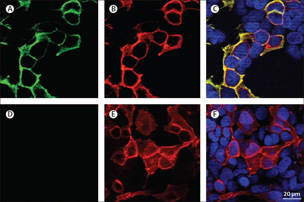
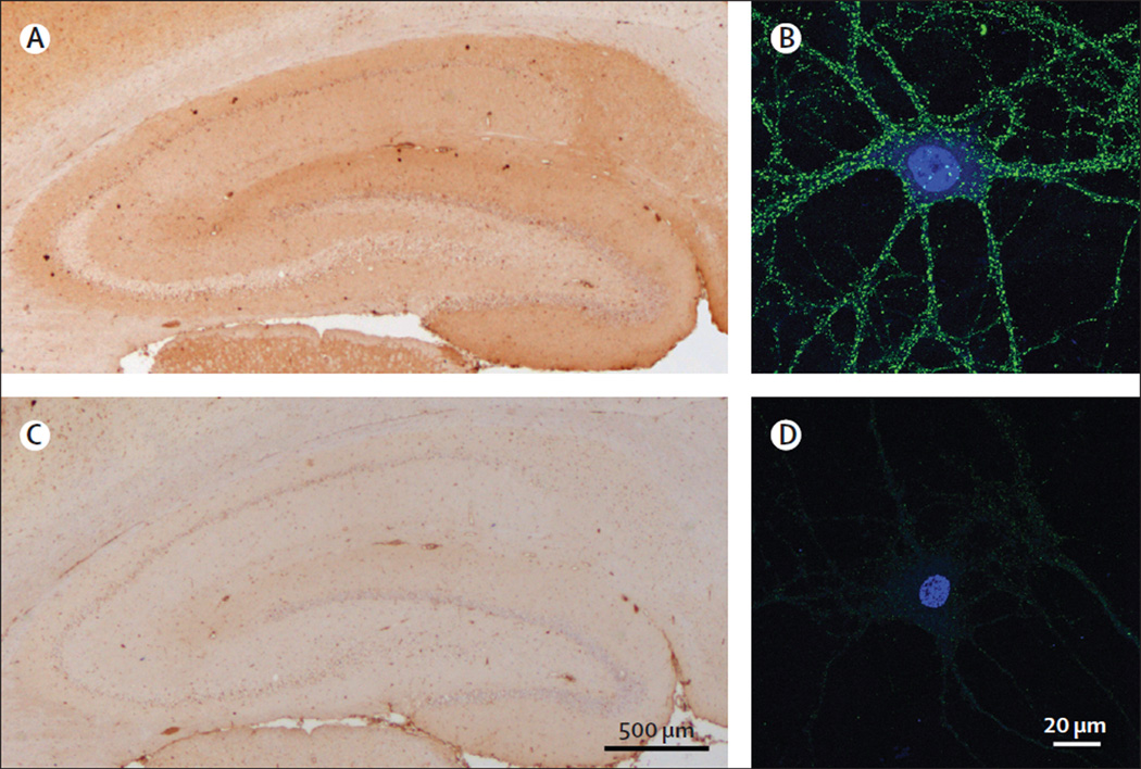
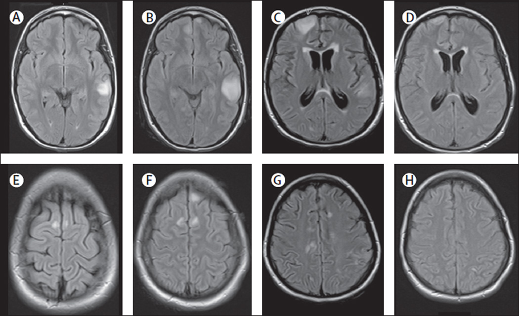
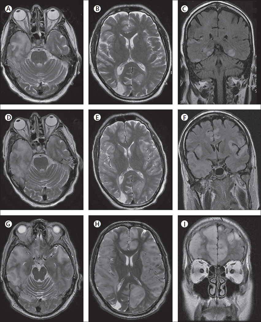
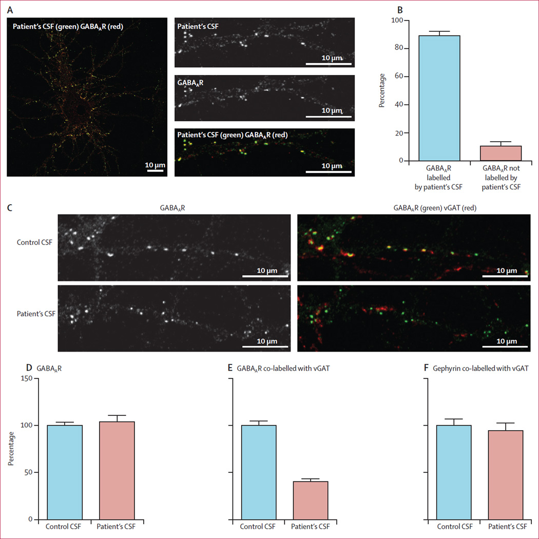
Findings: Neuronal cell-membrane immunoprecipitation with serum of two index patients revealed GABAA receptor sequences. Cell-based assay with HEK293 expressing α1/β3 subunits of the GABAA receptor showed high titre serum antibodies (>1:160) and CSF antibodies in six patients. All six patients (age 3-63 years, median 22 years; five male patients) developed refractory status epilepticus or epilepsia partialis continua along with extensive cortical-subcortical MRI abnormalities; four patients needed pharmacologically induced coma. 12 of 416 control patients with other diseases, but none of the healthy controls, had low-titre GABAA receptor antibodies detectable in only serum samples, five of them also had GAD-65 antibodies. These 12 patients (age 2-74 years, median 26.5 years; seven male patients) developed a broader spectrum of symptoms probably indicative of coexisting autoimmune disorders: six had encephalitis with seizures (one with status epilepticus needing pharmacologically induced coma; one with epilepsia partialis continua), four had stiff-person syndrome (one with seizures and limbic involvement), and two had opsoclonus-myoclonus. Overall, 12 of 15 patients for whom treatment and outcome were assessable had full (three patients) or partial (nine patients) response to immunotherapy or symptomatic treatment, and three died. Patients' antibodies caused a selective reduction of GABAA receptor clusters at synapses, but not along dendrites, without altering NMDA receptors and gephyrin (a protein that anchors the GABAA receptor).

Interpretation: High titres of serum and CSF GABAA receptor antibodies are associated with a severe form of encephalitis with seizures, refractory status epilepticus, or both. The antibodies cause a selective reduction of synaptic GABAA receptors. The disorder often occurs with GABAergic and other coexisting autoimmune disorders and is potentially treatable.

Funding: The National Institutes of Health, the McKnight Neuroscience of Brain Disorders, the Fondo de Investigaciones Sanitarias, Fundació la Marató de TV3, the Netherlands Organisation for Scientific Research (Veni-incentive), the Dutch Epilepsy Foundation.


Conflict of interest statement

Conflicts of interest

JD holds patents for the use of Ma2 and NMDAR as autoantibody tests, and has filed patents for the use of DPPX, GABAAR, and GABABR as diagnostic tests. JD and PS-S receive research grant support from Euroimmun. PS-S has filed a patent for the use of DNER as diagnostic test. MJT received a travel grant for Lecturing in India from Sun Pharma, India. The rest of the authors have no conflicts of interest.

Figures

Figure 1
Figure 1. Reactivity with brain tissue and neuron cultures of the CSF of patients with GABAAR or GABABR antibodies
The CSF of patient 2 showed extensive and diffuse immunostaining of the neuropil of cortical and subcortical regions (A; see the appendix for higher magnifications of selected brain regions). This pattern of brain and cerebellar staining is similar to that produced by the CSF of a patient with GABABR antibodies (C). However, patient 2 was negative for GABABR antibodies in a specific cell-based assay (data not shown). These findings suggested the presence of antibodies against a novel neuronal cell-surface antigen, which was confirmed in cultures of live rat hippocampal neurons (B). The CSF of the patient with GABABR antibodies also reacted with the neuronal cell surface, as expected (D). E and F show a similar study using CSF of a control patient without neuronal cell-surface antibodies. In B, D, and F the nucleus of the neurons was counterstained with DAPI. In A, C, and E the tissue was counterstained with haematoxylin.
Figure 2
Figure 2. Reactivity of a patient’s serum with live HEK293 cells expressing GABAAR
Reactivity of live HEK293 cells expressing human a1/b3 subunits of the GABAAR with a patient’s serum (A) and a monoclonal antibody against the a1 subunit (B). Merged reactivities (C). A similar assay with serum from a control individual is shown in (D–F). The nuclei of the cells are shown with DAPI in C and F. Note the specific reactivity of patient’s antibodies with cells expressing GABAAR and the co-localisation with the reactivity of the commercial antibody.
Figure 3
Figure 3. Abrogation of serum antibody reactivity with brain and cultures of neurons after GABAAR immunoabsorption
Panels A and B show the reactivity of a patient’s serum after immunoabsorption with non-transfected HEK293 cells. C and D show that this reactivity is abolished after the serum has been immunoabsorbed with HEK293 cells expressing the a1/β3 subunits of the GABAAR.
Figure 4
Figure 4. MRI findings in index patient 1
On day 3 of admission, the MRI of this 16-year-old girl showed multiple cortical-subcortical abnormalities with increased FLAIR and T2 signal involving the left temporal lobe and frontal parasagittal regions (A, E). On day 10, a repeat MRI showed an increase of the size of the temporal lesion and a new cortical lesion in the left frontal lobe (B, F). Repeat MRIs on days 22 and 48 did not show substantial changes (data not shown). Another MRI done 4 months after disease onset showed many new multifocal abnormalities and diffuse atrophy and increase of the size of the ventricles (C, G). A repeat MRI 2 months later, 6 months after symptom onset, showed substantial improvement and resolution of the abnormalities as well as improvement of the ventricular dilatation (D, H).
Figure 5
Figure 5. MRI findings in index patient 2
On day 2 of admission, the MRI of this patient showed multiple areas of FLAIR and T2 signal abnormality predominantly involving cortical regions (A–C), without oedema, mass effect, or contrast enhancement (data not shown), but with blurring of the grey-white matter junction. On day 14, repeated MRI showed interval increase of the cortical-subcortical involvement, with oedema in the right temporal lobe (D–F). Subsequent MRIs showed a pronounced worsening of these abnormalities now extensively involving cortical and subcortical regions (G–I).
Figure 6
Figure 6. Effect of patient’s antibodies on the density of GABAAR clusters in cultures of hippocampal neurons
Live 14-day-in-vitro cultures of dissociated rat hippocampal neurons were stained with patient’s CSF containing GABAAR antibodies (green), then fixed and stained with commercial GABAAR antibodies (red; A). Quantification of colocalisation between patient’s CSF antibodies and the commercial GABAAR antibody shows that 89% (SE 3%) of receptors labelled by patient’s antibodies were colabelled with the commercial antibody against GABAAR (B). In a similar assay, neurons were incubated with patient’s CSF for 48 h and subsequently stained for postsynaptic GABAAR (green) and presynaptic vesicular GABA transporter (vGAT) (red; C). The synaptic GABAARs (shown as yellow clusters in control conditions) were greatly reduced after treatment with patient’s CSF (C). The number of GABAAR clusters along dendrites of neurons treated with patient’s CSF is not different from neurons treated with control CSF (Mann-Whitney test p=0·6; D). The number of GABAARs localised in synapses, however, decreased significantly in neurons treated with a patient’s CSF compared with neurons treated with control CSF (40% [3%] compared with control as 100%; p<0·0001; E). Patients’ CSF did not affect the clusters of post-synaptic gephyrin colabelled with presynaptic vGAT when compared with the effects of control CSF (p=0·5; F).

Comment in

References

    1. Dalmau J. Status epilepticus due to paraneoplastic and nonparaneoplastic encephalitides. Epilepsia. 2009;50(suppl 12):58–60. - PubMed
    1. Wong-Kisiel LC, McKeon A, Wirrell EC. Autoimmune encephalopathies and epilepsies in children and teenagers. Can J Neurol Sci. 2012;39:134–144. - PubMed
    1. Quek AM, Britton JW, McKeon A, et al. Autoimmune epilepsy: clinical characteristics and response to immunotherapy. Arch Neurol. 2012;69:582–593. - PMC - PubMed
    1. Dalmau J, Tuzun E, Wu HY, et al. Paraneoplastic anti-N-methyl-D-aspartate receptor encephalitis associated with ovarian teratoma. Ann Neurol. 2007;61:25–36. - PMC - PubMed
    1. Lai M, Hughes EG, Peng X, et al. AMPA receptor antibodies in limbic encephalitis alter synaptic receptor location. Ann Neurol. 2009;65:424–434. - PMC - PubMed

Publication types

MeSH terms