. 2014 Feb 6;94(2):209-22.
doi: 10.1016/j.ajhg.2013.12.015. Epub 2014 Jan 23.

A founder mutation in PET100 causes isolated complex IV deficiency in Lebanese individuals with Leigh syndrome

Affiliations

A founder mutation in PET100 causes isolated complex IV deficiency in Lebanese individuals with Leigh syndrome

Sze Chern Lim et al. Am J Hum Genet. .

Abstract

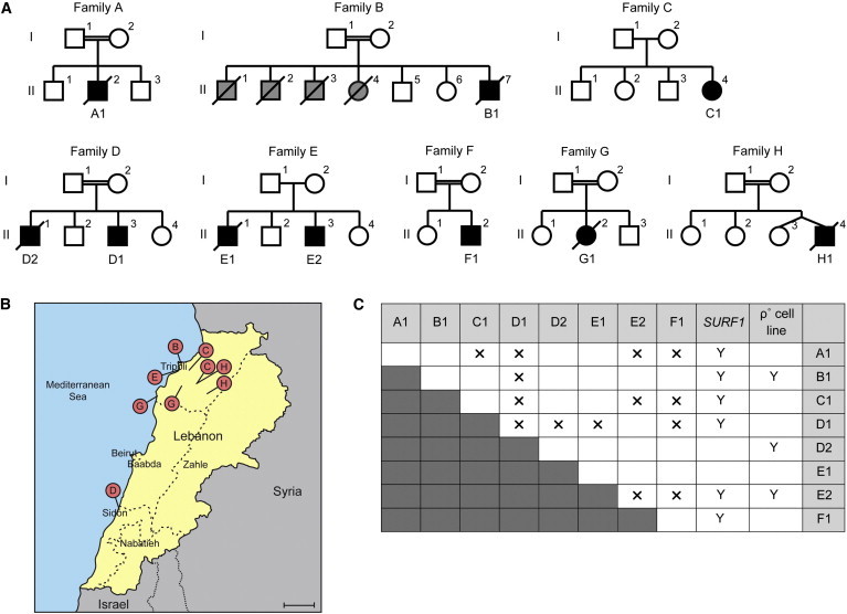
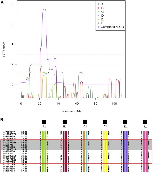
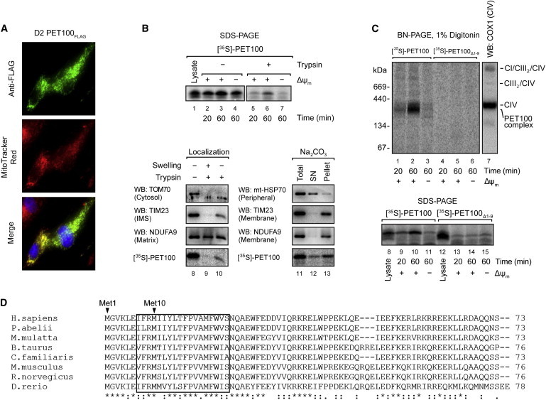
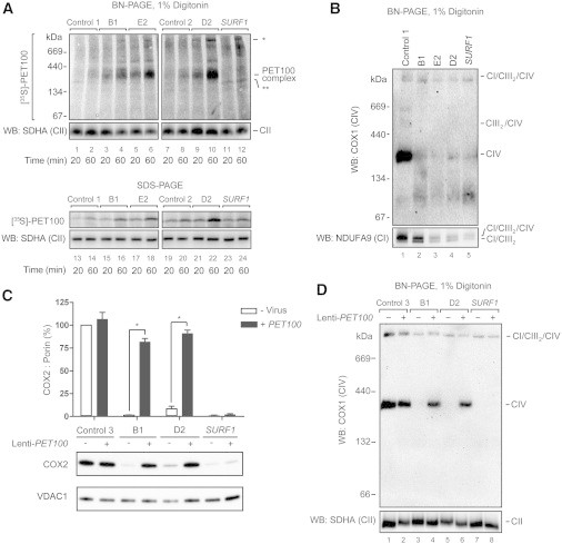
Leigh syndrome (LS) is a severe neurodegenerative disorder with characteristic bilateral lesions, typically in the brainstem and basal ganglia. It usually presents in infancy and is genetically heterogeneous, but most individuals with mitochondrial complex IV (or cytochrome c oxidase) deficiency have mutations in the biogenesis factor SURF1. We studied eight complex IV-deficient LS individuals from six families of Lebanese origin. They differed from individuals with SURF1 mutations in having seizures as a prominent feature. Complementation analysis suggested they had mutation(s) in the same gene but targeted massively parallel sequencing (MPS) of 1,034 genes encoding known mitochondrial proteins failed to identify a likely candidate. Linkage and haplotype analyses mapped the location of the gene to chromosome 19 and targeted MPS of the linkage region identified a homozygous c.3G>C (p.Met1?) mutation in C19orf79. Abolishing the initiation codon could potentially still allow initiation at a downstream methionine residue but we showed that this would not result in a functional protein. We confirmed that mutation of this gene was causative by lentiviral-mediated phenotypic correction. C19orf79 was recently renamed PET100 and predicted to encode a complex IV biogenesis factor. We showed that it is located in the mitochondrial inner membrane and forms a ∼300 kDa subcomplex with complex IV subunits. Previous proteomic analyses of mitochondria had overlooked PET100 because its small size was below the cutoff for annotating bona fide proteins. The mutation was estimated to have arisen at least 520 years ago, explaining how the families could have different religions and different geographic origins within Lebanon.


Figures

Figure 1
Figure 1
Ten PET100 Individuals from Eight Families of Lebanese Ancestry (A) Pedigrees of the eight Lebanese families. Double horizontal line indicates consanguinity. Squares and circles represent males and females, respectively. Diamonds indicate gender is unknown. Black symbols represent affected individuals and diagonal lines denote deceased individuals. Grey symbols represent children who died without investigation and remain undiagnosed. (B) A map of Lebanon with red pins marking the regions of origin of the parents in families B, C, D, E, G, and H. Scale bar represents 20 km. (C) Summary of the complementation analysis based on galactose sensitivity of fused cell lines. “A1–F1” and “SURF1” indicate fibroblast cell lines from the Lebanese LS and SURF1 individuals, respectively. 143B ρ° cell line does not contain mtDNA. Y and X indicate positive and negative complementation in galactose media, respectively.
Figure 2
Figure 2
Linkage and Haplotype Anlayses of Affected Individuals from Families A–F (A) The contribution of each family to the combined hLOD score peak on chromosome 19. LOD scores of individual families were plotted in black, red, green, blue, orange, and dark aqua, respectively. The combined hLOD score was plotted in purple. (B) Haplotypes for each affected individuals in the region under the chromosome 19 linkage peak. “1” and “2” are arbitrary labels to indicate the two different alleles of each SNP. Different colors indicate alleles inherited from different ancestors. Some markers used for linkage analysis are not displayed for conciseness. All affected individuals were homozygous by state from 23.14 to 26.89 cM (red box) and shared identical homozygous alleles from 23.14 to 25.05 cM (shaded in gray).
Figure 3
Figure 3
Mitochondrial Localization and Assembly of PET100 (A) D2 fibroblasts expressing PET100FLAG were stained with MitoTracker Red (red), fixed, and immunostained for the FLAG epitope (green). Nuclei were labeled with Hoechst (blue). Colocalization of the expressed PET100FLAG in the mitochondria (yellow) is shown in the merged image. (B) Top: The presence of membrane potential (ΔΨm) was required for the import of 35S-labeled PET100 into isolated HEK293T mitochondria, protecting it from digestion by externally added trypsin. Untreated input lysate is shown for comparison. 10% of input lysate used in the import was loaded in lane 1. Bottom: [35S]PET100 imported into HEK293T mitochondria was digested by trypsin only after hypo-osmotic mitochondrial swelling (left) and remained in the membrane (Pellet) fraction after alkaline extraction (Na2CO3) (right). Abbreviations are as follows: SN, supernatant; WB, immunoblotting. (C) BN-PAGE (top) or SDS-PAGE (bottom) followed by phosphorimaging showed that [35S]PET100Δ1-9 was incapable of assembly into the ∼300 kDa complex (lanes 4–6) and was not imported into mitochondria (lanes 13–15). Abbreviations are as follows: CI, complex I; CIII, complex III; CIV, complex IV. (D) Sequence alignment of human PET100 with its homologs in seven additional vertebrate species. The PET100 in the Lebanese LS individuals (if present) was predicted to lack the first nine amino acid residues, which are highly conserved in vertebrate species. Asterisk (), colon (:), and period (.) indicate that the amino acids are identical, strongly similar, and weakly similar, respectively, across the aligned species. The transmembrane domain predicted from the human protein is boxed.
Figure 4
Figure 4
The Import of 35S-Labeled PET100 or Overexpression of PET100 cDNA in Fibroblasts from Affected Individuals (A) Samples treated with trypsin and analyzed by BN-PAGE (top) or SDS-PAGE (bottom) and phosphorimaging showed that [35S]PET100 was imported (lanes 13–22) and assembled (lanes 1–10) into the ∼300 kDa complex with an increased efficiency in the fibroblast mitochondria of affected individuals. In SURF1 fibroblast mitochondria, [35S]PET100 was imported (lanes 23–24) but not assembled into the ∼300 kDa complex (lanes 11–12) and instead, assembled into a ∼250 kDa complex (∗∗). [35S]PET100 additionally assembled into a number of very high molecular weight complexes in mitochondria of affected individuals (). Immunoblot detection of SDHA, a complex II (CII) subunit, was used as loading control. (B) BN-PAGE and immunoblotting showed that CIV holoenzyme was barely detectable in the fibroblasts of affected individuals. The PET100 individuals also had CI/CIII2 supercomplex and a small amount of CI/CIII2/CIV supercomplex. (C) Lentiviral-mediated overexpression of wild-type PET100 restored COX2 levels in the PET100 individuals but not the SURF1 individuals (SURF1). Top: Bar graph showing protein levels of COX2 relative to VDAC1 (loading control) normalized to control, before or after transduction. The bars: mean of three biological replicates. Error bars: ± 1 standard error of the mean (SEM). p < 0.001. Bottom: A representative blot of SDS-PAGE immunoblot analysis of COX2 and VDAC1 from whole cell lysates before (-) and after (+) transduction. (D) Lentiviral-mediated overexpression of wild-type PET100 restored CIV assembly in the PET100 individuals but not in the SURF1 individual.
Figure 5
Figure 5
Assembly of mtDNA-Encoded Subunits in D2 Mitochondria (A) Assembly of newly synthesized mtDNA-encoded subunits into OXPHOS complexes was studied by pulse/chase labeling with [35S]methionine/cysteine in whole cells, followed by isolation of mitochondria and analysis by BN-PAGE and phosphorimaging. The CIV holoenzyme was not assembled in D2 mitochondria. The bottom panel shows the steady-state levels of mature complexes IV and I by immunoblotting. Abbreviations are as follows: CIVI, complex IV subcomplex; CV, complex V. (B) 2D-PAGE analysis of labeled mtDNA-encoded subunits from D2 revealed a blocked entry of COX1, COX2, and COX3 into the CIV holoenzyme, the stalling of these subunits in various subcomplexes, and an increased turnover of COX2 and COX3. The bottom panel shows the steady-state levels of mature complexes IV and II by immunoblotting. The positions of mtDNA-encoded CIV subunits COX1, COX2, and COX3 are indicated.

References

    1. Leigh D. Subacute necrotizing encephalomyelopathy in an infant. J. Neurol. Neurosurg. Psychiatry. 1951;14:216–221. - PMC - PubMed
    1. Tiranti V., Hoertnagel K., Carrozzo R., Galimberti C., Munaro M., Granatiero M., Zelante L., Gasparini P., Marzella R., Rocchi M. Mutations of SURF-1 in Leigh disease associated with cytochrome c oxidase deficiency. Am. J. Hum. Genet. 1998;63:1609–1621. - PMC - PubMed
    1. Zhu Z., Yao J., Johns T., Fu K., De Bie I., Macmillan C., Cuthbert A.P., Newbold R.F., Wang J., Chevrette M. SURF1, encoding a factor involved in the biogenesis of cytochrome c oxidase, is mutated in Leigh syndrome. Nat. Genet. 1998;20:337–343. - PubMed
    1. Dahl H.-H.M. Getting to the nucleus of mitochondrial disorders: identification of respiratory chain-enzyme genes causing Leigh syndrome. Am. J. Hum. Genet. 1998;63:1594–1597. - PMC - PubMed
    1. Kirby D.M., Thorburn D.R. Approaches to finding the molecular basis of mitochondrial oxidative phosphorylation disorders. Twin Res. Hum. Genet. 2008;11:395–411. - PubMed

Publication types

MeSH terms