. 2014 Feb 6;94(2):295-302.
doi: 10.1016/j.ajhg.2014.01.001. Epub 2014 Jan 23.

Heterozygous loss-of-function mutations in YAP1 cause both isolated and syndromic optic fissure closure defects

Collaborators, Affiliations

Heterozygous loss-of-function mutations in YAP1 cause both isolated and syndromic optic fissure closure defects

Kathleen A Williamson et al. Am J Hum Genet. .

Abstract

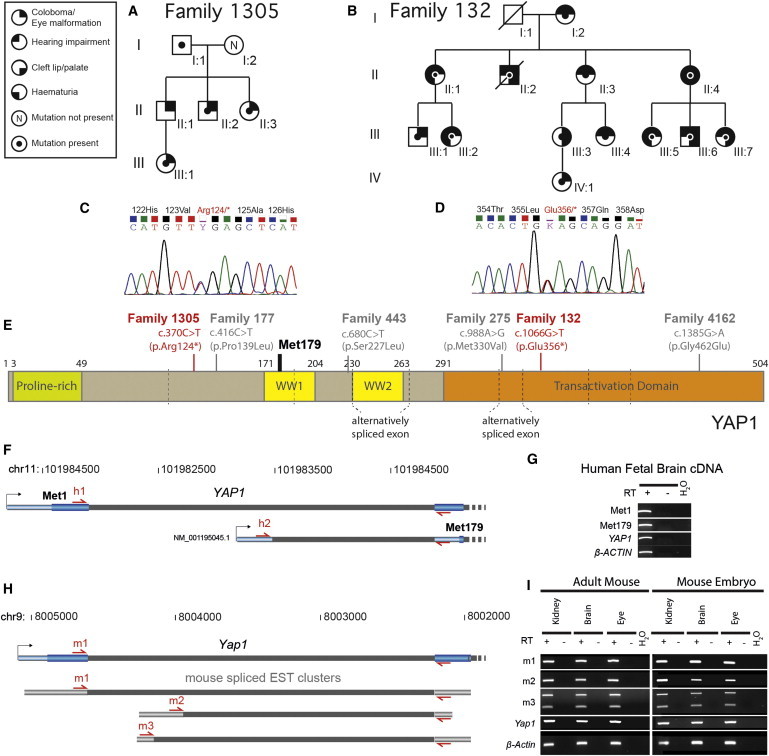
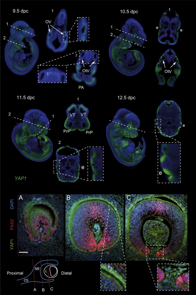
Exome sequence analysis of affected individuals from two families with autosomal-dominant inheritance of coloboma identified two different cosegregating heterozygous nonsense mutations (c.370C>T [p.Arg124*] and c. 1066G>T [p.Glu356*]) in YAP1. The phenotypes of the affected families differed in that one included no extraocular features and the other manifested with highly variable multisystem involvement, including hearing loss, intellectual disability, hematuria, and orofacial clefting. A combined LOD score of 4.2 was obtained for the association between YAP1 loss-of-function mutations and the phenotype in these families. YAP1 encodes an effector of the HIPPO-pathway-induced growth response, and whole-mount in situ hybridization in mouse embryos has shown that Yap1 is strongly expressed in the eye, brain, and fusing facial processes. RT-PCR showed that an alternative transcription start site (TSS) in intron 1 of YAP1 and Yap1 is widely used in human and mouse development, respectively. Transcripts from the alternative TSS are predicted to initiate at codon Met179 relative to the canonical transcript (RefSeq NM_001130145). In these alternative transcripts, the c.370C>T mutation in family 1305 is within the 5' UTR and cannot result in nonsense-mediated decay (NMD). The c. 1066G>T mutation in family 132 should result in NMD in transcripts from either TSS. Amelioration of the phenotype by the alternative transcripts provides a plausible explanation for the phenotypic differences between the families.


Figures

Figure 1
Figure 1
Position and Consequences of YAP1 Mutations (A) Pedigree structure of family 1305 (F1305). The symbol key is shown on the left. DNA was available from three affected (II:2, II:3, and III:1) and two unaffected (I:1 and I:2) individuals. (B) An abridged version of the published pedigree of family 132 (F132, Ravine et al.17) shows affected individuals from whom DNA samples were available. (C and D) Representative chromatograms of the confirmatory Sanger sequencing that was performed in all individuals from whom DNA was available in families 1305 and 132. (E) A diagram of the domain structure of YAP1. The amino acid numbers indicating the beginning and end of each domain are shown above relative to isoform 1 (P46937, UniProt). The positions of the exon junctions are indicated by the vertical gray dashed lines. The positions of the nonsense mutations are shown in red text, and the probably nonpathogenic missense mutations are shown in gray text. The probable initiating methionine used in transcripts from the alternative transcription start site (TSS) is indicated by Met179. (F and H) Diagrams of the 5′ end of the gene models for the canonical and alternative TSSs in human and mouse, respectively. In both species, the most 5′ TSS is canonical and encodes transcripts with an open reading frame that uses the initiation methionine Met1. The TSS in intron 1 of the canonical transcript splices to the same exon 2, but in this transcript, the initiating methionine is equivalent to Met179 of the canonical transcript. (G and I) A gel photograph shows the results of RT-PCR using primers indicated by the red arrows on the cognate gene models and the tissues indicated above each lane. This shows that the alternative TSS was present in all examined tissues.
Figure 2
Figure 2
Developmental Expression of YAP1 and Yap1 during Mouse Development The top panel shows optical-projection-tomography images of Yap1 expression detected by whole-mount in situ hybridization on mouse embryos as 9.5, 10.5, 11.5, and 12.5 dpc. At each stage, a 3D reconstruction of the whole embryo is presented on the left. The dashed and numbered white lines indicate the position and angle of the digital sections that are shown to the right of the 3D representation for each stage. The boxed white sections indicate regions that are presented at higher magnification. The abbreviations for the highlighted tissues are as follows: OV, optic vesicle; OtV, otic vesicle; PA, pharyngeal arch; e, eye; PrP, fusing primary palate; and VT, ventral telencephalon. Yap1 expression is indicated in green. The bottom panel shows immunohistochemical staining of sections from the mouse embryonic eye at 10.5 dpc with antibodies specific to YAP1 (green) and PAX2 (red). Nuclear staining was performed with DAPI. A diagram below indicates the position of the sections along the proximodistal axis of the developing eye. Section A is through the optic stalk (OS); sections B and C both show the neural retina (NR) and lens (L). Higher-magnification images of boxed areas from sections B and C indicate nuclear staining for YAP1 in the RPE and in the periocular mesenchyme between the optic fissure margins, respectively.

References

    1. Morrison D., FitzPatrick D., Hanson I., Williamson K., van Heyningen V., Fleck B., Jones I., Chalmers J., Campbell H. National study of microphthalmia, anophthalmia, and coloboma (MAC) in Scotland: investigation of genetic aetiology. J. Med. Genet. 2002;39:16–22. - PMC - PubMed
    1. Chang L., Blain D., Bertuzzi S., Brooks B.P. Uveal coloboma: clinical and basic science update. Curr. Opin. Ophthalmol. 2006;17:447–470. - PubMed
    1. Hornby S.J., Adolph S., Gilbert C.E., Dandona L., Foster A. Visual acuity in children with coloboma: clinical features and a new phenotypic classification system. Ophthalmology. 2000;107:511–520. - PubMed
    1. Daufenbach D.R., Ruttum M.S., Pulido J.S., Keech R.V. Chorioretinal colobomas in a pediatric population. Ophthalmology. 1998;105:1455–1458. - PubMed
    1. Mihelec M., Abraham P., Gibson K., Krowka R., Susman R., Storen R., Chen Y., Donald J., Tam P.P., Grigg J.R. Novel SOX2 partner-factor domain mutation in a four-generation family. Eur. J. Hum. Genet. 2009;17:1417–1422. - PMC - PubMed

Publication types

MeSH terms