. 2014 May;15(5):485-95.
doi: 10.1016/j.jpain.2014.01.490. Epub 2014 Jan 23.

Activation of cutaneous immune responses in complex regional pain syndrome

Affiliations

Activation of cutaneous immune responses in complex regional pain syndrome

Frank Birklein et al. J Pain. 2014 May.

Abstract

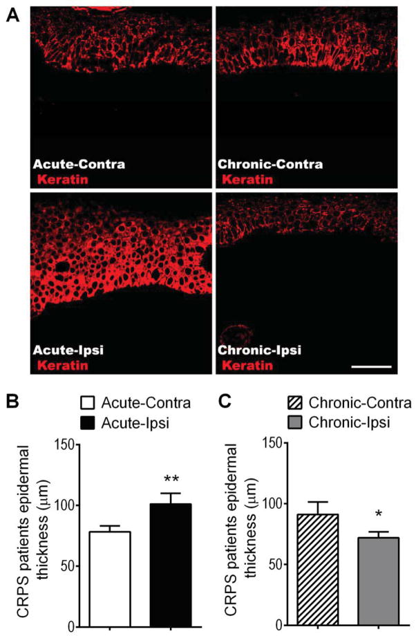
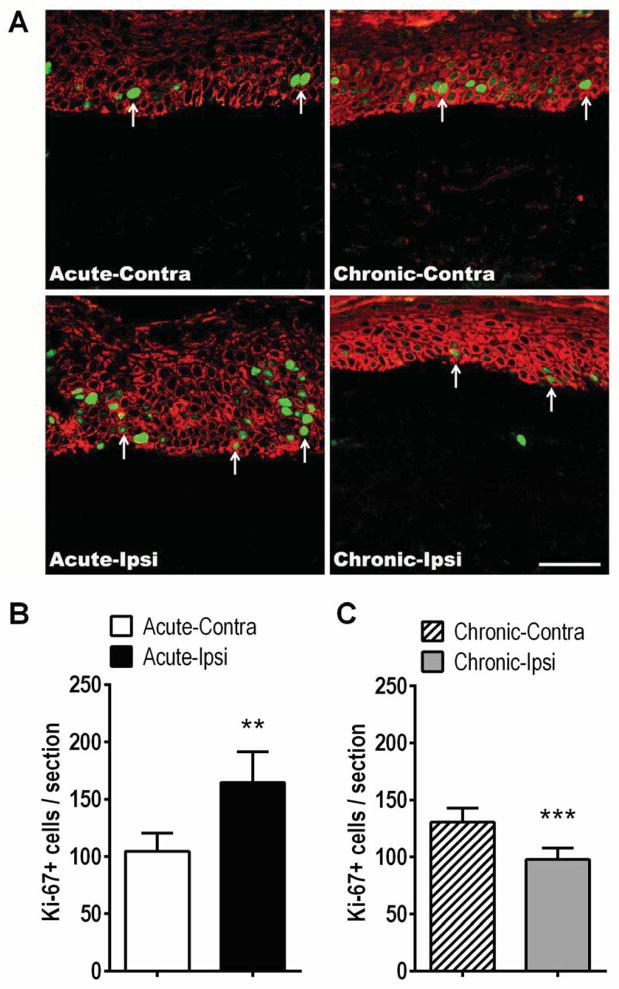
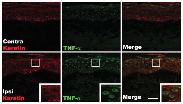
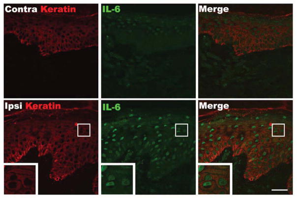
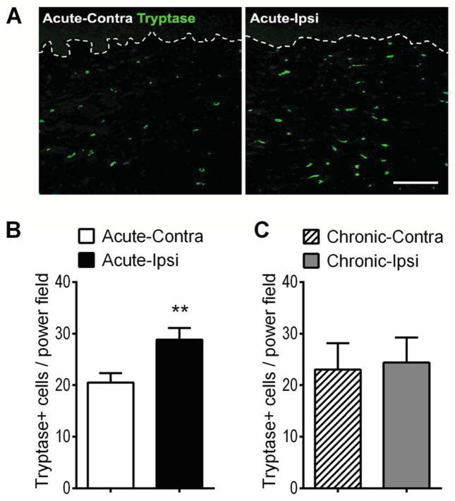
The pathogenesis of complex regional pain syndrome (CRPS) is unresolved, but tumor necrosis factor alpha (TNF-α) and interleukin-6 (IL-6) are elevated in experimental skin blister fluid from CRPS-affected limbs, as is tryptase, a marker for mast cells. In the rat fracture model of CRPS, exaggerated sensory and sympathetic neural signaling stimulate keratinocyte and mast cell proliferation, causing the local production of high levels of inflammatory cytokines leading to pain behavior. The current investigation used CRPS patient skin biopsies to determine whether keratinocyte and mast cell proliferation occur in CRPS skin and to identify the cellular source of the up-regulated TNF-α, IL-6, and tryptase observed in CRPS experimental skin blister fluid. Skin biopsies were collected from the affected skin and the contralateral mirror site in 55 CRPS patients and the biopsy sections were immunostained for keratinocyte, cell proliferation, mast cell markers, TNF-α, and IL-6. In early CRPS, keratinocytes were activated in the affected skin, resulting in proliferation, epidermal thickening, and up-regulated TNF-α and IL-6 expression. In chronic CRPS, there was reduced keratinocyte proliferation, leading to epidermal thinning in the affected skin. Acute CRPS patients also had increased mast cell accumulation in the affected skin, but there was no increase in mast cell numbers in chronic CRPS.

Perspective: The results of this study support the hypotheses that CRPS involves activation of the innate immune system, with keratinocyte and mast cell activation and proliferation, inflammatory mediator release, and pain.

Keywords: Complex regional pain syndrome; immunology; keratinocytes; mast cells; pain.


Conflict of interest statement

Disclosures: The authors do not have financial or other relationships that might lead to conflict of interest.

Figures

Figure 1
Figure 1
Epidermal thickness changes in the affected skin of patients suffering from acute and chronic CRPS. (A) Immunostaining for keratin protein (red, a keratinocyte marker) in skin sections obtained from patients with acute (< 3 months duration, left panels) and chronic (> 3 months duration, right panels) CRPS. (A) Panels exhibit confocal images from an acute CRPS patient’s contralateral hand (Acute-Contra, upper left panel), the same patient’s ipsilateral CRPS affected hand (Acute-Ipsi, lower left panel), a chronic CRPS patient’s contralateral hand (Chronic-Contra, upper right panel), and the same patient’s ipsilateral CRPS affected hand (Chronic-Ipsi, lower right panel). Scale bar = 50 μm. (B) There was a 29% increase in epidermal thickness in the affected skin of patients with acute CRPS (101 ± 9 μm, n = 20), as compared to the contralateral healthy limb (78 ± 5 μm). (C) In chronic CRPS patients the inverse was true, there was a 21% decrease in epidermal thickness in the affected skin (72 ± 5 μm, n = 26), as compared to the contralateral healthy limb (91 ± 10 μm). Although the epidermal thickness in the contralateral healthy skin of the acute and chronic CRPS patient cohorts differed, this did not reach significance and may be a reflection of the different average ages between the acute (53.7 ± 2.9) and chronic (45.6 ± 2.0) cohorts (B,C). *P < 0.05, ***P < 0.001 for ipsi vs. contra values. Contra: contralateral, Ipsi: ipsilateral.
Figure 2
Figure 2
Keratinocyte proliferation changes in the affected skin of patients suffering from acute and chronic CRPS. (A) Immunostaining for keratin protein (red, a keratinocyte marker) and Ki-67 (green, a marker of DNA synthesis) in skin sections obtained from patients with acute (< 3 months duration, left panels) and chronic (> 3 months duration, right panels) CRPS. (A) Left panels are representative confocal microscopy images from an acute CRPS patient’s contralateral hand (Acute-Contra, upper left panel) and the same patient’s ipsilateral CRPS affected hand (Acute-Ipsi, lower left panel). The right panels are representative images of a chronic CRPS patient’s contralateral hand (Chronic-Contra, upper right panel), and the same patient’s ipsilateral CRPS affected hand (Chronic-Ipsi, lower right panel). Arrows: Ki-67 positive keratinocytes. Scale bar = 50 μm. (B) There was a 57% increase in Ki-67 positive keratinocyte numbers in the affected skin of patients with acute CRPS (165 ± 27), as compared to the contralateral healthy limb (105 ± 16, n = 18). (C) In chronic CRPS patients the inverse was true, there was a 25% decrease in Ki-67 labeled keratinocytes in the affected skin (98 ± 10), as compared to the contralateral healthy limb (131 ± 12, n = 25). Although the contralateral healthy skin Ki-67 labeled keratinocyte counts in the acute patients (105 ± 16) and the chronic patients (131 ± 12) differed, this did not reach significance and may be a reflection of the different average ages between the acute (53.1 ± 2.7) and chronic (45.7 ± 1.9) cohorts (Fig. 2B,C) **P < 0.01 for ipsi vs. contra values. Contra: contralateral, Ipsi: ipsilateral.
Figure 3
Figure 3
Fluorescence photomicrographs showing co-localization of keratin immunostaining in keratinocytes (red) with immunostaining for TNF-α (green) in skin biopsy sections from a patient with acute CRPS. Compared with the contralateral healthy skin (upper panels), TNF-α expression was up-regulated in the cytoplasm of epidermal keratinocytes of the ipsilateral CRPS affected skin (lower panels). The insets are enlargements of the boxed regions. Scale bar = 40μm. Contra: contralateral, Ipsi: ipsilateral.
Figure 4
Figure 4
Fluorescence photomicrographs showing co-localization of keratin immunostaining in keratinocytes (red) with immunostaining for IL-6 (green) in skin biopsy sections from a patient with acute CRPS. Compared with the contralateral healthy skin (upper panels), IL-6 expression was up-regulated in the cytoplasm of epidermal keratinocytes in the ipsilateral CRPS affected skin. The insets are enlargements of the boxed regions. Scale bar = 40μm. Contra: contralateral, Ipsi: ipsilateral.
Figure 5
Figure 5
Mast cell accumulation changes in the affected skin of patients suffering from acute and chronic CRPS. (A) Immunostaining for tryptase positive cells (green, mast cell marker) in the dermis of the skin sections obtained from a patient with acute CRPS. The dotted line denotes the basement membrane at the dermal-epidermal border. Scale bar = 200μm. (B) There was a 41% increase in tryptase positive mast cell numbers at the affected skin (29 ± 2) of patients with acute CRPS (< 3 months duration), compared to the contralateral healthy limb (21 ± 2, n=20). (C) In chronic CRPS (> 3 months duration) patients no differences were observed between the tryptase positive mast cell numbers in the affected skin (24 ± 5) and the contralateral healthy skin (23 ± 5, n=10). **P < 0.01 for ipsi vs. contra values. Contra: contralateral, Ipsi: ipsilateral.
Figure 6
Figure 6
Proposed model for the integration of primary afferent and sympathetic nervous system signaling with mast cell and keratinocyte innate immune responses in supporting CRPS-related nociception. Cutaneous primary sensory afferents release SP and CGRP in the epidermis. These neuropeptides then diffuse through the interstitial space to bind and activate their cognate receptors on the keratinocyte cell surface, the SP NK1-R and the CGRP receptor dimer complex of CRLR and RAMP1. Keratinocyte NK1-R activation stimulates cellular proliferation, SP expression and secretion, NK1-R expression, and stimulates TNFα expression and secretion. SP also binds to NK1-Rs on the mast cell surface, resulting in mast cell accumulation, activation, degranulation, and release of tryptase, histamine, cytokines, proteases and eicosanoids, all of which might sensitize nociceptive neurons in the skin. Similarly, activation of the keratinocyte CGRP receptor dimer complex stimulates keratinocyte proliferation, CGRP expression and secretion, RAMP1 receptor expression, and up-regulates TNFα expression and secretion. Sympathetic neurons release NE into the interstitial space, which binds and activates β2-ARs on the keratinocyte cell surface, resulting in IL-6 expression and secretion. Keratinocyte secreted inflammatory cytokines can directly activate their cognate receptors expressed on cutaneous sensory afferent neurons, with subsequent pain sensitization. CGRP, calcitonin gene-related peptide; SP, substance P; NK1-R, neurokinin 1 receptor; CRL-R, calcitonin receptor-like receptor; RAMP1 receptor activity-modifying protein; NE, norepinephrine; β2-AR, β2 adrenergic receptor; IL-6, interleukin-6; IL-6R, IL-6 receptor; TNF-α, tumor necrosis factor α; TNF-α R, TNF-α receptor.

References

    1. Bernateck M, Karst M, Gratz KF, Meyer GJ, Fischer MJ, Knapp WH, Koppert W, Brunkhorst T. The first scintigraphic detection of tumor necrosis factor-alpha in patients with complex regional pain syndrome type 1. Anesthesia and analgesia. 2010;110:211–215. - PubMed
    1. Bernateck M, Rolke R, Birklein F, Treede RD, Fink M, Karst M. Successful intravenous regional block with low-dose tumor necrosis factor-alpha antibody infliximab for treatment of complex regional pain syndrome 1. Anesthesia and analgesia. 2007;105:1148–1151. - PubMed
    1. Birklein F, Riedl B, Sieweke N, Weber M, Neundorfer B. Neurological findings in complex regional pain syndromes--analysis of 145 cases. Acta Neurol Scand. 2000;101:262–269. - PubMed
    1. Bruehl S. An update on the pathophysiology of complex regional pain syndrome. Anesthesiology. 2010;113:713–725. - PubMed
    1. Carroll I, Clark JD, Mackey S. Sympathetic block with botulinum toxin to treat complex regional pain syndrome. Annals of neurology. 2009;65:348–351. - PMC - PubMed

Publication types

MeSH terms