. 2014 May;26(5):868-79.
doi: 10.1016/j.cellsig.2014.01.014. Epub 2014 Jan 22.

Ezrin expression and cell survival regulation in colorectal cancer

Affiliations

Ezrin expression and cell survival regulation in colorectal cancer

Premila D Leiphrakpam et al. Cell Signal. 2014 May.

Abstract

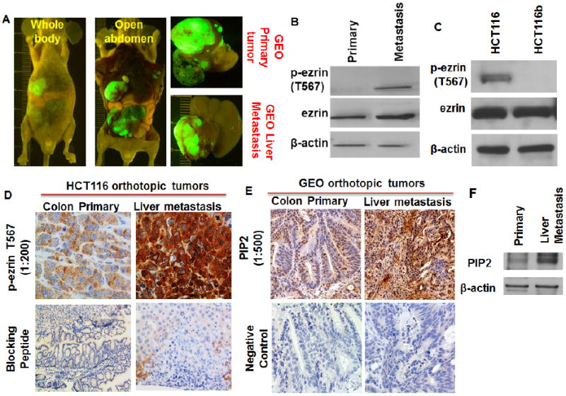
Colorectal cancer (CRC) is the second largest cause of cancer deaths in the United States. A key barrier that prevents better outcomes for this type of cancer as well as other solid tumors is the lack of effective therapies against the metastatic disease. Thus there is an urgent need to fill this gap in cancer therapy. We utilized a 2D-DIGE proteomics approach to identify and characterize proteins that are differentially regulated between primary colon tumor and liver metastatic deposits of the IGF1R-dependent GEO human CRC xenograft, orthotopically implanted in athymic nude mice that may serve as potential therapeutic targets against CRC metastasis. We observed increased expression of ezrin in liver metastasis in comparison to the primary colonic tumor. Increased ezrin expression was further confirmed by western blot and microarray analyses. Ezrin, a cytoskeletal protein belonging to Ezrin-Radixin-Moesin (ERM) family plays important roles in cell motility, invasion and metastasis. However, its exact function in colorectal cancer is not well characterized. Establishment of advanced GEO cell lines with enhanced liver-metastasizing ability showed a significant increase in ezrin expression in liver metastasis. Increased phosphorylation of ezrin at the T567 site (termed here as p-ezrin T567) was observed in liver metastasis. IHC studies of human CRC patient specimens showed an increased expression of p-ezrin T567 in liver metastasis compared to the primary tumors of the same patient. Ezrin modulation by siRNA, inhibitors and T567A/D point mutations significantly downregulated inhibitors of apoptosis (IAP) proteins XIAP and survivin that have been linked to increased aberrant cell survival and metastasis and increased cell death. Inhibition of the IGF1R signaling pathway by humanized recombinant IGF1R monoclonal antibody MK-0646 in athymic mouse subcutaneous xenografts resulted in inhibition of p-ezrin T567 indicating ezrin signaling is downstream of the IGF1R signaling pathway. We identified increased expression of p-ezrin T567 in CRC liver metastasis in both orthotopically implanted GEO tumors as well as human patient specimens. We report for the first time that p-ezrin T567 is downstream of the IGF1R signaling and demonstrate that ezrin regulates cell survival through survivin/XIAP modulation.

Keywords: 2D-DIGE proteomics; Cell survival signaling; Colorectal cancer metastasis; Ezrin; IGF1R signaling; XIAP and survivin.


Conflict of interest statement

Competing interests

The authors declare that they have no competing interests.

Figures

Figure 1
Figure 1. Proteomic analysis identifies Ezrin upregulation in CRC liver metastasis
(A) IGF1R-dependent GEO cells were used to generate primary colon and liver metastatic tumors by orthotopic implantation techniques as described in the Material and methods section. These tumor tissues were further utilized for the proteomics analysis. (B) 2D-DIGE image showing differential protein expression between primary colon tumor and liver metastases. The primary colon and liver metastatic tumor tissue lysates were labeled with Cy3 (green) and Cy5 (red) fluorescent dyes respectively. The images of the cy3 and cy5 labeled primary and liver metastatic tumor protein samples are generated by laser scanning. The white circle depicts protein spot number 2. (C) The individual primary tumor and liver metastasis gel images and merged image of protein spot number 2 showing greater intensity in the liver metastatic tumor lysate. (D) The 3D-peak view of protein spot number 2 in primary and liver metastatic tumors analyzed by the DeCyder software. The protein spot number 2 was identified as ezrin by MALDI-TOF mass spectrometry.
Figure 2
Figure 2
The individual 2D-DIGE gel images of GEO primary colon carcinoma and liver metastasis tumors and overlay merged image.
Figure 3
Figure 3. Elevated levels of p-ezrin T567 in liver metastasis
(A) Western blot analysis comparing GEO primary colon and liver metastatic tumors showing increased ezrin and p-ezrin T567 protein expression in the liver metastasis. (B) IHC analysis of the GEO primary colon and liver metastatic tumors showing increased p-ezrin T567 staining in the liver metastasis. The increased expression of p-ezrin T567 is specifically localized to the membrane as demonstrated by its ring-like appearance of the liver metastatic cells. (C) Densitometry analysis of the IHC images of GEO primary tumors and liver metastasis by Image J followed by statistical analysis showing an increased expression of p-ezrin T567 in liver metastasis specimen. (D) IGF1R-dependent CBS cells were used to generate primary colon and liver metastatic tumors by orthotopic implantation techniques as described in the Material and methods section. These tumor tissues were further utilized for the western blot analysis. (E) Western blot analysis comparing CBS primary colon and liver metastatic tumors showing increased p-ezrin T567 protein expression in the liver metastasis. (F) Highly metastatic GEO cells that lacks functional TGFβ tumor suppressor signaling shows high ezrin and p-ezrin T567 expression in vitro. In contrast, poorly metastatic GEORI cells [14] stably transfected with TGFβ Receptor Type I (TGFβRI) to restore functional TGFβ tumor suppressor signaling shows complete loss of p-ezrin T567 expression and a reduction in total ezrin expression. (*=P<0.001).
Figure 4
Figure 4
(A) Another set of IGF1R-dependent GEO cells were used to generate primary colon and liver metastatic tumors by orthotopic implantation techniques as described in the Material and methods section. These tumor tissues were further utilized for the western blot analysis. (B) Western blot analysis comparing GEO primary colon and liver metastatic tumors showing increased p-ezrin T567 protein expression in the liver metastasis. (C) Western blot analysis comparing highly metastatic HCT116 [16] and poorly metastatic iso-clonal variant HCT116b cells showing increased p-ezrin T567 in the HCT116 cells. (D) IHC analysis of the HCT116 primary colon and liver metastatic tumors showing increased p-ezrin T567 staining in the liver metastasis. (E, F) IHC and western blot analysis of the GEO primary colon and liver metastatic tumors showing increased PIP2 staining in the liver metastasis.
Figure 5
Figure 5. Ezrin network in colorectal cancer metastasis signaling
The intuitive web-based analytical tool Ingenuity Pathway Analysis (IPA) was used to generate plausible colorectal cancer metastasis signaling networks.
Figure 6
Figure 6. IHC studies on ezrin and p-ezrin T567 in human CRC patient specimens
(A) NCI colon tissue microarray (colon TMA) analysis of ezrin in normal colon and stage IV primary colon carcinoma obtained from CRC patient specimens. (B) Densitometry analysis of the IHC images of colon TMA comparing normal colon and stage IV primary colon carcinoma by Image J followed by statistical analysis showing an increased expression of ezrin in stage IV colon carcinoma specimen. (C) IHC staining of p-ezrin T567 in colon TMAs showing negligible staining in both normal colon and stage IV colon carcinoma specimen. (D) IHC analysis of the human CRC stage IV colon carcinoma and liver metastatic tumors obtained from the same patient showing increased p-ezrin T567 staining in the liver metastasis. Similar to the p-ezrin T567 staining in the GEO orthotopically implanted liver metastatic tumors, the increased p-ezrin T567 is specifically localized to the cellular membrane of the liver metastatic cells. (E) Densitometry analysis of the IHC images of the human CRC stage IV colon carcinoma and liver metastatic tumors obtained from the same patient by Image J followed by statistical analysis showing an increased expression of p-ezrin T567 in liver metastasis specimen. (**=P<0.01).
Figure 7
Figure 7. Ezrin expression is correlated with enhanced liver metastasizing ability
(A) IGF1R-dependent GEO Met6 cells were generated by serial passaging of the liver metastatic tumor tissues as discussed in the Material and methods section. Comparison of the orthotopically implanted GEO cells (mentioned here as GEO Control) and GEO Met6 cells showed remarkable differences in their tumor formation. (B) GEO Control and GEO Met6 primary colonic and liver metastatic tumor images. (C) Comparison of the approximate number of days needed to develop liver metastasis in the GEO and GEO Met6 xenograft implanted animals. (D) TUNEL assay of GEO and GEO Met6 liver metastatic tumor tissues showing a significant decrease in cell death in the GEO Met6 liver metastatic tumor tissues as observed by reduction in TUNEL staining. (E) Densitometry analysis of the TUNEL staining of GEO and GEO Met6 liver metastatic tumor tissues by Image J followed by statistical analysis showing a decrease in TUNEL staining in the GEO Met6 liver metastasis specimen. (F) Western blot analysis showing a significant increase in ezrin protein expression in the GEO Met6 liver metastasis tumors compared to the GEO Met6 primary tumors. (*=P<0.001).
Figure 8
Figure 8. Ezrin regulates aberrant cell survival in CRC cells
(A) Ezrin siRNA knockdown in GEO cells (termed as GEO Ezrin siRNA) shows a reduction in the inhibitor of apoptosis (IAP) family proteins XIAP and survivin. (B) DNA fragmentation assay of GEO and GEO Ezrin siRNA cells showing significant increase in cell death in vitro. (C) Ezrin siRNA knockdown in CBS cells (termed as CBS Ezrin siRNA) shows a reduction in XIAP and survivin expression. (D) DNA fragmentation assay of CBS and CBS Ezrin siRNA cells showing significant increase in cell death in vitro. (E) Ezrin inhibitor NS668394 treatment dephosphorylates p-ezrin T567 and decreases XIAP expression. (F) NS668394 treatment induces 7.5-fold increase in cell death in GEO cells. (G) DNA fragmentation assay showing increased cell death with ezrin T567A mutation compared to T567D transfection on GEO cells. (*=P<0.001).
Figure 9
Figure 9. Regulation of p-ezrin T567 by IGF1R signaling
(A) Inhibition of the IGF1R signaling pathway by human recombinant monoclonal antibody MK-0646 in athymic mice IGF1R-dependent GEO xenograft lead to inhibition of p-ezrin T567. However, the total ezrin protein remained unchanged. (B) IHC analysis of GEO control and MK-0646 treated xenograft showing a decrease in staining intensity of p-ezrin T567 in the MK-0646 treated xenograft. (C) Treatment of IGF1R-kinase inhibitor OSI-906 on GEO cells in vitro leads to loss of ezrin phosphorylation at T567 site without change in total ezrin. (D) Ezrin IP studies showing decrease in p-Serine following IGF1R inhibition by OSI-906 and increase following IGF1R activation for 1hour (E) Ezrin IP following site-directed mutagenesis showing decrease in p-Serine in dominant negative ezrin S66A expressing GEO cells. In contrast, constitutive active ezrin S66D expression increases p-Serine on ezrin IP pull down. Ezrin S66A/D mutation also shows loss of XIAP and survivin with S66A and increase with S66D ezrin mutation. (F) Increased cell death observed with ezrin S66A mutation compared to S66D indicative of its role in cell survival (* P<0.001)

References

    1. Nguyen DX, Bos PD, Massague J. Nat Rev Cancer. 2009;9(4):274–284. - PubMed
    1. Markowitz SD, Bertagnolli MM. N Engl J Med. 2009;361(25):2449–2460. - PMC - PubMed
    1. Vasan RS. Circulation. 2006;113(19):2335–2362. - PubMed
    1. Imielinski M, Cha S, Rejtar T, Richardson EA, Karger BL, Sgroi DC. Mol Cell Proteomics. 2012;11(6) M111 014910. - PMC - PubMed
    1. Chung CH, Levy S, Chaurand P, Carbone DP. Crit Rev Oncol Hematol. 2007;61(1):1–25. - PubMed

Publication types

MeSH terms