. 2014 Mar:68:89-97.
doi: 10.1016/j.yjmcc.2014.01.006. Epub 2014 Jan 23.

BDNF secretion by human pulmonary artery endothelial cells in response to hypoxia

Affiliations

BDNF secretion by human pulmonary artery endothelial cells in response to hypoxia

Martin Helan et al. J Mol Cell Cardiol. 2014 Mar.

Abstract

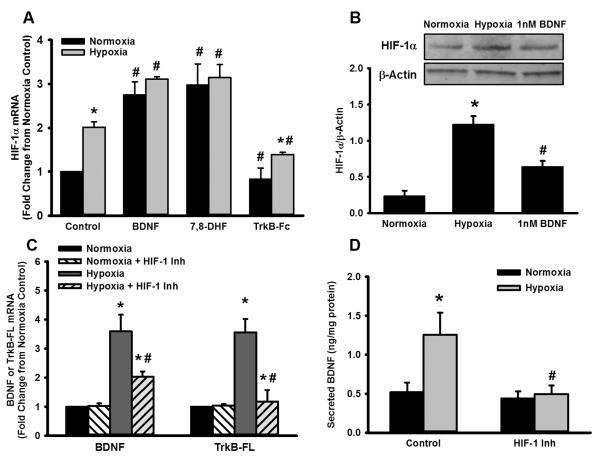
Within human pulmonary artery, neurotrophin growth factors [NTs; e.g. brain-derived neurotrophic factor (BDNF)] and their high-affinity receptors (tropomyosin-related kinase; Trk) and low-affinity receptors p75 neurotrophin receptor (p75NTR) have been reported, but their functional role is incompletely understood. We tested the hypothesis that BDNF is produced by human pulmonary artery endothelial cells (PAECs). In the context of hypoxia as a risk factor for pulmonary hypertension, we examined the effect of hypoxia on BDNF secretion and consequent autocrine effects on pulmonary endothelium. Initial ELISA analysis of circulating BDNF in 30 healthy human volunteers showed that 72 h exposure to high altitude (~11,000 ft, alveolar PO2 = 100 mmHg) results in higher BDNF compared to samples taken at sea level. Separately, in human PAECs exposed for 24h to normoxia vs. hypoxia (1-3% O2), ELISA of extracellular media showed increased BDNF levels. Furthermore, quantitative PCR of PAECs showed 3-fold enhancement of BDNF gene transcription with hypoxia. In PAECs, BDNF induced NO production (measured using an NO-sensitive fluorescent dye DAF2-DA) that was significantly higher under hypoxic conditions, an effect also noted with the TrkB agonist 7,8-DHF. Importantly, hypoxia-induced NO was blunted by neutralization of secreted BDNF using the chimeric TrkB-Fc. Both hypoxia and BDNF increased iNOS (but not eNOS) mRNA expression. In accordance, BDNF enhancement of NO in hypoxia was not blunted by 50 nM L-NAME (eNOS inhibition) but substantially lower with 100 μM L-NAME (eNOS and iNOS inhibition). Hypoxia and BDNF also induced expression of hypoxia inducible factor 1 alpha (HIF-1α), a subunit of the transcription factor HIF-1, and pharmacological inhibition of HIF-1 diminished hypoxia effects on BDNF expression and secretion, and NO production. These results indicate that human PAECs express and secrete BDNF in response to hypoxia via a HIF-1-regulated pathway.

Keywords: Hypoxia inducible factor 1; Neurotrophin; Nitric oxide; Tropomyosin related kinase; eNOS; iNOS.


Figures

Figure 1
Figure 1
Effect of hypoxia on circulating and pulmonary artery endothelial cell (PAEC)-derived brain derived neurotrophic factor (BDNF) in humans. (A) ELISA for BDNF in peripheral blood serum samples from healthy individuals was performed before (control) and following exposure to 15% hypoxia for 72 h (see methods for details). In most individuals, hypoxia significantly increased serum BDNF. (B) A similar in vitro ELISA of supernatants from human PAECs exposed to <5% hypoxia for 24h showed increased secretion of BDNF following hypoxia, even when corrected for protein content due to any potential increase in cell number due to hypoxia. Values are means ± SE (n=30 in (A) and 5 in (B)). *Significant difference from normoxia (p<0.05).
Figure 2
Figure 2
Hypoxia-BDNF interactions in PAEC expression of BDNF and its receptors. Measurement of mRNA levels for BDNF, its high affinity receptor tropomyosin related kinase (TrkB, truncated T1 and full length FL forms) and low affinity receptor p75NTR in PAECs under normoxia vs. 24h hypoxia showed that hypoxia augments both BDNF (A) and TrkB-FL (C; in relation to the T1 isoform). In contrast, p75NTR expression was unchanged (D). Hypoxia also increased BDNF protein expression in PAEC lysates (B). Interestingly, acting via TrkB, BDNF caused its own upregulation (A and B) as well as that of TrkB (C) as evidenced the effect of exogenous human recombinant BDNF or activation of TrkB only using the flavanoid agonist 7,8-DHF. These effects of the BDNF/TrkB system were substantially enhanced in the presence of hypoxia. Neutralization of extracellular (presumably PAEC-secreted) BDNF using the chimeric TrkB-Fc protein blunted PAEC expression of TrkB under both hypoxic conditions. Values are means ± SE (n=7 patient samples). *Significant difference from normoxia, # significant effect of BDNF, 7,8-DHF or TrkB-Fc (p<0.05).
Figure 3
Figure 3
Role of HIF-1α in hypoxia-induced BDNF expression and activity. mRNA analysis (A) and Western blots (B) showed that 24h hypoxia increased PAEC expression of HIF-1α. Interestingly, in both normoxia and hypoxia-exposed PAECs, BDNF also upregulated expression of HIF-1α, with a relatively greater effect in hypoxia (A). A similar effect was observed with the TrkB agonist, while in contrast, neutralization of extracellular BDNF suppressed hypoxia-induced HIF-1α levels. In terms of the role of HIF-1α in hypoxia effects on BDNF and TrkB per se, pharmacological inhibition of HIF-1 (using EMD 400083, see methods) substantially blunted hypoxia enhancement of both BDNF and TrkB mRNA (C), and in accordance, blunted hypoxia-induced enhancement of BDNF secretion (D). Values are means ± SE (n=5 patient samples). *Significant difference from normoxia, # significant effect of BDNF, 7,8-DHF, TrkB-Fc or HIF-1 inhibitor (p<0.05).
Figure 4
Figure 4
Hypoxia-BDNF interactions in nitric oxide (NO) production by PAECs. NO production in PAECs was measured using diaminofluorescein imaging (see methods). Baseline measurements represented effects of 24h exposures such as hypoxia or inhibitors, while fluorescence responses to BDNF were measured in real-time (A). In PAECs exposed to normoxia vs. hypoxia, exogenous BDNF increased NO generation in normoxia, and potentiated the enhancing effects of hypoxia on NO levels (A) especially at 1 nM that lies in the physiological range. Pre-treatment of PAECs with TrkB-Fc blunted the increase in baseline fluorescence that occurs with hypoxia, while BDNF-induced NO production was also reduced (C). Pharmacologically inhibiting HIF-1 causes a decrease in NO, and blunts the effects of BDNF, strongly suggesting a role for this transcription factor in hypoxia-mediated BDNF signaling (D). To distinguish between the potential roles of iNOS vs. eNOS, the inhibitor L-NAME at lower concentration (50 nM; for eNOS; E) and higher concentration (100 μM; for iNOS; F) was used. Inhibition of eNOS only slightly blunted the enhanced NO production by hypoxia while blocking iNOS substantially blunted the effects of hypoxia on NO. Values in bar graphs are means ± SE (n=5 patient samples). *Significant difference from normoxia, # significant effect of BDNF, TrkBFc, HIF-1 inhibitor, or L-NAME (p<0.05).
Figure 5
Figure 5
Role of BDNF in hypoxia effects on eNOS and iNOS in PAECs. With 24h hypoxia, eNOS expression was not significantly affected (A), while iNOS expression substantially increased (B). Exposure to BDNF substantially increased iNOS expression, which was further enhanced in hypoxia (B). Neutralization of BDNF with TrkB-Fc as well as the HIF-1 inhibitor both eliminated increases in iNOS expression, suggesting a link between BDNF and HIF-1 in iNOS regulation. In contrast, eNOS expression was substantially less affected by disrupting BDNF or HIF1 signaling (A). Values are means ± SE (n=4 patient samples). *Significant difference from normoxia, # significant effect of BDNF, 7,8-DHF, TrkB-Fc or HIF-1 inhibitor (p<0.05).
Figure 6
Figure 6
Putative mechanisms for hypoxia- and BDNF-induced effects in PAECs. Expression levels of iNOS, and the arginases 1 and 2 were increased by 24h of hypoxia or by BDNF (A). Perturbation of BDNF signaling via TrkB-Fc or of HIF-1 activation via inhibitor blunts increases iNOS (A, B) and arginases 1 (A, C) and 2 (A, D). Values are means ± SE (n=4 patient samples). *Significant difference from normoxia, # significant effect of BDNF, TrkB-Fc or HIF-1 inhibitor (p<0.05).
Figure 7
Figure 7
Schematic of BDNF-hypoxia interactions in PAECs. Hypoxia can increase circulating BDNF, where PAECs may be a potential source. Hypoxia, acting via HIF-1 can enhance BDNF secretion as well as TrkB expression, which in turn, can increase HIF1α even under normoxia, thus priming PAECs to respond to hypoxia. Both BDNF/TrkB and hypoxia can increase NO production via iNOS.

References

    1. Frumkin LR. The pharmacological treatment of pulmonary arterial hypertension. Pharmacol Rev. 2012;64:583–620. - PubMed
    1. Tajsic T, Morrell NW. Smooth muscle cell hypertrophy, proliferation, migration and apoptosis in pulmonary hypertension. Compr Physiol. 2011;1:295–317. - PubMed
    1. Sakao S, Tatsumi K, Voelkel NF. Reversible or irreversible remodeling in pulmonary arterial hypertension. Am J Respir Cell Mol Biol. 2010;43:629–34. - PMC - PubMed
    1. Morrell NW, Adnot S, Archer SL, Dupuis J, Jones PL, MacLean MR, et al. Cellular and molecular basis of pulmonary arterial hypertension. J Am Coll Cardiol. 2009;54:S20–31. - PMC - PubMed
    1. Humbert M, Gerry Coghlan J, Khanna D. Early detection and management of pulmonary arterial hypertension. Eur Respir Rev. 2012;21:306–12. - PMC - PubMed

Publication types

MeSH terms

LinkOut - more resources