. 2014 Mar 15;387(2):167-78.
doi: 10.1016/j.ydbio.2014.01.010. Epub 2014 Jan 21.

Dkk2/Frzb in the dermal papillae regulates feather regeneration

Affiliations

Dkk2/Frzb in the dermal papillae regulates feather regeneration

Qiqi Chu et al. Dev Biol. .

Abstract

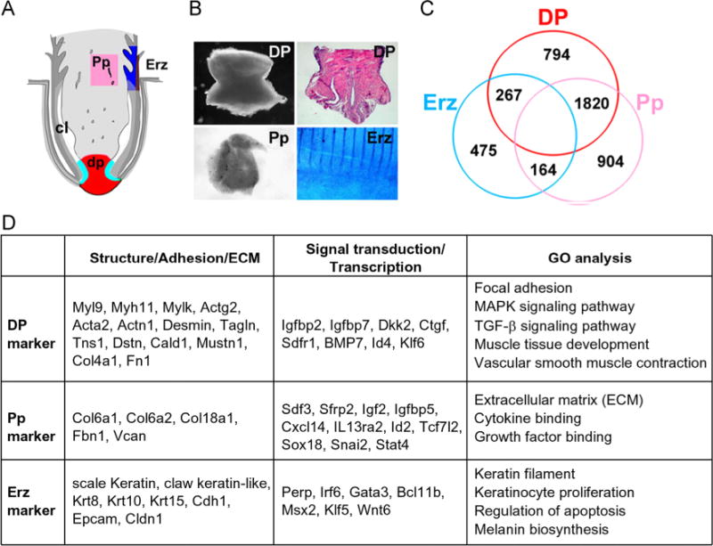
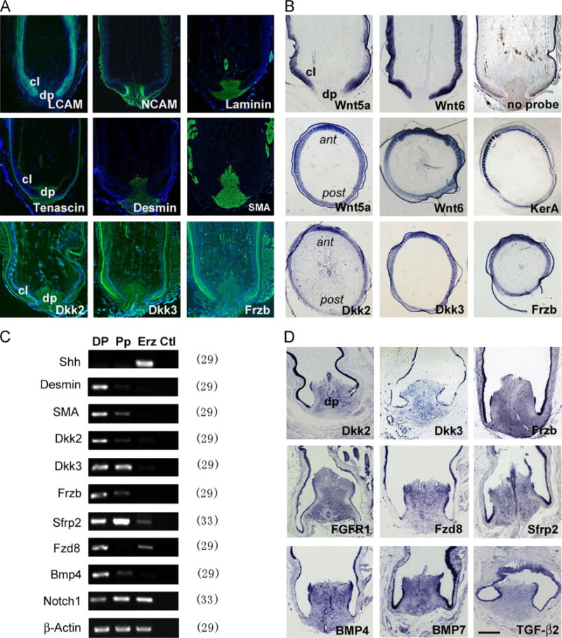
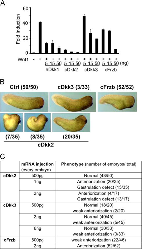
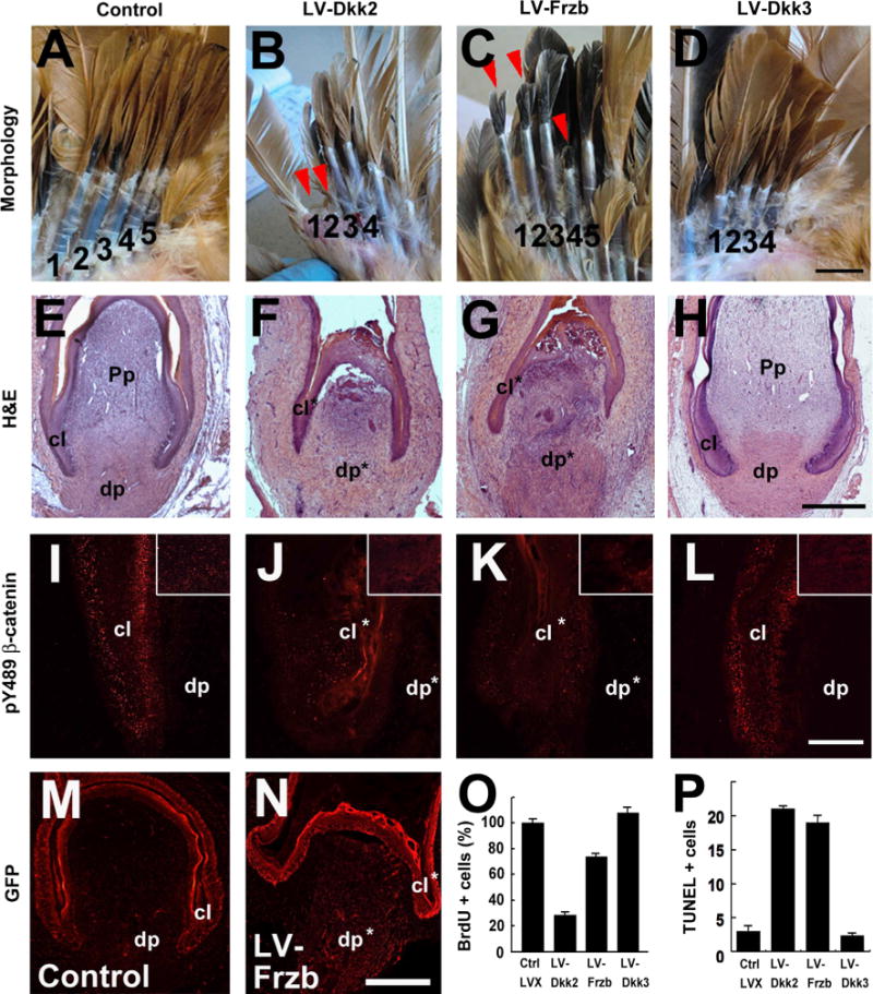
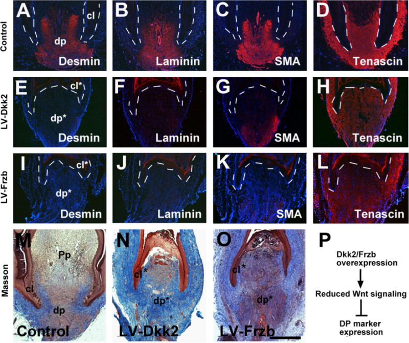
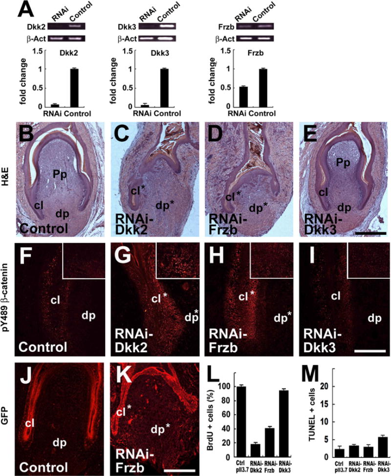
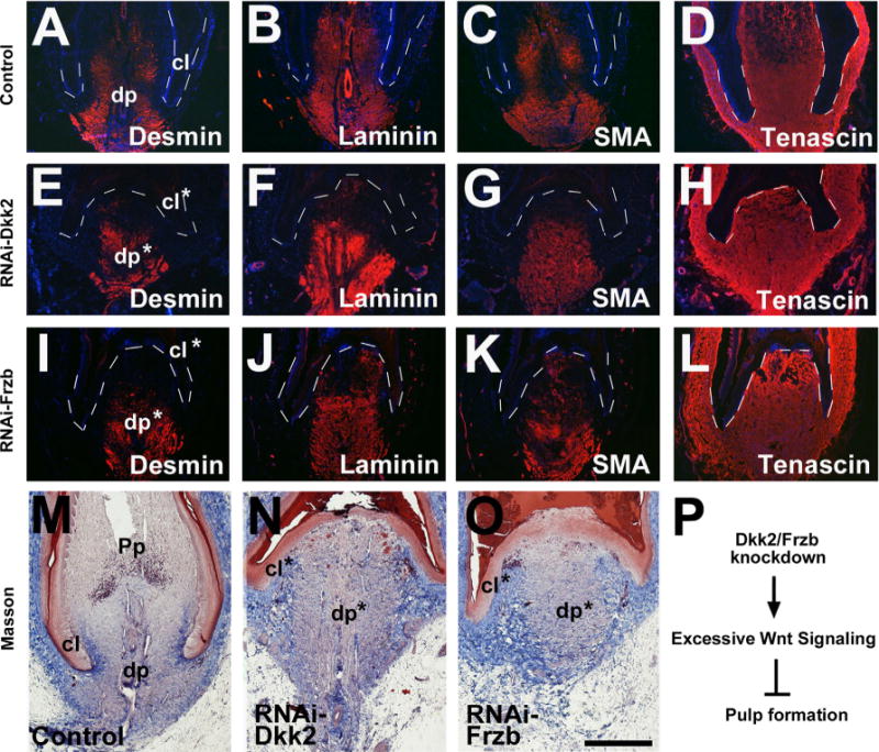
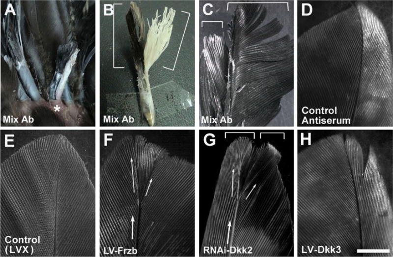
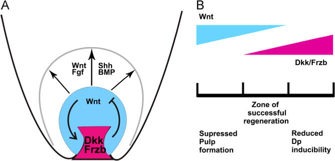
Avian feathers have robust growth and regeneration capability. To evaluate the contribution of signaling molecules and pathways in these processes, we profiled gene expression in the feather follicle using an absolute quantification approach. We identified hundreds of genes that mark specific components of the feather follicle: the dermal papillae (DP) which controls feather regeneration and axis formation, the pulp mesenchyme (Pp) which is derived from DP cells and nourishes the feather follicle, and the ramogenic zone epithelium (Erz) where a feather starts to branch. The feather DP is enriched in BMP/TGF-β signaling molecules and inhibitors for Wnt signaling including Dkk2/Frzb. Wnt ligands are mainly expressed in the feather epithelium and pulp. We find that while Wnt signaling is required for the maintenance of DP marker gene expression and feather regeneration, excessive Wnt signaling delays regeneration and reduces pulp formation. Manipulating Dkk2/Frzb expression by lentiviral-mediated overexpression, shRNA-knockdown, or by antibody neutralization resulted in dual feather axes formation. Our results suggest that the Wnt signaling in the proximal feather follicle is fine-tuned to accommodate feather regeneration and axis formation.

Keywords: Axis formation; Dermal papillae; Dkk2/Dkk3/Frzb; Feather follicle; Regeneration.


Figures

Fig.1
Fig.1
Gene expression profiling in the feather follicle. (A) Diagram and (B) examples showing the feather structure and the dissection process. H&E showing the structure of the DP. Erz was illustrated based upon DAPI staining. The stripes in the Erz sample are feather branches. dp, dermal papilla; Pp, pulp; Erz, ramogenic zone feather epithelium. (C) Venn diagram showing differentially expressed genes among DP, Pp and Erz. (D) Lists of highly expressed genes in each compartment that could serve as “markers”. The gene abbreviations are according to the NCBI listings. Gene ontology (GO) analysis results are also shown.
Fig. 2
Fig. 2
Gene expression analysis in the feather follicle. (A) Marker gene expression shown by immunofluorescence (green). LCAM marks the feather epithelium. NCAM marks the DP/dermal sheath and weakly the feather branching epithelium. Laminin marks the DP and vessel walls. Tenascin marks the DP/dermal sheath. Desmin is more DP specific, and SMA marks the DP and vessel walls. Dkk2/Dkk3/Frzb is enriched in the DP, presents in the pulp but less in the epithelium. Some unspecific staining is found in the keratinized feather sheath. (B) Expression of Wnt ligands and inhibitors in the feather follicles shown by in situ hybridization. Notice Wnt5a and Wnt6 appear primarily in the epithelium. A gradient distribution pattern is detected for Wnt5a/Wnt6, Dkk2/Dkk3/Frzb and feather keratin A in the Erz region. A control staining with no probe is also shown. (C) Semi-quantitative RT-PCR and (D) in situ hybridization analysis of gene expression in the feather follicles. No template reactions are used as control for PCR analysis, and equal amount of RNAs are monitored by β-Actin gene expression. The number after each gene indicates PCR cycles. dp, dermal papilla; cl, collar; ant, anterior where the rachis locates; post, posterior as opposite to the rachis position. Bar = 1 mm in A and B, and 0.5 mm in D (shown in D).
Fig. 3
Fig. 3
Dkk2/Frzb antagonizes Wnt signaling. (A) Wnt reporter assay in HEK 293T cells. Super-TOPFLASH, a Wnt responsive reporter was co-transfected into HEK293T cells together with Wnt1 and other plasmids as indicated. hDkk1 (human Dkk1) was used as a positive control. Fold induction of Wnt reporter activity is shown. The numbers for each gene indicates the amount of DNA transfected; total amount of DNA transfected in each well was 150 ng, adjusted with pCS2+ plasmid. (B) Anteriorization of Xenopus embryos by injected mRNAs as indicated. mRNAs of chicken Dkk2 (1 ng per embryo), Dkk3 (6 ng per embryo) or Frzb (2 ng per embryo) were injected at 4-cell stage. The numbers of embryos with indicated phenotypes are also shown. (3/33) stands for 3 out of 33 injected embryos showed indicated phenotype. Control animals were injected with 250 pg mRNA of preprolectin gene. (C) Summary of the Xenopus injection experiment.
Fig. 4
Fig. 4
Overexpression of Dkk2/Frzb, but not Dkk3, disrupts feather regeneration. (A–D) Representative samples of lentivirus (LV) mediated gene overexpression in the feather follicle. A control virus carrying GFP only was also shown. Four to five wing flight feather follicles in their resting phase were plucked to induce regeneration and infected with the virus, and photographed 3 weeks afterwards. Delayed feather regeneration was indicated by arrow heads. (E–H) H&E analysis of virus-infected feather follicles 4 days post-infection. (I–L) pY489 β-catenin antibody staining (red spots) showing the reduced Wnt signaling after Dkk2/Frzb overexpression. The epithelia in the collar regions are shown, and the DP regions are shown in inserts. (M and N) Anti-GFP immunostaining (red) showing lentiviral-mediated GFP expression in the feather follicles. (O and P) Quantification of BrdU and TUNEL staining results in the feather follicles. The dp, Pp and collar (cl) are labeled; * indicates the disrupted structures. Bar = 1 cm (A–D, shown in D), 1 mm (E–H, shown in H; M and N, shown in N), and 200 μm (I–L, shown in L).
Fig. 5
Fig. 5
Overexpression of Dkk2/Frzb, but not Dkk3, reduces DP marker gene expression. (A–L) DP marker gene expression showing reduced Desmin/Laminin/SMA, but not Tenascin in LV-Dkk2 and LV-Frzb transduced feathers. An empty pLVX virus was used as control. Samples were collected at day 4 post-infection. For a comparison of staining intensity, equal exposure time was used when taken pictures. (M–O) Masson staining showing altered DP characteristics in Dkk2/Frzb overexpressed follicles. (P) Summary of the events after Dkk2/Frzb overexpression. Dashed lines indicate the epithelial–mesenchymal borders. The dp and collar (cl) are labeled; *indicates the disrupted structures. Bar = 1 mm (shown in O).
Fig. 6
Fig. 6
RNAi knockdown of Dkk2/Frzb, but not Dkk3, disrupts feather regeneration. (A) Efficiency of shRNA tested in DF-1 cells. Semi-quantitative RT-PCR and qPCR results were shown. A construct targeting a random sequence was used as control. Endogenous mRNA levels were quantified. (B–E) Representative samples of lentiviral-mediated shRNA knockdown in the feather follicles 4 days post-infection. A shRNA virus targeting a random sequence was used as control. (F–I) pY489 β-catenin antibody staining (red spots) showing the increased Wnt signaling after Dkk2/Frzb knockdown. The epithelia in the collar regions are shown, and the DP regions are shown in inserts. (J–K) Virus expression monitored by GFP staining (red). (L and M) Quantification of cell proliferation (BrdU staining) and apoptosis (TUNEL) in the feather follicles. The dp, Pp and collar (cl) are labeled; *indicates the disrupted structures. Bar = 1 mm (B–E, shown in E; J and K, shown in K), and 200 μm (F–I, shown in I).
Fig. 7
Fig. 7
RNAi knockdown of Dkk2/Frzb, but not Dkk3, maintains DP properties but reduces pulp formation. (A–L) RNAi-Dkk2/Frzb maintains DP marker gene expression. A virus targeting a random sequence was used as control. Samples were collected at day 4 post-infection. For a comparison of staining intensity, equal exposure time was used when taken pictures. (M–O) Masson staining showing Dkk2/Frzb knockdown retains DP characteristics but reduces pulp formation. (P) Summary of the events after Dkk2/Frzb knockdown. Dashed lines indicate the epithelial–mesenchymal borders. The dp and collar (cl) are labeled; * indicates the disrupted structures. Bar = 1 mm (shown in O).
Fig. 8
Fig. 8
Dkk2/Frzb regulates feather axis formation. (A–D) Micro-bead mediated antibody delivery into the feather follicle induces two feathers from a single follicle (marked by*). Two feather vanes were formed in 20% (3/15) of the cases. Control antiserum delivery caused no abnormality in the feather (n = 5). (E–H) Lentiviral-mediated overexpression of Frzb or RNAi knockdown of Dkk2 produced two feather axes, and to a less extent, two feather vanes in 20% of the cases (3/15). No abnormality was produced by an empty viral vector pLVX transduction in the control feather follicles (n = 10), or LV-Dkk3 transduced feather follicles (n = 10). Bar = 1 cm (shown in H).
Fig. 9
Fig. 9
Wnt/Dkk regulates feather growth and regeneration. (A) The feather DP expresses high levels of Wnt inhibitors including Dkk2 and Frzb. These inhibitors interact with the mostly epithelial/pulp Wnt signaling, and regulate feather regeneration and axis formation. Other signaling molecules are also involved in various stages of feather growth and regeneration, including FGF/BMP./Shh etc. (B) The Wnt signaling must be properly balanced to promote successful feather regeneration. When Dkk2/Frzb is overexpressed (reduced Wnt signaling), there is less DP property; when Dkk2/Frzb is knockdown (excessive Wnt signaling), pulp formation is reduced. Both will lead to delayed feather regeneration.

Similar articles

Cited by

References

    1. Andl T, Reddy ST, Gaddapara T, Millar SE. Wnt signals are required for the initiation of hair follicle development. Dev Cell. 2002;2:643–653. - PubMed
    1. Chang CH, Jiang TX, Lin CM, Burrus LW, Chuong CM, Widelitz RB. Distinct Wnt members regulates the hierachical morphogenesis of skin regions (spinal tract) and individual feathers. Mech Dev. 2004;121:157–171. - PMC - PubMed
    1. Chodankar R, Chang CH, Yue Z, Suksaweang S, Burrus L, Chuong CM, Widelitz RB. Shift of localized growth zones contributes to skin appendage morphogenesis: role of the Wnt/beta-catenin pathway. J Invest Dermatol. 2003;120:19–26. - PMC - PubMed
    1. Clavel C, Grisanti L, Zemla R, Rezza A, Barros R, Sennett R, Mazloom AR, Chung CY, Cai X, Cai CL, Pevny L, Nicolis S, Ma’ayan A, Rendl M. Sox2 in the dermal papilla niche controls hair growth by fine-tuning BMP signaling in differentiating hair shaft progenitors. Dev Cell. 2012;23:981–994. - PMC - PubMed
    1. Das RM, Van Hateren NJ, Howell GR, Farrell ER, Bangs FK, Porteous VC, Manning EM, McGrew MJ, Ohyama K, Sacco MA, Halley PA, Sang HM, Storey KG, Placzek M, Tickle C, Nair VK, Wilson SA. A robust system for RNA interference in the chicken using a modified microRNA operon. Dev Biol. 2006;294:554–563. - PubMed

Publication types

MeSH terms

Substances