. 2014 Apr:61:125-31.
doi: 10.1016/j.bone.2014.01.004. Epub 2014 Jan 23.

Microgravity control of autophagy modulates osteoclastogenesis

Affiliations

Microgravity control of autophagy modulates osteoclastogenesis

Yuvaraj Sambandam et al. Bone. 2014 Apr.

Abstract

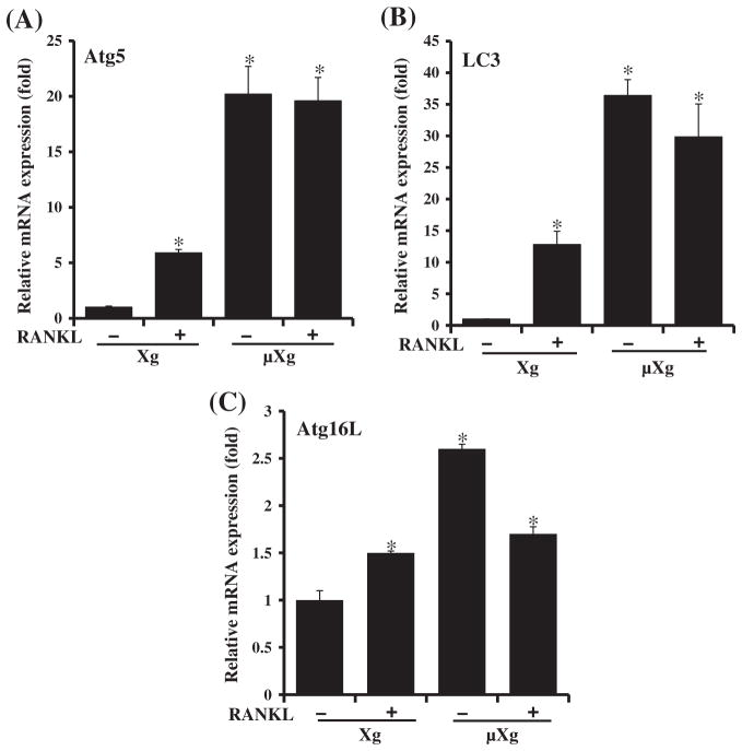
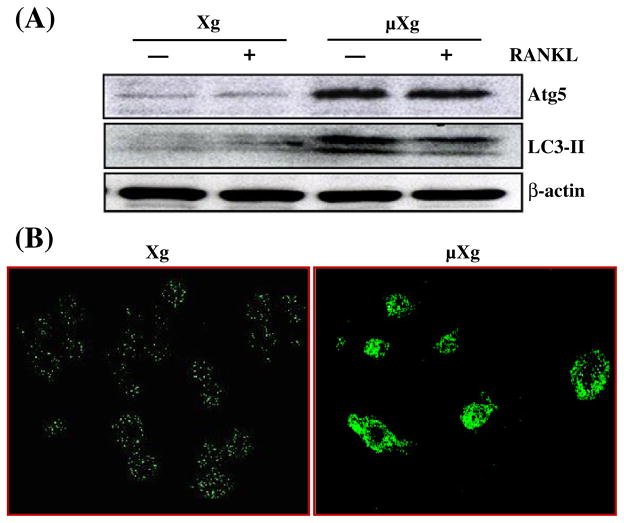
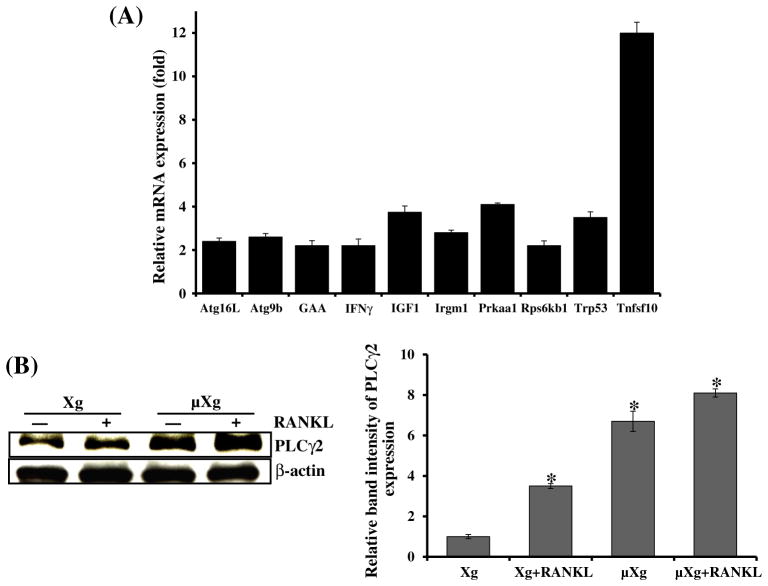
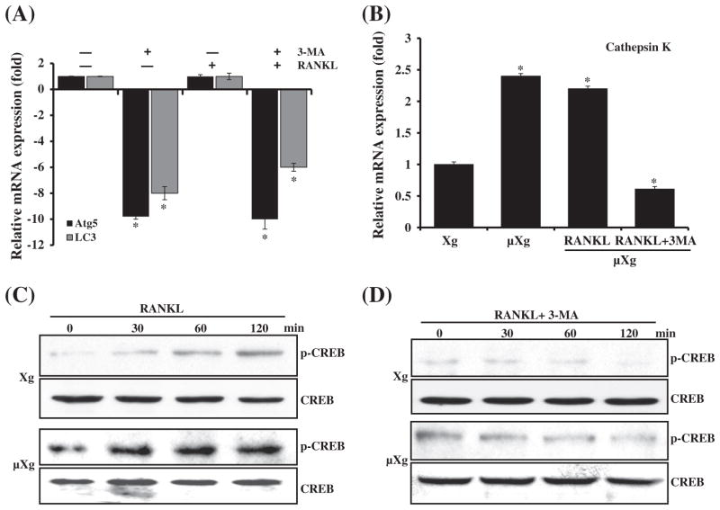
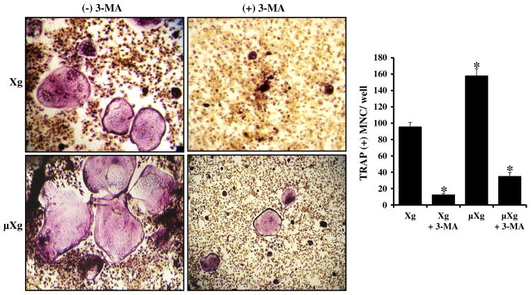
Evidence indicates that astronauts experience significant bone loss during space mission. Recently, we used the NASA developed rotary cell culture system (RCCS) to simulate microgravity (μXg) conditions and demonstrated increased osteoclastogenesis in mouse bone marrow cultures. Autophagy is a cellular recycling process of nutrients. Therefore, we hypothesize that μXg control of autophagy modulates osteoclastogenesis. Real-time PCR analysis of total RNA isolated from mouse bone marrow derived non-adherent cells subjected to modeled μXg showed a significant increase in autophagic marker Atg5, LC3 and Atg16L mRNA expression compared to ground based control (Xg) cultures. Western blot analysis of total cell lysates identified an 8.0-fold and 7.0-fold increase in the Atg5 and LC3-II expression, respectively. Confocal microscopy demonstrated an increased autophagosome formation in μXg subjected RAW 264.7 preosteoclast cells. RT(2) profiler PCR array screening for autophagy related genes identified that μXg upregulates intracellular signaling molecules associated with autophagy, autophagosome components and inflammatory cytokines/growth factors which coregulate autophagy in RAW 264.7 preosteoclast cells. Autophagy inhibitor, 3-methyladenine (3-MA) treatment of mouse bone marrow derived non-adherent mononuclear cells showed a significant decrease in μXg induced Atg5 and LC3 mRNA expression in the presence or absence of RANK ligand (RANKL) stimulation. Furthermore, RANKL treatment significantly increased (8-fold) p-CREB transcription factor levels under μXg as compared to Xg cultures and 3-MA inhibited RANKL increased p-CREB expression in these cells. Also, 3-MA suppresses μXg elevated osteoclast differentiation in mouse bone marrow cultures. Thus, our results suggest that μXg induced autophagy plays an important role in enhanced osteoclast differentiation and could be a potential therapeutic target to prevent bone loss in astronauts during space flight missions.

Keywords: Autophagy; Microgravity; NASA; Osteoclast; Rotary cell culture system (RCCS).


Conflict of interest statement

The authors declare no conflict of interests.

Figures

Fig. 1
Fig. 1
Real-time RT-PCR analysis of autophagy marker Atg5, LC3 and Atg16L mRNA expression. Ground based control (Xg) and microgravity (μXg) subjected mouse bone marrow non-adherent cells were treated with or without RANKL (75 ng/ml) for 24 h. Total RNA isolated were subjected to real-time RT-PCR analysis using gene specific primers for (A) Atg5, (B) LC3 and (C) Atg16L. The mRNA expression was normalized with respect to GAPDH amplification. Each bar represents the mean ± SD of three independent experiments. *Significant (P < 0.05) difference when compared to ground based control without RANKL treatment.
Fig. 2
Fig. 2
μXg modulation of autophagosome formation. (A) Western blot analysis of Atg5 and LC3-II expression. Ground based control (Xg) and μXg subjected mouse bone marrow non-adherent cells were treated with or without RANKL (75 ng/ml) for 24 h. Total cell lysates were subjected to Western blot for Atg5 and LC3-II. β-actin expression served as control. (B) Autophagosome formation in preosteoclast cells. RAW 264.7 cells were cultured in Xg and μXg conditions for 24 h and autophagosomes were visualized by confocal microscopy analysis using anti-LC3-II antibody.
Fig. 3
Fig. 3
(A) RT2 Profiler PCR array analysis for autophagy related gene expression. RAW 264.7 cells were cultured in Xg and μXg conditions for 24 h. Total RNA isolated from these cells was screened for autophagy related genes by real-time PCR in triplicate studies as described in the Materials and methods section. (B) PLCγ2 expression in mouse bone marrow derived non-adherent cells under μXg conditions. Cells were cultured in μXg for 24 h and stimulated with or without RANKL (75 ng/ml) for 24 h. Total cell lysates were subjected to western blot analysis using anti-PLCγ2 antibody. β-actin expression served as control. The band intensity was quantified by the National Institutes of Health ImageJ program, and PLCγ2 expression was normalized with β-actin expression in these cells. The values are expressed as mean ± SD for three independent experiments. *Significant (P < 0.05) difference when compared to ground based control without RANKL treatment.
Fig. 4
Fig. 4
(A) Autophagy inhibitor 3-methyladenine (3-MA) suppresses Atg5 and LC3 expression. Mouse non-adherent bone marrow cells were cultured in μXg for 24 h and treated with or without RANKL (75 ng/ml) and 3-MA (2 mM) for 24 h. Total RNA isolated were subjected to real-time RT-PCR analysis using gene specific primers for (A) Atg5 and LC3. (B) Cathepsin K gene expression. mRNA expression was normalized with respect to β-actin amplification. Each bar represents the mean ± SD of three independent experiments. *Significant (P < 0.05) difference when compared to ground based control without RANKL treatment. (C & D) RANKL stimulation and 3-MA inhibition of CREB activation under Xg and μXg conditions. Mouse non-adherent bone marrow cells were cultured in μXg for 24 h and treated with RANKL (75 ng/ml) in the presence and absence of 3-MA (2 mM) for different time points (0–120 min). Total cell lysate obtained from these samples was subjected to western blot analysis for p-CREB expression. Total CREB expression served as control.
Fig. 5
Fig. 5
Autophagy inhibitor (3-MA) inhibits μXg elevated OCL formation. Mouse bone marrow derived non-adherent cells were cultured with RANKL (75 ng/ml) and M-CSF (10 ng/ml) for 7 days in the presence and absence of 3-MA (2 mM) under Xg and μXg conditions. TRAP-positive multinucleated osteoclasts formed at the end of the culture period were scored. *Significant (P < 0.05) difference when compared to ground based control.

References

    1. Carmeliet G, Bouillon R. The effect of microgravity on morphology and gene expression of osteoblasts in vitro. FASEB J. 1999;13(Suppl):S129–34. - PubMed
    1. Sibonga JD. Spaceflight-induced bone loss: is there an osteoporosis risk? Curr Osteoporos Rep. 2013;11:92–8. - PubMed
    1. Garber MA, McDowell DL, Hutton WC. Bone loss during simulated weightlessness: a biomechanical and mineralization study in the rat model. Aviat Space Environ Med. 2000;71:586–92. - PubMed
    1. Lang TF, Leblanc AD, Evans HJ, Lu Y. Adaptation of the proximal femur to skeletal reloading after long-duration spaceflight. J Bone Miner Res. 2006;21:1224–30. - PubMed
    1. Cavanagh PR, Licata AA, Rice AJ. Exercise and pharmacological countermeasures for bone loss during long-duration space flight. Gravit Space Biol Bull. 2005;18:39–58. - PubMed

Publication types

LinkOut - more resources