MiR-196a exerts its oncogenic effect in glioblastoma multiforme by inhibition of IκBα both in vitro and in vivo
- PMID: 24463357
- PMCID: PMC3984554
- DOI: 10.1093/neuonc/not307
MiR-196a exerts its oncogenic effect in glioblastoma multiforme by inhibition of IκBα both in vitro and in vivo
Abstract
Background: Recent studies have revealed that miR-196a is upregulated in glioblastoma multiforme (GBM) and that it correlates with the clinical outcome of patients with GBM. However, its potential regulatory mechanisms in GBM have never been reported.
Methods: We used quantitative real-time PCR to assess miR-196a expression levels in 132 GBM specimens in a single institution. Oncogenic capability of miR-196a was detected by apoptosis and proliferation assays in U87MG and T98G cells. Immunohistochemistry was used to determine the expression of IκBα in GBM tissues, and a luciferase reporter assay was carried out to confirm whether IκBα is a direct target of miR-196a. In vivo, xenograft tumors were examined for an antiglioma effect of miR-196a inhibitors.
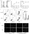
Results: We present for the first time evidence that miR-196a could directly interact with IκBα 3'-UTR to suppress IκBα expression and subsequently promote activation of NF-κB, consequently promoting proliferation of and suppressing apoptosis in GBM cells both in vitro and in vivo. Our study confirmed that miR-196a was upregulated in GBM specimens and that high levels of miR-196a were significantly correlated with poor outcome in a large cohort of GBM patients. Our data from human tumor xenografts in nude mice treated with miR-196 inhibitors demonstrated that inhibition of miR-196a could ameliorate tumor growth in vivo.
Conclusions: MiR-196a exerts its oncogenic effect in GBM by inhibiting IκBα both in vitro and in vivo. Our findings provide new insights into the pathogenesis of GBM and indicate that miR-196a may predict clinical outcome of GBM patients and serve as a new therapeutic target for GBM.
Keywords: IκBα; apoptosis; glioblastoma; miR-196a; tumor growth.
Figures


Similar articles
-
Hypoxia-inducible miR-196a modulates glioblastoma cell proliferation and migration through complex regulation of NRAS.Cell Oncol (Dordr). 2021 Apr;44(2):433-451. doi: 10.1007/s13402-020-00580-y. Epub 2021 Jan 19. Cell Oncol (Dordr). 2021. PMID: 33469841
-
miR-129-5p targets Wnt5a to block PKC/ERK/NF-κB and JNK pathways in glioblastoma.Cell Death Dis. 2018 Mar 12;9(3):394. doi: 10.1038/s41419-018-0343-1. Cell Death Dis. 2018. PMID: 29531296 Free PMC article.
-
Downregulation of ZMYND11 induced by miR-196a-5p promotes the progression and growth of GBM.Biochem Biophys Res Commun. 2017 Dec 16;494(3-4):674-680. doi: 10.1016/j.bbrc.2017.10.098. Epub 2017 Oct 21. Biochem Biophys Res Commun. 2017. PMID: 29066350
-
Transcription factors in glioblastoma - Molecular pathogenesis and clinical implications.Biochim Biophys Acta Rev Cancer. 2022 Jan;1877(1):188667. doi: 10.1016/j.bbcan.2021.188667. Epub 2021 Dec 8. Biochim Biophys Acta Rev Cancer. 2022. PMID: 34894431 Review.
-
Functional importance of glucose transporters and chromatin epigenetic factors in Glioblastoma Multiforme (GBM): possible therapeutics.Metab Brain Dis. 2023 Jun;38(5):1441-1469. doi: 10.1007/s11011-023-01207-5. Epub 2023 Apr 24. Metab Brain Dis. 2023. PMID: 37093461 Review.
Cited by
-
miR-196 acts as a tumor suppressor in osteosarcoma by targeting HOXA9.Int J Clin Exp Pathol. 2018 Sep 1;11(9):4579-4584. eCollection 2018. Int J Clin Exp Pathol. 2018. Retraction in: Int J Clin Exp Pathol. 2021 Jun 15;14(6):794. PMID: 31949856 Free PMC article. Retracted.
-
MicroRNA Functions in Brite/Brown Fat - Novel Perspectives towards Anti-Obesity Strategies.Comput Struct Biotechnol J. 2014 Sep 18;11(19):101-5. doi: 10.1016/j.csbj.2014.09.005. eCollection 2014 Sep. Comput Struct Biotechnol J. 2014. PMID: 25408843 Free PMC article. Review.
-
Hypoxia-inducible miR-196a modulates glioblastoma cell proliferation and migration through complex regulation of NRAS.Cell Oncol (Dordr). 2021 Apr;44(2):433-451. doi: 10.1007/s13402-020-00580-y. Epub 2021 Jan 19. Cell Oncol (Dordr). 2021. PMID: 33469841
-
Identification of Deregulated miRNAs and mRNAs Involved in Tumorigenesis and Detection of Glioblastoma Patients Applying Next-Generation RNA Sequencing.Pharmaceuticals (Basel). 2025 Mar 19;18(3):431. doi: 10.3390/ph18030431. Pharmaceuticals (Basel). 2025. PMID: 40143207 Free PMC article.
-
Identifying Novel Glioma-Associated Noncoding RNAs by Their Expression Profiles.Int J Genomics. 2017;2017:2312318. doi: 10.1155/2017/2312318. Epub 2017 Sep 12. Int J Genomics. 2017. PMID: 29138748 Free PMC article.
References
Publication types
MeSH terms
Substances
LinkOut - more resources
Full Text Sources
Other Literature Sources
Medical

