. 2014 Feb;124(2):730-41.
doi: 10.1172/JCI70812. Epub 2014 Jan 27.

NRF2-mediated Notch pathway activation enhances hematopoietic reconstitution following myelosuppressive radiation

NRF2-mediated Notch pathway activation enhances hematopoietic reconstitution following myelosuppressive radiation

Jung-Hyun Kim et al. J Clin Invest. 2014 Feb.

Abstract

A nuclear disaster may result in exposure to potentially lethal doses of ionizing radiation (IR). Hematopoietic acute radiation syndrome (H-ARS) is characterized by severe myelosuppression, which increases the risk of infection, bleeding, and mortality. Here, we determined that activation of nuclear factor erythroid-2-related factor 2 (NRF2) signaling enhances hematopoietic stem progenitor cell (HSPC) function and mitigates IR-induced myelosuppression and mortality. Augmenting NRF2 signaling in mice, either by genetic deletion of the NRF2 inhibitor Keap1 or by pharmacological NRF2 activation with 2-trifluoromethyl-2'-methoxychalone (TMC), enhanced hematopoietic reconstitution following bone marrow transplantation (BMT). Strikingly, even 24 hours after lethal IR exposure, oral administration of TMC mitigated myelosuppression and mortality in mice. Furthermore, TMC administration to irradiated transgenic Notch reporter mice revealed activation of Notch signaling in HSPCs and enhanced HSPC expansion by increasing Jagged1 expression in BM stromal cells. Administration of a Notch inhibitor ablated the effects of TMC on hematopoietic reconstitution. Taken together, we identified a mechanism by which NRF2-mediated Notch signaling improves HSPC function and myelosuppression following IR exposure. Our data indicate that targeting this pathway may provide a countermeasure against the damaging effects of IR exposure.


Figures

Figure 1
Figure 1. Augmenting NRF2 signaling by genetic disruption of Keap1 improves HSPC function under steady-state conditions.
(A) mRNA expression of Keap1 and NRF2-regulated genes Nqo1 and Gclm in BM cells isolated from poly(I:C)-treated MxCre-Keap1flox/flox mice and floxed controls. *P < 0.05; **P < 0.01; ***P < 0.001. RFC, relative fold change. (BF) Flow cytometric analysis (B), percentage (C), and absolute number of LSK cells (D). Percentage (E) and absolute number (F) of HSCs and MPP cells in BM from MxCre-Keap1flox/flox mice and floxed controls following poly(I:C) treatment under baseline conditions. MxCre-Keap1–/– is represented as Keap1–/– in the graph legends. *P < 0.05, **P < 0.01, and ***P < 0.001 compared with respective floxed controls (n = 10–12). (G) CFU/200 LSK cells from MxCre-Keap1–/– mice and floxed controls. *P < 0.05; **P < 0.01. BFU-E, erythroid burst-forming units; M-CFU, megakaryocyte CFU.
Figure 2
Figure 2. Augmenting NRF2 signaling enhances BM engraftment.
(A) Schematic of experiment depicted in B and C. Sorted LSK cells from poly(I:C)-treated MxCre-Keap1flox/flox or floxed controls were transplanted into recipient mice with 250,000 competitor cells. (B and C) Flow cytometric analysis (B) and percentage of donor cell in PB at 6, 8, and 14 weeks following BMT (C). (D) Schematic of experiment depicted in E and F. Mice were treated daily with poly(I:C) for 7 days after BMT. (E) Percentage of donor cells in PB following serial BMT (first and second BMT). (F) Donor-derived granulocyte (GR1+), B cell (B220+), and T cell (THY1.2+) lineages in PB of recipient mice after the second transplantation. (G) Schematic of ex vivo experiment depicted in H and I. BM stromal cells isolated from tamoxifen-treated CMVCre-Keap1flox/flox or floxed mice were plated as feeder cells. Five hundred sorted LSK cells were cocultured with BM stromal cells. LSK progeny cells harvested on day 7 were stained and analyzed. (H) Expression of Keap1, Nqo1, and Gclm genes in BM stromal cells (CD45TER119) isolated from CMVCre-Keap1–/– mice and floxed control mice. (I) Total number of LSK cells on day 7. (J and K) Percentage of donor cells (CD45.2) in the PB (J) and BM (LIN) (K) of TMC- or vehicle-administered recipient mice (n = 10–15/group) after BMT. CD45.2 BM cells (500,000 cells) were transferred into irradiated recipient WT mice. The recipient mice were orally administered vehicle (Veh) or TMC 6 times, once every 48 hours, beginning 1 hour after BMT (n = 10–15/group). *P < 0.05, **P < 0.01, ***P < 0.001.
Figure 3
Figure 3. NRF2-null mice show increased sensitivity to TBI-induced mortality and myelosupression.
(A and B) Thirty-day survival rate of Nrf2–/– and Nrf2+/+ mice exposed to (A) 7.0 Gy and (B) 7.25 Gy TBI. (C and D) Differential blood counts (C) (wbc, neutrophils [Ne], lymphocytes [Ly], and monocytes [Mo]) and LSK cell populations (D) in Nrf2+/+ and Nrf2–/– mice on day 23 after TBI (6.9 Gy). *P < 0.05 compared with Nrf2–/– mice.
Figure 4
Figure 4. Administration of NRF2 activator TMC mitigates TBI-induced mortality in mice in an NRF2-dependent manner.
(AC) Relative gene expression of Nqo1, Gclm, or Nrf2 in (A) CD45+LIN HSPCs, (B) CD45 stromal cells, and (C) CD34 LSK cells isolated from BM of irradiated mice. Mice were treated with vehicle or TMC 24 hours after TBI and every 48 hours after 6.9 Gy radiation. *P < 0.05; ***P < 0.001. (D and E) Thirty-day survival rates of mice administered TMC or vehicle 6 times (once every 48 hours), beginning 24 hours after 6.9 Gy (D) and 7.1 Gy (E) TBI. (F) Thirty-day survival rates of mice administered TMC or vehicle 6 times (once every 48 hours), beginning 1, 6, or 24 hours after 7.3 Gy TBI. (G) Thirty-day survival rates of Nrf2–/– mice administered TMC or vehicle 6 times (once every 48 hours), beginning 24 hours after 6.9 Gy TBI.
Figure 5
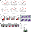
Figure 5. Administration of NRF2 activator TMC mitigates TBI-induced myelosuppression in mice.
Mice exposed to 6.9 Gy TBI were administered 6 doses of vehicle or TMC (once every 48 hours), beginning 24 hours after TBI. At each time period indicated, a cohort of mice (n = 5–8) were sacrificed and PB was isolated. (A and B) Differential blood cell counts (wbc, rbc, neutrophils, platelets [Plt], and hematocrit [Hct]) in TMC- or vehicle-administered mice at the indicated time period (A) and on day 23 after TBI (B). *P < 0.05; **P < 0.01; ***P < 0.001. Data are representative of two independent experiments. (C) Image of blood samples isolated from TMC- or vehicle-treated mice on day 23 following TBI. (D) H&E-stained bones of TMC- or vehicle-administered mice on day 23 after TBI. Scale bars: 1 mm. (E) Analysis and image of bacterial CFU in PB of TMC- or vehicle-administered mice on days 18–23 after TBI (n = 16–25). **P < 0.001 compared with vehicle-treated mice.
Figure 6
Figure 6. Administration of NRF2 activator TMC enhances expansion of HSPCs in TBI-exposed mice.
(A) Analysis of LSK, HSC (LSK CD48CD150+), and MPP (LSK CD48CD150) cell populations in the BM of TMC- or vehicle-administered mice on days 3, 14, and 21 after TBI. *P < 0.05 and **P < 0.01 compared with vehicle-treated mice (n = 15). (B) Flow cytometric analysis of KI67 markers in LSK cells isolated from BM of TMC- or vehicle-treated mice on day 23 after TBI. Data represent the mean ± SD of the percentage of KI67+ LSK cells. *P < 0.05 compared with vehicle-treated mice. (C) Flow cytometric analysis of GSH and ROS levels in LSK cells isolated from TMC- or vehicle-treated mice on day 3 after TBI. Data represent the MFI ± SD. *P < 0.05 compared with vehicle-treated mice. **P < 0.01; ***P < 0.001. (D) Schematic of experiments depicted in EG. BM cells isolated from vehicle- or TMC-treated mice 24 hours after the last dose (day 12) was injected into lethally irradiated recipient mice together with competitive CD45.1 cells. (E and F) Flow cytometric analysis showing percentage of donor-derived CD45.2 PB cells in recipient mice 6 weeks after BMT. (G) Percentage of donor CD45.2 cell–derived granulocyte (GR1+), B cell (B220+), and T cell (THY1.2+) lineages in the PB of recipient mice 6 weeks after BMT. *P < 0.05; **P < 0.01.
Figure 7
Figure 7. Administration of NRF2 activator TMC expands HSPCs by activating Notch1 signaling in irradiated mice.
(A) Relative mRNA expression of Notch1 and Hes1 in BM cells isolated from unirradiated MxCre-Keap1flox/flox mice and floxed control mice following poly(I:C) treatment. **P < 0.01; ***P < 0.001. (B) Relative mRNA expression of Notch1 and Hes1 in BM cells isolated from unirradiated mice treated with vehicle or TMC. Mice were given 6 doses (once every 48 hours) of TMC or vehicle, beginning 24 hours after TBI. *P < 0.05. (C) Jag1 mRNA expression in BM cells isolated from vehicle- or TMC-treated mice on days 2, 7, 13, and 20 following TBI. *P < 0.05 and **P < 0.01 compared with day-0 vehicle-treated mice (0 Gy radiation). (D and E) Immunohistochemical analysis of GFP expression in bone sections (D) and GFP+ LSK cells (E) in the BM of irradiated (6.9 Gy) TNR mice treated with vehicle or TMC beginning 24 hours after TBI (6 times, once every 48 hours) on day 14. (F) Absolute number of LSK cells in the BM of irradiated mice treated with TMC, vehicle, and/or GSI on day 12 following TBI. Mice were injected with 4 doses (1 dose/day) of GSI, beginning 24 hours after the first dose of TMC. *P < 0.05 (D and E).
Figure 8
Figure 8. TMC enhanced expansion of HSPCs ex vivo by upregulating JAG1 in BM stromal cells in an NRF2-dependent manner.
(A) Expression of Nqo1 and Gclm in BM stromal cells isolated from Nrf2+/+ or Nrf2–/– unirradiated mice 12 hours after exposure to TMC (1 μM) or DMSO. Data represent the mean ± SD. ***P < 0.001 and *P < 0.05 compared with vehicle-treated mice. (B) Jag1 mRNA expression in Nrf2+/+ and Nrf2–/– stromal cells isolated from BM 12 hours after exposure to increasing doses of TMC or DMSO in vitro. ***P < 0.001. (C) LSK cells isolated from irradiated mice were cocultured with TMC- (1 μM) or vehicle-exposed Nrf2+/+ or Nrf2–/– BM stromal cells, and the total number of LSK cells was enumerated on day 7. ***P < 0.001. (D) LSK cells were cocultured with TMC- (1 μM) or vehicle-exposed BM stromal cells in the presence or absence of anti-JAG1 antibody, and the total number of LSK cells was enumerated on day 7. LSK and BM stromal cells were isolated from irradiated mice. For neutralization of JAG1, TMC- or vehicle-treated BM stromal cells were incubated with anti-JAG1 antibody prior to incubation with CD34 LSK cells. *P < 0.05; **P < 0.01.

Comment in

References

    1. Williams JP, et al. Animal models for medical countermeasures to radiation exposure. Radiat Res. 2010;173(4):557–578. doi: 10.1667/RR1880.1. - DOI - PMC - PubMed
    1. Johnson SM, et al. Mitigation of hematologic radiation toxicity in mice through pharmacological quiescence induced by CDK4/6 inhibition. J Clin Invest. 2010;120(7):2528–2536. doi: 10.1172/JCI41402. - DOI - PMC - PubMed
    1. Mauch P, et al. Hematopoietic stem cell compartment: acute and late effects of radiation therapy and chemotherapy. Int J Radiat Oncol Biol Phys. 1995;31(5):1319–1339. doi: 10.1016/0360-3016(94)00430-S. - DOI - PubMed
    1. Geiger H, et al. Pharmacological targeting of the thrombomodulin-activated protein C pathway mitigates radiation toxicity. Nat Med. 2012;18(7):1123–1129. doi: 10.1038/nm.2813. - DOI - PMC - PubMed
    1. Guinan EC, et al. Bactericidal/permeability-increasing protein (rBPI21) and fluoroquinolone mitigate radiation-induced bone marrow aplasia and death. Sci Transl Med. 2011;3(110):110ra118. - PMC - PubMed

Publication types

MeSH terms