. 2014 Feb;124(2):543-52.
doi: 10.1172/JCI71858. Epub 2014 Jan 27.

Platelet factor 4 limits Th17 differentiation and cardiac allograft rejection

Platelet factor 4 limits Th17 differentiation and cardiac allograft rejection

Guanfang Shi et al. J Clin Invest. 2014 Feb.

Abstract

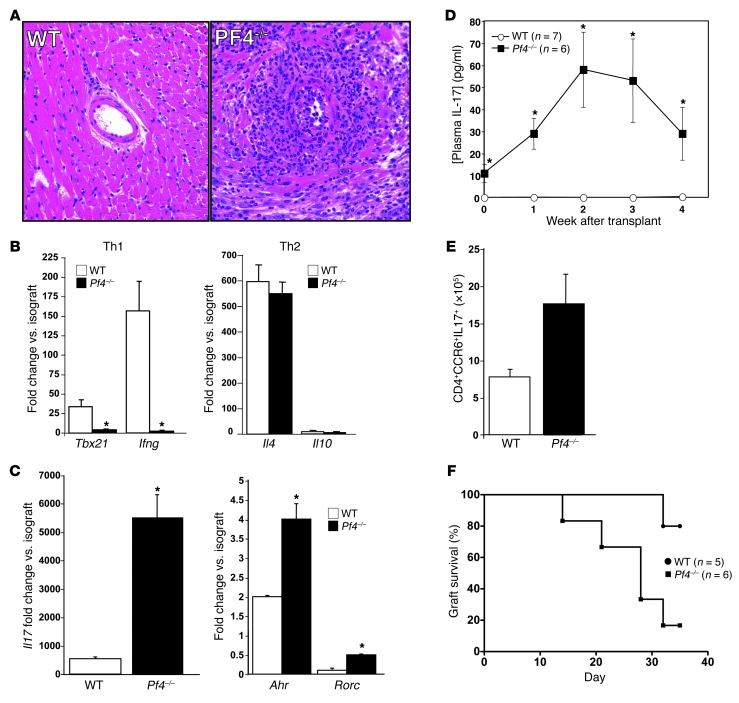
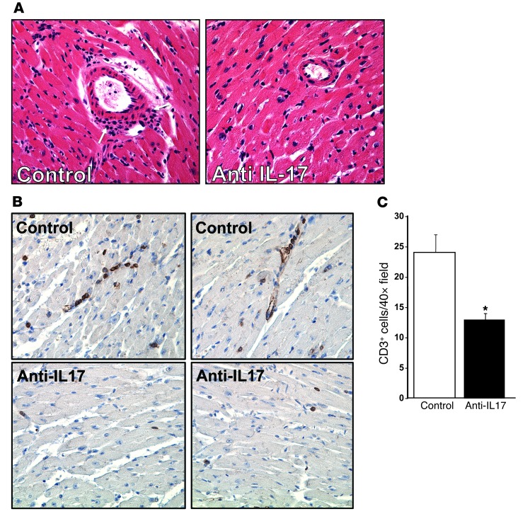
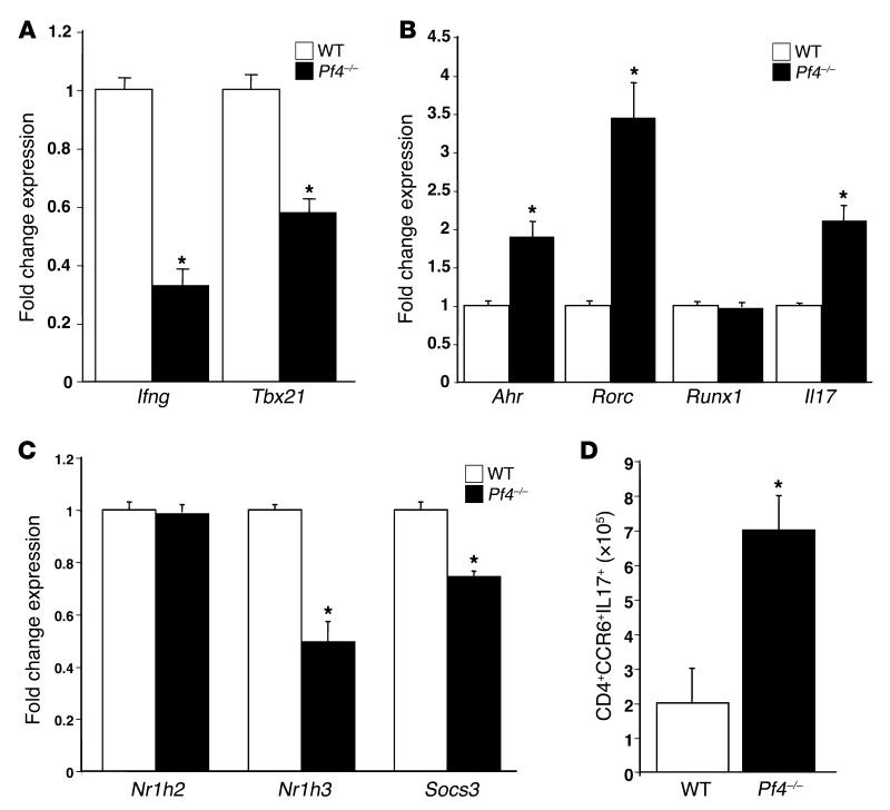
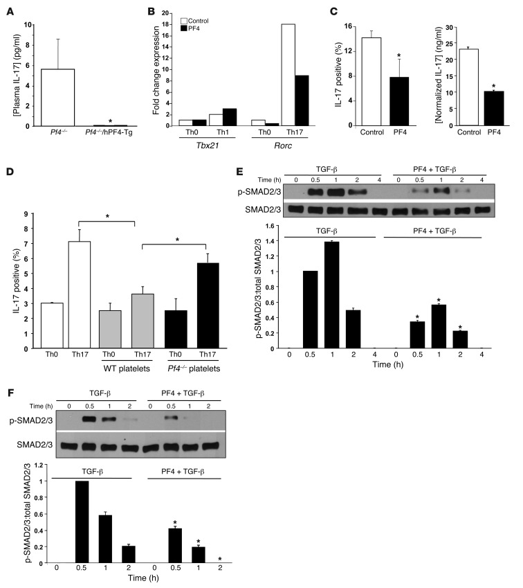
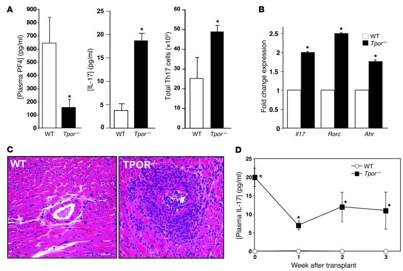
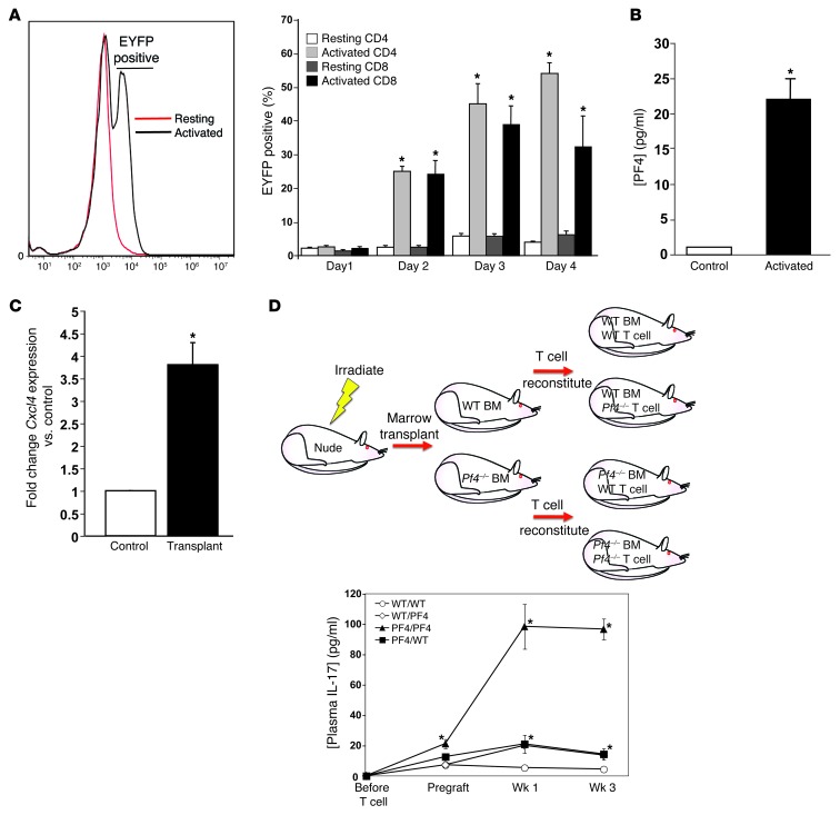
Th cells are the major effector cells in transplant rejection and can be divided into Th1, Th2, Th17, and Treg subsets. Th differentiation is controlled by transcription factor expression, which is driven by positive and negative cytokine and chemokine stimuli at the time of T cell activation. Here we discovered that chemokine platelet factor 4 (PF4) is a negative regulator of Th17 differentiation. PF4-deficient and platelet-deficient mice had exaggerated immune responses to cardiac transplantation, including increased numbers of infiltrating Th17 cells and increased plasma IL-17. Although PF4 has been described as a platelet-specific molecule, we found that activated T cells also express PF4. Furthermore, bone marrow transplantation experiments revealed that T cell-derived PF4 contributes to a restriction in Th17 differentiation. Taken together, the results of this study demonstrate that PF4 is a key regulator of Th cell development that is necessary to limit Th17 differentiation. These data likely will impact our understanding of platelet-dependent regulation of T cell development, which is important in many diseases, in addition to transplantation.


Figures

Figure 1
Figure 1. Pf4–/– mice have an exaggerated Th17 response to cardiac transplantation.
(A) Hearts from BM12 mice were transplanted into WT or Pf4–/– mice and harvested 35 days later. Pf4–/– mice had extensive inflammatory infiltrates (representative histology). (B) Heart grafts from Pf4–/– mice had decreased expression of mRNA representative of a Th1 infiltrate, no change in Th2, and (C) increased Th17-associated mRNA (n = 5–7; mean ± SD; *P < 0.01 vs. isograft). (D) Pf4–/– mice had increased plasma IL-17 and (E) increased Th17 cells in the spleen after cardiac allograft (5–7; mean ± SD. *P < 0.01 vs. WT). (F) Heart transplants in Pf4–/– mice had shorter time to loss of transplant heartbeat. WT and Pf4–/– were transplanted and time to rejection determined by loss of palpable transplant heartbeat (P < 0.03, log-rank test).
Figure 2
Figure 2. IL-17 is the major mediator of graft rejection response in Pf4–/– mice.
(A) IL-17 neutralizing antibody reduced inflammatory infiltrates. Pf4–/– mice were given cardiac allografts and treated with control IgG or IL-17 neutralizing antibody. Original magnification, ×20. Two weeks after transplant, grafts were removed, and histology was performed (arrows indicate inflammatory infiltrate), as well as (B) immunohistochemistry for CD3+ T cells (representative sections). (C) CD3+ cell quantification. CD3+ cells per ×40 field (n = 4; data represent mean ± SD; *P < 0.01 vs. control).
Figure 3
Figure 3. PF4 maintains normal Th differentiation.
CD4+ T cells were isolated from WT and Pf4–/– mice, and qRT-PCR was performed. Pf4–/– CD4+ T cells had (A) decreased expression of Th1, (B) increased expression of Th17, and (C) decreased expression of negative regulators of Th17 differentiation (n = 4; mean ± SD; *P < 0.01 vs. WT). (D) Pf4–/– mouse inguinal lymph nodes also had increased Th17 cells (n = 4; mean ± SD. *P < 0.01 vs. WT).
Figure 4
Figure 4. PF4 has a direct effect on limiting Th17 differentiation.
(A) Transgenic expression of PF4 normalizes plasma IL-17 in Pf4–/– mice (n = 5–7, ± SD; *P < 0.01 vs. Pf4–/–). (B) PF4 directly inhibits Th17 differentiation in vitro. CD4+ T cells were cultured in Th0, Th1, or Th17 conditions in the presence of control or PF4 (1 μg/ml). After 3 days representative Th1 (Tbx21) and Th17 (Rorc) gene expression was determined (representative of 3 separate experiments with the same expression pattern). (C) T cells were cultured in Th17 conditions in the presence of buffer or PF4. IL-17–positive cells (flow cytometry) and IL-17 in the supernatant (ELISA) were measured (n = 4; mean ± SD. *P < 0.03 vs control). (D) Platelet PF4 limits Th17 differentiation. Naive CD4+ T cells were incubated in Th0 or Th17 conditions with buffer, WT,or Pf4–/– platelets. IL-17–positive T cells were quantified 4 days later. WT, but not Pf4–/– platelets, inhibited Th17 differentiation (n = 4; mean ± SD, *P < 0.03). (E and F) PF4 blocks TGF-β–induced Smad 2/3 phosphorylation. Jurkat T cells (E) or naive mouse CD4+ T cells (F) were incubated with control buffer, TGF-β (2 ng/ml), or TGF-β and PF4 (1 μg/ml). p-Smad2/3 was determined by Western blot and quantified by densitometry (n = 3; mean ± SD, *P < 0.01 vs. TGF-β only).
Figure 5
Figure 5. Platelet-deficient mice have increased Th17 differentiation.
(A) Pf4 mice have greatly reduced plasma PF4, increased plasma IL-17, and increased Th17 cells (n = 5; mean ± SD. *P < 0.01 vs. WT). (B) Th17 gene expression is increased in CD4+ T cells from Pf4 mice (n = 4; mean ± SD. *P < 0.01 vs. WT. (C) Inflammatory cell infiltrates after transplantation were increased in Pf4 mice compared with WT control mice (representative histology). Original magnification, ×20. (D) Plasma IL-17 does not increase after transplant in Pf4 mice (n = 5; mean ± SEM. *P < 0.01 vs. WT).
Figure 6
Figure 6. Activated T cell–derived PF4 limits Th17 differentiation.
(A) T cells were isolated from Pf4-Cre+ Rosa26 flox-stop-flox mice and cultured in resting or activated conditions (anti-CD3 and anti-CD28, 1 μg/ml). EYFP-positive CD4+ and CD8+ T cells were quantified by flow cytometry (n = 4; mean ± SD. *P < 0.01 vs. Resting). (B) Stimulated T cells produce PF4. ELISA for PF4 in 4 day cell culture supernatants (n = 4; mean ± SD. *P < 0.01 vs. Control). (C) PF4 expression is induced in T cells in vivo. Mice were given cardiac transplants, and after 7 days CD4+ cells were isolated to measure Cxcl4 expression relative to control mice by qRT-PCR (n = 4; mean ± SD; *P < 0.01 vs. Control). (D) T cell PF4 can compensate partially for a lack of platelet PF4. Nude mice were given WT or Pf4–/– T cell–depleted bone marrow transplants, and reconstituted with either WT or Pf4–/– T cells. Mice were then given cardiac transplants, and plasma IL-17 was measured (n = 4; mean ± SD; *P < 0.05 vs. WT/WT).

Comment in

References

    1. Boilard E, et al. Platelets amplify inflammation in arthritis via collagen-dependent microparticle production. Science. 2010;327(5965):580–583. doi: 10.1126/science.1181928. - DOI - PMC - PubMed
    1. Gawaz M, Favaloro EJ. Platelets, inflammation and cardiovascular diseases. New concepts and therapeutic implications. Semin Thromb Hemost. 2010;36(2):129–130. doi: 10.1055/s-0030-1251495. - DOI - PubMed
    1. Gawaz M, Langer H, May AE. Platelets in inflammation and atherogenesis. J Clin Invest. 2005;115(12):3378–3384. doi: 10.1172/JCI27196. - DOI - PMC - PubMed
    1. Smyth SS, et al. Platelet functions beyond hemostasis. J Thromb Haemost. 2009;7(11):1759–1766. doi: 10.1111/j.1538-7836.2009.03586.x. - DOI - PubMed
    1. Ombrello C, Block RC, Morrell CN. Our expanding view of platelet functions and its clinical implications. J Cardiovasc Transl Res. 2010;3(5):538–546. doi: 10.1007/s12265-010-9213-7. - DOI - PMC - PubMed

Publication types