. 2014 Feb;124(2):785-800.
doi: 10.1172/JCI72017. Epub 2014 Jan 27.

Requirement of enhanced Survival Motoneuron protein imposed during neuromuscular junction maturation

Requirement of enhanced Survival Motoneuron protein imposed during neuromuscular junction maturation

Shingo Kariya et al. J Clin Invest. 2014 Feb.

Abstract

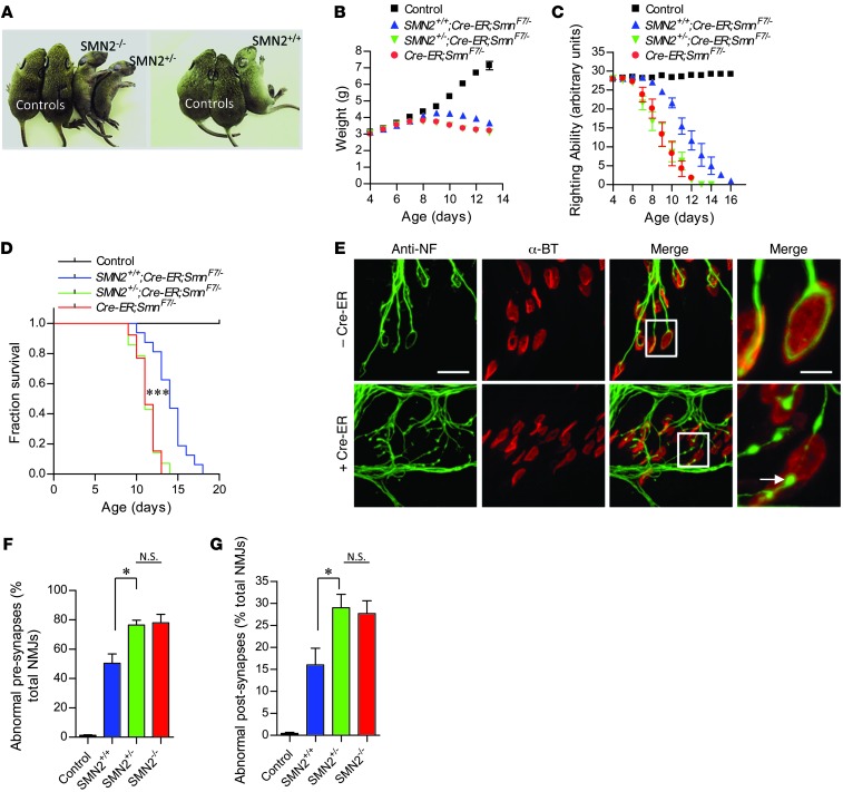
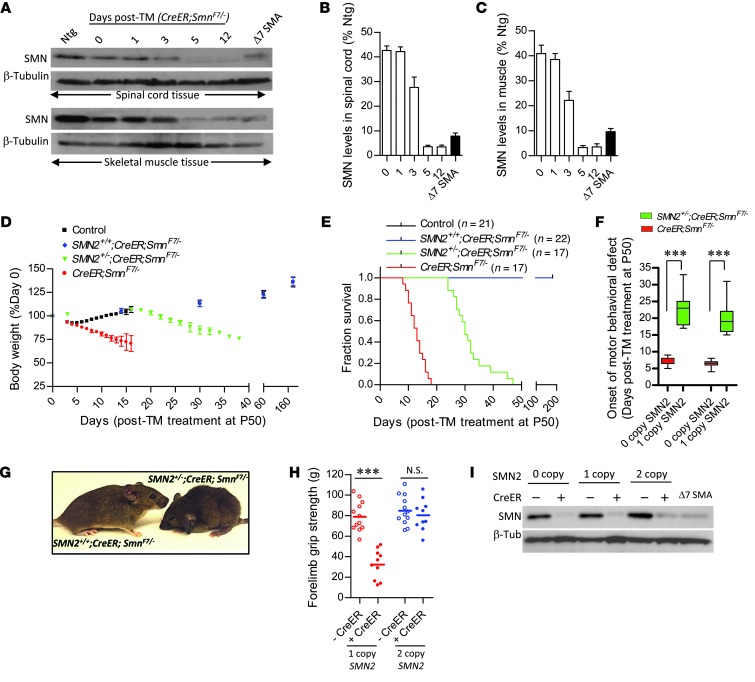
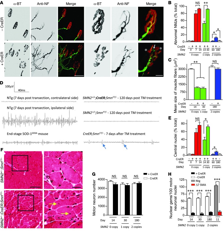
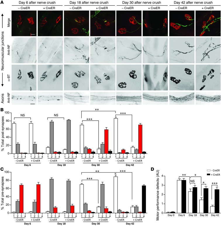
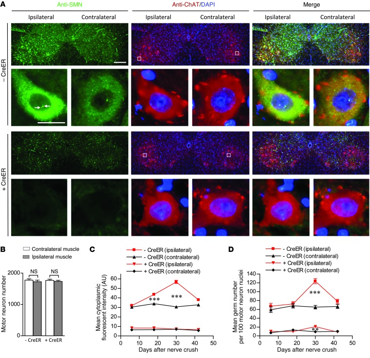
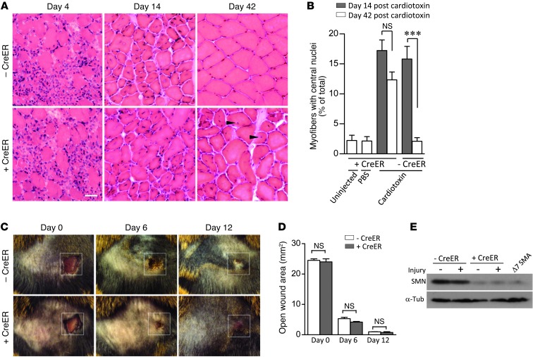
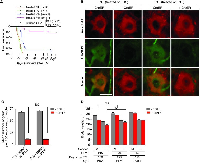
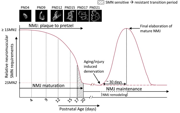
Spinal muscular atrophy is a common motor neuron disease caused by low survival motoneuron (SMN), a key protein in the proper splicing of genes. Restoring the protein is therefore a promising therapeutic strategy. Implementation of this strategy, however, depends on defining the temporal requirements for SMN. Here, we used controlled knockdown of SMN in transgenic mice to determine the precise postnatal stage requirements for this protein. Reducing SMN in neonatal mice resulted in a classic SMA-like phenotype. Unexpectedly, depletion of SMN in adults had relatively little effect. Insensitivity to low SMN emerged abruptly at postnatal day 17, which coincided with establishment of the fully mature neuromuscular junction (NMJ). Mature animals depleted of SMN eventually exhibited evidence of selective neuromuscular pathology that was made worse by traumatic injury. The ability to regenerate the mature NMJ in aged or injured SMN-depleted mice was grossly impaired, a likely consequence of the inability to meet the surge in demand for motoneuronal SMN that was seen in controls. Our results demonstrate that relative maturity of the NMJ determines the temporal requirement for the SMN protein. These observations suggest that the use of potent but potentially deleterious SMN-enhancing agents could be tapered in human patients once the neuromuscular system matures and reintroduced as needed to enhance SMN for remodeling aged or injured NMJs.


Figures

Figure 1
Figure 1. Severe neuromuscular disease in mice depleted of SMN during neonatal life.
(A) Characteristic paralytic phenotype of P11 0-copy (SMN2–/–), 1-copy (SMN2+/–), or 2-copy (SMN2+/+) CreER;SmnF7/– mutants treated with TM at 4 days of age. Decline in (B) body weight, (C) righting ability, and (D) survival during the first 2 weeks following SMN depletion characterizes the onset and progression of neuromuscular disease in the mutants. Two SMN2 copies partially mitigate disease, slowing the loss of body weight, improving motor performance, and enhancing survival. n ≥ 10 (B and C) and n ≥ 13 (D) mice of each genotype for analyses. ***P < 0.0001, log-rank test. (EG) Evidence of abnormal NMJs in 0, 1-, or 2-copy SMN2 mice depleted of SMN protein. NMJs in E are from 2-copy SMN2 mutants but are illustrative of the defects seen in all 3 cohorts of SMA mice. Arrow depicts NF-containing varicosity at nerve terminal. NMJ analysis was conducted on n ≥ 900 synapses from n ≥ 3 mice of each genotype. *P < 0.05, 1-way ANOVA. Note that in every case, controls are 2-copy SMN2;SmnF7/– mice lacking the CreER transgene. Scale bars: 38 μm; 10 μm (inset).
Figure 2
Figure 2. An SMN-resistant state is acquired during adult life.
(A) Western blot analysis of SMN in tissues of inducible mice demonstrates dramatic depletion of protein 5 days following TM administration. Quantification of protein levels in (B) spinal cord and (C) skeletal muscle of treated mutants at the various time points examined. n ≥ 3 mice analyzed for each time point. (D) Loss of body weight and (E) early mortality of SMN-depleted mutants harboring 0 or 1 but not 2 SMN2 copies. (F) A delay in the onset of disease in mice bearing 1 versus 0 copies of the SMN2 gene. ***P < 0.001, t test, n ≥ 10 mice. (G) Representative image of 1- and 2-copy SMN2 inducible mutants 30 days following TM administration depict an alert, relatively healthy SMN2+/+ mouse compared with its hunched and somewhat disheveled looking hemizygous (SMN2+/–) littermate. (H) Two- but not one SMN2 copy in SMN-inducible mice protects against loss of forelimb strength. ***P < 0.001, t test, n ≥ 12 mice of each genotype. (I) Western blot depicting robust and equivalent depletion of SMN protein in the various cohorts of inducible mutants.
Figure 3
Figure 3. Age-dependent emergence of NMJ and muscle pathology in SMN-depleted mutants.
(A) Immunostains of NMJs in the gastrocnemius muscle of end-stage 0- or 1-copy SMN2 inducible mutants revealed profound abnormalities including AChR cluster fragmentation (asterisks) and imperfect overlap between pre- and postsynaptic regions (arrow). Depicted are NMJs from a TM-administered CreER;SmnF7/– mouse and a control without the CreER transgene. Scale bars: 100 μm; 30 μm (detail). (B) Quantification of NMJ defects in 0-, 1-, and 2-copy SMN2-inducible mutants. *P < 0.05; **P < 0.01. n = 300 NMJs from each of 3 mice of each genotype, 1-way ANOVA. (C) Normal muscle fiber size in the gastrocnemius of homozygous SMN2 but not hemizygous or 0-copy SMN2 inducible mice is indicative of relatively normal innervations in the presence of 2 SMN2 copies. **P < 0.01, 1-way ANOVA. n > 50 fibers from each of 3 or more mice of each genotype. (D) Representative EMG traces recorded from the gastrocnemius muscle of 2-copy SMN2-inducible mutants and relevant controls. No evidence of denervation was detected. Shown are fibrillation potentials (arrows), a sign of denervation in CreER;SmnF7/– mutants, end-stage SOD-1G93A ALS model mice and animals in which the sciatic nerve was transected. (E) Extent of centrally nucleated myofibers in the various cohorts of SMN-inducible mutants. Statistics calculated as in C. (F) H&E stains of gastrocnemius muscle in transverse section are illustrative of a primary myopathy in the SMN-inducible mice; arrows indicate central nuclei. Scale bars: 100 μm; 30 μm (inset). Quantification of (G) motor neurons and (H) motor neuronal gems failed to provide evidence of cell loss despite profound SMN depletion. Motor neurons and gems in each of 3 mice of the various genotypes were quantified. ***P < 0.001, t test.
Figure 4
Figure 4. An increased SMN requirement during NMJ maturation.
(A) Immunostains of the NMJs and distal sciatic nerve axons of treated 2-copy SMN2;SmnF7/– mutants with or without the CreER transgene at 4 time points following nerve crush. Scale bars: 25 μm. Morphological analysis of (B) post- and (C) presynaptic specializations of the mice during axon regeneration and NMJ remodeling. n = 1500 NMJs from 5 mice of each genotype. (D) An assessment of motor performance following nerve crush indicates a significant difference in recovery between mutants expressing normal and disease-causing levels of SMN. n ≥ 6 mice tested for the analysis. *P < 0.05; **P < 0.01; ***P < 0.001, 1-way ANOVA.
Figure 5
Figure 5. An elevated demand for motor neuronal SMN as disrupted NMJs remodel and mature.
(A) Immunostains of spinal cord sections from TM-treated 2-copy SMN2;SmnF7/– mice with or without the CreER transgene 30 days following nerve crush. An elevation in the number of gems (arrows) and cytoplasmic SMN is evident in ipsilateral motor neurons of the CreER spinal cord. Scale bars: 240 μm; 30 μm (detail). (B) Quantification of motor neurons in the cords of the mice failed to reveal evidence of cell loss. n ≥ 3 mice of each genotype, t tests. However, (C) cytoplasmic SMN levels and (D) nuclear gems of the ipsilateral motor neurons rose following the nerve crush. Especially evident is the increase which peaks at day 30 after the crush in the CreER-negative animals. Cytoplasmic SMN staining in at least 150 motor neurons from 3 mice of each cohort was assessed; ≥ 300 nuclei were examined for gems. **P < 0.01; ***P < 0.001, 1-way ANOVA for the analyses.
Figure 6
Figure 6. An enhanced requirement for the SMN protein following tissue injury is restricted to the neuromuscular system.
(A) H&E stains of the gastrocnemii muscles of TM-treated 2-copy SMN2;SmnF7/– mice with or without CreER reveals an impaired ability of SMN-depleted muscle to fully recover from cardiotoxin injury — see central nuclei (arrowheads) in CreER positive muscle. Scale bar: 30 μm. (B) Quantification of centrally nucleated myofibers in the 2 sets of mice at days 14 and 42 after -cardiotoxin injection. ***P < 0.001, 1-way ANOVA; muscle fibers from n ≥ 5 mice were examined. (C) Recovery from a surface wound to the skin is not compromised by low SMN. Original magnification, ×1. (D) Assessment of open wound area in the presence of absence of normal SMN failed to reveal differences at any of the time points examined. n ≥ 3, t test. (E) Western blot analysis of SMN from the wound area did not indicate an increase in protein relative to the contralateral area.
Figure 7
Figure 7. An SMN refractory state emerges concomitant with the establishment of the fully mature neuromuscular synapse.
(A) Kaplan-Meier curves indicate a dramatic improvement in survival if SMN depletion is effected in mice of P15 or greater. (B) Immunostains of motor neurons from P12 and P15 TM-treated mice indicate equivalent low levels of SMN, confirming the efficiency of SMN depletion at the 2 ages. Scale bar: 30 μm. (C) Quantification of nuclear gems in motor neurons of the 2 sets of mice did not reveal a difference, confirming equally effective SMN depletion in the 2 cohorts. n ≥ 400 motor neuron nuclei from n = 3 mice of each genotype examined, 1-way ANOVA. (D) Analysis of body weights of P15, P21, and P50 TM-treated 2 SMN2 copy SmnF7/– mice with or without CreER indicates small but significant improvement in the phenotypes of mutants depleted of SMN later than P15. *P < 0.05; **P < 0.01. n ≥ 4 mice of each genotype, t test.
Figure 8
Figure 8. A model encapsulating the postnatal requirement for the SMN protein in the murine model.
The requirement for the protein is adequately met by 1 or more SMN1 copies or SMN2 copies that, between them, express equivalent levels of the protein. This requirement is at its greatest during the neonatal period encompassing NMJ refinement and maturation. It eventually falls by approximately P20, a time point defined by the establishment of the fully mature neuromuscular synapse (see photomicrographs), to levels that are satisfied by 2 SMN2 copies. In the approximately 3-day window prior to this time point, mice become relatively resistant to low SMN. Injury of the NMJ during aging or trauma is accompanied by a surge in demand for the protein, specifically in tissues of the neuromuscular system, which is not adequately met by 2 SMN2 copies. The enhanced requirement appears to peak as the NMJ matures and brings about, in SMN-depleted mutants, an inability to fully repair the neuromuscular synapse. Thus, SMN is thought to play 2 related roles at the neuromuscular synapse — its initial maturation and its continued maintenance. Note: figure not drawn to scale.

Comment in

Similar articles

Cited by

References

    1. Lefebvre S, et al. Identification and characterization of a spinal muscular atrophy-determining gene. Cell. 1995;80(1):155–165. doi: 10.1016/0092-8674(95)90460-3. - DOI - PubMed
    1. Monani UR. Spinal muscular atrophy: a deficiency in a ubiquitous protein; a motor neuron-specific disease. Neuron. 2005;48(6):885–896. doi: 10.1016/j.neuron.2005.12.001. - DOI - PubMed
    1. Burghes AH, Beattie CE. Spinal muscular atrophy: why do low levels of survival motor neuron protein make motor neurons sick? Nat Rev Neurosci. 2009;10(8):597–609. - PMC - PubMed
    1. Lorson CL, Hahnen E, Androphy EJ, Wirth B. A single nucleotide in the SMN gene regulates splicing and is responsible for spinal muscular atrophy. Proc Natl Acad Sci U S A. 1999;96(11):6307–6311. doi: 10.1073/pnas.96.11.6307. - DOI - PMC - PubMed
    1. Monani UR, et al. A single nucleotide difference that alters splicing patterns distinguishes the SMA gene SMN1 from the copy gene SMN2. Hum Mol Genet. 1999;8(7):1177–1183. doi: 10.1093/hmg/8.7.1177. - DOI - PubMed

Publication types

Substances