. 2014 Feb 27;506(7489):498-502.
doi: 10.1038/nature12907. Epub 2014 Jan 19.

A discrete genetic locus confers xyloglucan metabolism in select human gut Bacteroidetes

Affiliations

A discrete genetic locus confers xyloglucan metabolism in select human gut Bacteroidetes

Johan Larsbrink et al. Nature. .

Abstract

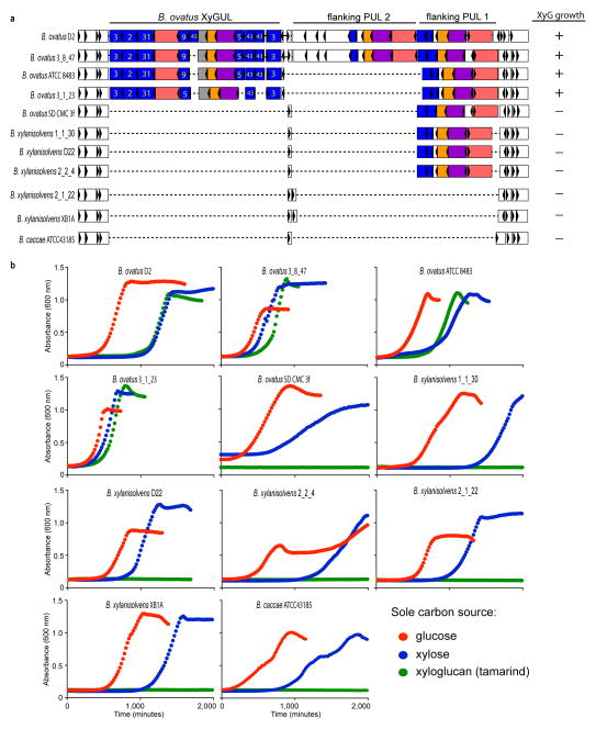
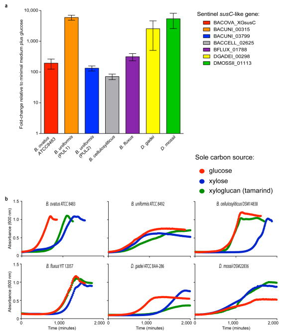
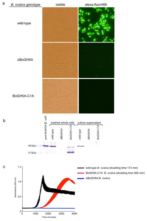
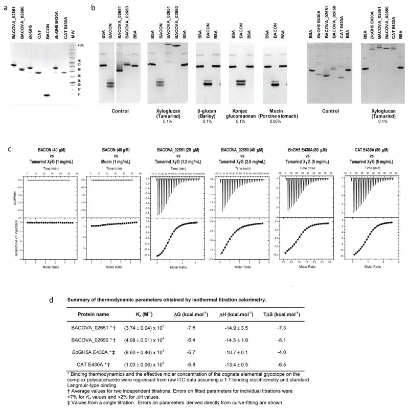
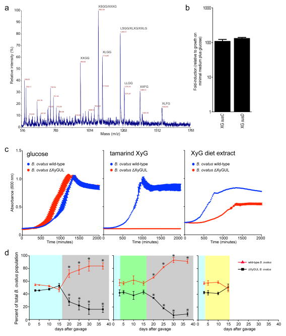
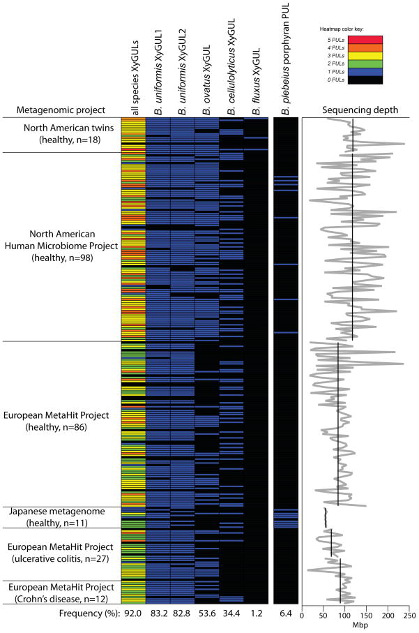
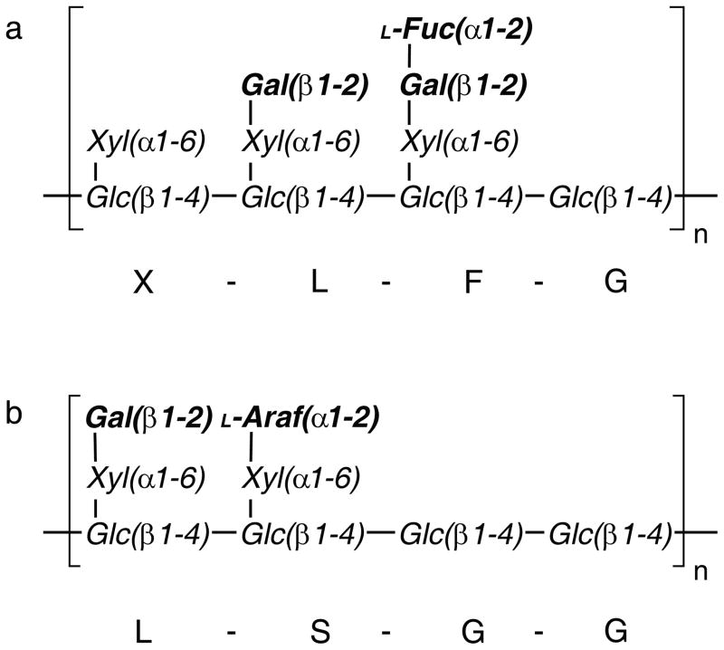
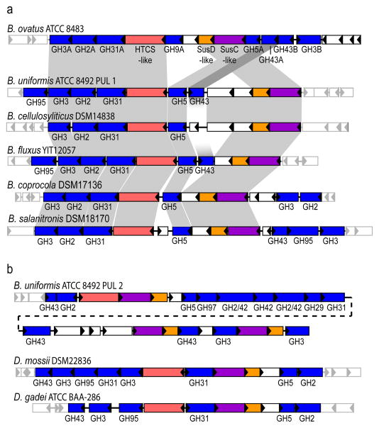
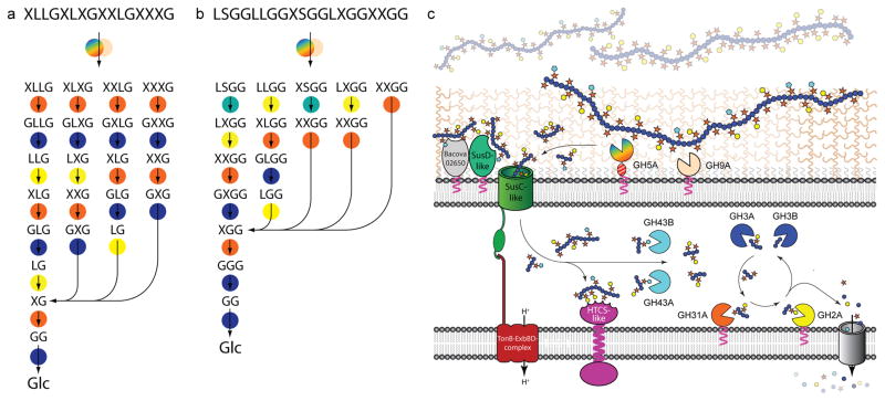
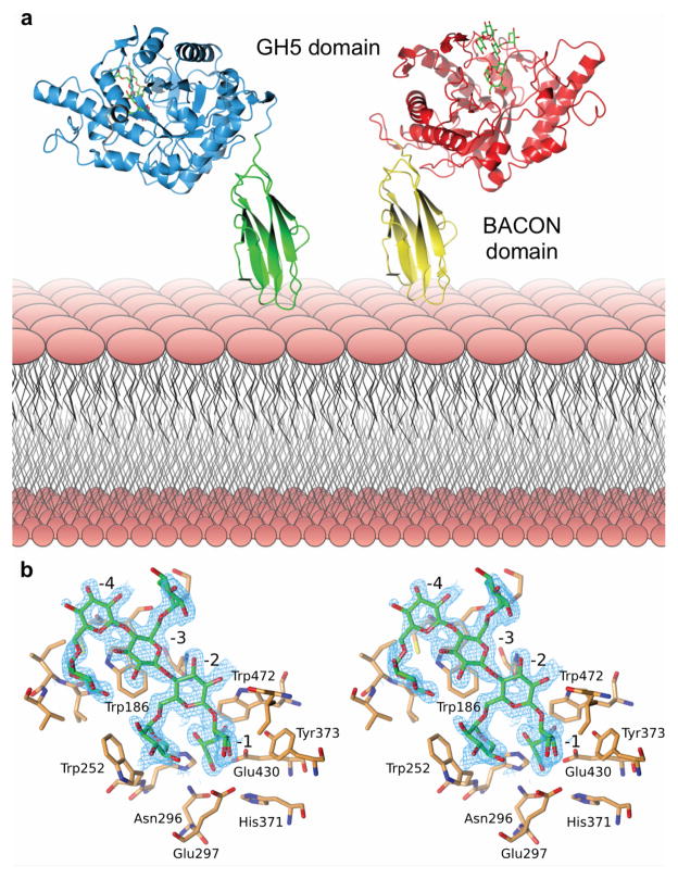
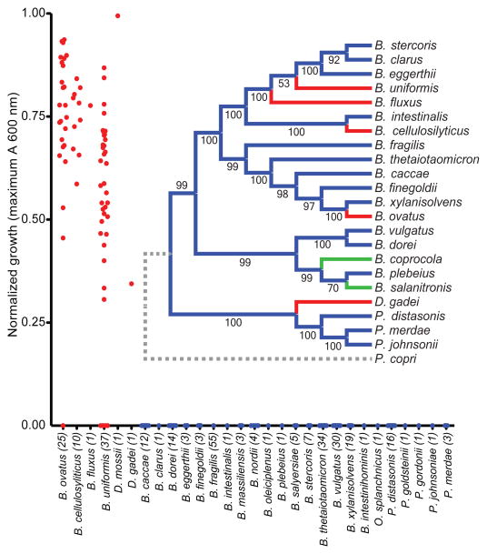
A well-balanced human diet includes a significant intake of non-starch polysaccharides, collectively termed 'dietary fibre', from the cell walls of diverse fruits and vegetables. Owing to the paucity of alimentary enzymes encoded by the human genome, our ability to derive energy from dietary fibre depends on the saccharification and fermentation of complex carbohydrates by the massive microbial community residing in our distal gut. The xyloglucans (XyGs) are a ubiquitous family of highly branched plant cell wall polysaccharides whose mechanism(s) of degradation in the human gut and consequent importance in nutrition have been unclear. Here we demonstrate that a single, complex gene locus in Bacteroides ovatus confers XyG catabolism in this common colonic symbiont. Through targeted gene disruption, biochemical analysis of all predicted glycoside hydrolases and carbohydrate-binding proteins, and three-dimensional structural determination of the vanguard endo-xyloglucanase, we reveal the molecular mechanisms through which XyGs are hydrolysed to component monosaccharides for further metabolism. We also observe that orthologous XyG utilization loci (XyGULs) serve as genetic markers of XyG catabolism in Bacteroidetes, that XyGULs are restricted to a limited number of phylogenetically diverse strains, and that XyGULs are ubiquitous in surveyed human metagenomes. Our findings reveal that the metabolism of even highly abundant components of dietary fibre may be mediated by niche species, which has immediate fundamental and practical implications for gut symbiont population ecology in the context of human diet, nutrition and health.


Conflict of interest statement

The authors declare no competing financial interests.

Figures

Figure ED1
Figure ED1
Evolution of the genomic region containing the XyGUL and corresponding growth on XyG as a sole carbon source. a. Genomic organisation of 11 representative strains from 3 different species of Bacteroides. b. Growth of these strains on tamarind XyG with glucose and xylose as controls (average of n=2 growths per strain). The observation that one B. ovatus strain (SD CMC 3f) lacks a corresponding XyGUL, as do all B. xylanisolvens (the closest cultured relative of B. ovatus), suggests that the XyGUL entered B. ovatus after it diverged. Also note that two other unrelated flanking PULs show variable presence at this locus, suggesting that it is a ‘hotspot’ for PUL evolution.
Figure ED2
Figure ED2
Activation of putative XyGULs in Bacteroidetes isolates via growth on XyG. a. Sentinel (ref. 7) susC-like gene expression (n=3, expression measurements from separately grown cultures relative to a minimal medium plus glucose control). b. Growth profiles of the corresponding strains in medium containing tamarind XyG with glucose and xylose as controls (average of n=2 growths per strain). Error bars represent standard errors of the mean.
Figure ED3
Figure ED3
Cell-surface localisation of BoGH5A and effect of localisation on B. ovatus growth. a. staining of fixed wild-type and mutant B. ovatus strains. b. Western blot indicating that BoGH5A is still produced in the C1A lipidation site mutant, albeit in multiple degraded forms. c. Growth of wild-type (positive control), ΔBoGH5A (negative control) and BoGH5A-C1A strains on tamarind XyG. The BoGH5A-C1A mutant exhibits ~2.6-fold slower exponential growth than the wild-type. Vertical error bars on each curve indicate the standard deviation of the mean (n=3 replicates). The residual growth ability of the BoGH5A-C1A strain, despite mis-localization, is unlikely to be explained by the presence of BoGH5A enzyme accumulation in the supernatant, which was only detected by Western blot for wild-type bacteria expressing BoGH5A on the cell surface. Detection of BoGH5A in panels a and b was achieved with a rabbit polyclonal antibody raised against the recombinant protein produced in E. coli. Panels a nd b show representative data two experiments each that yielded very similar results.
Figure ED4
Figure ED4
Non-catalytic interaction of BoGH5A variants, SusD-like Bacova_02651 and Bacova_02650 of the XyGUL with polysaccharides. a. SDS-PAGE of recombinant proteins (representative data from at least three preparations for each protein is shown). b. Affinity gel electrophoresis (representative data from at least two gels for each experimental condition). c. Isothermal titration calorimetry (ITC); the upper graph in each pair shows the raw heat during titration, while the lower graph shows the integrated heats after correction. d. Association constants and thermodynamic parameters obtained from ITC data. Bovine serum albumin (BSA) was used as non-interacting negative control protein. Other protein names were abbreviated as follows: BACON, residues Cys1-Tyr97 corresponding to the BACON domain of BoGH5A; Bacova_02651, SusD-like XyGUL gene product; Bacova_02650, SusE-positioned XyGUL gene product; BoGH5A E430A, full-length (Cys1 to Asn470) catalytic nucleophile mutant of BoGH5A; CAT E430A, catalytic nucleophile mutant of the BoGH5A catalytic domain only (Ser98 to Asn470). Reducing-sugar assays confirmed the catalytic mutants had no detectable hydrolytic activity on xyloglucan (data not shown), while an active variant (i.e., E430) of CAT had a 2-fold higher specific activity than the full-length, wild-type BoGH5A at saturating xyloglucan concentrations (0.5 – 3 mM).
Figure ED5
Figure ED5
Abundance of Bacteroides XyGULs in human from a survey of metagenomic sequencing data from a total of 250 adult human samples (211 healthy, 27 ulcerative colitis, 12 Crohn’s Disease; see Methods for references). Datasets were individually queried by BLAST using the entire XyGUL nucleotide sequence from each of the four Bacteroides species listed at the top (cf. Fig. 2) and a PUL involved in degrading the red algal polysaccharide porphyran. Each horizontal line represents the presence or absence of a hit in a single individual. The leftmost column summarizes the total XyGUL content in each person (annotated according to the color key in the upper right corner). The XyGUL frequency across all 250 samples is shown at the bottom for each condition. The graph at the far right illustrates the variation in sequencing depth for each sample/study: black lines show the average depth in megabasepairs (Mbp) for each study; the light gray line shows the depth for each individual sample.
Figure ED6
Figure ED6
Presence of the XyGUL confers a fitness advantage to B. ovatus in the presence of dietary xyloglucan, but only when other dietary polysaccharides are eliminated. a. MALDI analysis of BoGH5A-digested alkaline extract from a custom mouse diet that contained a large amount of xyloglucan from natural vegetable sources (equal amounts of cooked bell pepper, eggplant, tomato fruit and lettuce; see Methods), indicating the presence of both solanaceous (arabinogalacto)xyloglucan and (fucogalacto)xyloglucan. b. qPCR analysis of XyGUL sentinel gene (ref. 7) expression in wild-type B. ovatus grown on extracted polysaccharides from the XyG-rich custom diet, demonstrating that it significantly activates XyGUL expression over a glucose control (error bars show the standard deviation of the mean of three biological replicates for both growth conditions). c. In vitro growth of wild-type and ΔXyGUL B. ovatus strains in the polysaccharide extract from the XyG-rich diet, including glucose and tamarind XyG as positive and negative control substrates, respectively. Compared to growth on tamarind XyG (middle panel), the incomplete growth defect of ΔXyGUL mutant on the food extract (right panel) indicates that the food contains other polysaccharides that are accessible by B. ovatus. Vertical error bars on each curve indicate the standard deviation of the mean of 3 replicates. d. In vivo competition of wild-type and ΔXyGUL B. ovatus strains in mice consuming various amounts of dietary XyG. All mice were initially fed a synthetic diet containing glucose as the sole digestible carbohydrate for 1 week and then gavaged with a 7:3 ratio of ΔXyGUL:WT (based on independent culture optical densities, total of ~108 viable B. ovatus) and the communities were allowed to equilibrate for 3 days. Despite the initial ratio biased in favour of the ΔXyGUL strain, the communities equilibrated in the range 5:5 – 4:6, but thereafter remained stable while mice were maintained on the XyG-free diet (blue boxes in three competition plots). Subsequent to community stabilization, three different dietary regimens were analysed. Left panel: Mice were maintained on the control diet (glucose only, devoid of XyG) between days 5–37, but switched to water containing 0.25% purified XyG for days 15–37 (gray box); Middle panel: Mice were switched to a xyloglucan-rich, custom diet from natural food sources while simultaneously drinking water containing 0.25% purified XyG (green box). These mice were then switched to the glucose-only, XyG-free control diet while remaining on water containing 0.25% XyG (gray box); Right panel: Mice were switched to the XyG-rich diet between days 3–15 but given normal water (yellow box), these mice were not continued further on any dietary regimen. Maintenance on either XyG food/XyG water (middle panel) or XyG food only (right panel) does not exert a measurable fitness pressure on the competing WT and ΔXyGUL strains. However, when the complex natural food polysaccharides were withheld while 0.25% XyG was maintained in water, a clear fitness pressure was observed via the significant, sequential reduction of the ΔXyGUL mutant between 15–37 days. These data suggest that although the XyGUL confers an advantage to B. ovatus by broadening its substrate range to include XyG, the presence of alternative oligo- and polysaccharides (e.g., other hemicelluloses, pectins) in a complex vegetable-based diet is nonetheless sufficient to support strains lacking this locus in vivo. Each data point is the mean abundance of the indicated strain in 4 separate mice and error bars represent one standard deviation. Measurements conformed to a normal distribution based on the observation that 67% of all assay values were within one standard deviation of their respective means. Asterisks indicate statistically significant alterations (p < 0.01; student’s t test, one-tailed, paired) in strain abundance relative to the day 15 samples, which immediately preceded the diet switch aimed at isolating XyG as the sole exogenous polysaccharide.
Figure 1
Figure 1
Representative structures of XXXG- and XXGG-type xyloglucans. a. XXXG-type xyloglucans, comprised of a Glc4Xyl3 repeating motif with variable branch extensions (bold residues). Tamarind seed xyloglucan and primary cell wall xyloglucans (e.g. from lettuce leaves) are distinguished by the absence of fucose in the former. b. XXGG-type xyloglucans, comprised of Glc4Xyl2 repeating motif. These xyloglucans are common to solanaceous species (e.g. tomato) and are typified by branches extended with arabinofuranosyl residues. Standard single-letter abbreviations for designating backbone decorations are shown.
Figure 2
Figure 2
Structure of the B. ovatus xyloglucan utilization locus and evolution in the Bacteroidetes lineage. a. PULs with partial homology and synteny; homologous genes are connected by gray bars and flanking genes lacking synteny are shown as semi-transparent. b. PULs with partial homology, but lacking overall synteny. Extended Data Figure ED2 provides transcriptional evidence that each of these gene clusters is responsive to growth on XyG.
Figure 3
Figure 3
The concerted action of XyGUL gene products in the degradation of xyloglucans. Most probable sequential pathways for the hydrolysis of (galacto)xyloglucan (a) and (arabinogalacto)xyloglucans (b) based on enzyme kinetic data, product analysis, and selected gene knock-out studies (see Fig. 1 for XyG motif abbreviations). Enzymes are represented as circles, colour-coded as in panel c: Rainbow, endo-xyloglucanase BoGH5A; tan, endo-xyloglucanase BoGH9A; orange, α-xylosidase BoGH31A; turquoise α-L-arabinofuranosides BoGH43A and/or BoGH43B; yellow β-galactosidase BoGH2; dark blue β-glucosidases BoGH3A and/or BoGH3B. c. Model of enzyme localisation by analogy with the archetypal Sus locus and based on inference of N-terminal lipoprotein modification from protein sequence data.
Figure 4
Figure 4
Structural biology of BoGH5A. a. Tertiary structure; the two conformations observed in crystallo have been oriented relative to the N-terminal, membrane-anchored BACON domain (see also Supplementary Video V1). b. Wall-eyed stereo view of the binding of XXXG in the -4 to -1 subsites (see also Supplementary Video V2). The wireframe represents an unbiased 2Fo-Fc map (contoured at 0.3 electrons per Å3) obtained using phases calculated from the best model prior to the incorporation of any ligand in refinement.
Figure 5
Figure 5
Growth of different human gut-resident Bacteroidetes on xyloglucan. Each point represents the normalized maximum growth OD600 achieved by each strain after 4 d of growth in minimal medium with tamarind XG as the sole carbon source (average of n=2 independent growth analyses per strain). The inset shows a phylogenetic tree constructed from fully sequenced strains of the species shown; those labeled in red have the ability to grow on xyloglucan as sole carbon source and those labeled in green have PULs similar to B. ovatus XyGUL but were not tested for growth on XG. Bootstrap values based on 1000 replicate trees are indicated.

Comment in

References

    1. McDougall GJ, Morrison IM, Stewart D, Hillman JR. Plant cell walls as dietary fibre: Range, structure, processing and function. J Sci Food Agric. 1996;70:133–150.
    1. El Kaoutari A, Armougom F, Gordon JI, Raoult D, Henrissat B. The abundance and variety of carbohydrate-active enzymes in the human gut microbiota. Nat Rev Microbiol. 2013;11:497–504. doi: 10.1038/nrmicro3050. - DOI - PubMed
    1. Flint H, Scott K, Duncan S, Louis P, Forano E. Microbial degradation of complex carbohydrates in the gut. Gut Microbes. 2012;3:289–306. doi: 10.4161/gmic.19897. - DOI - PMC - PubMed
    1. Koropatkin NM, Cameron EA, Martens EC. How glycan metabolism shapes the human gut microbiota. Nat Rev Microbiol. 2012;10:323–335. doi: 10.1038/nrmicro2746. - DOI - PMC - PubMed
    1. Hoffman M, et al. Structural analysis of xyloglucans in the primary cell walls of plants in the subclass Asteridae. Carbohydr Res. 2005;340:1826–1840. doi: 10.1016/j.carres.2005.04.016. - DOI - PubMed

Publication types

MeSH terms

Associated data