. 2014 Mar 6;507(7490):124-128.
doi: 10.1038/nature12931. Epub 2014 Jan 22.

SRA- and SET-domain-containing proteins link RNA polymerase V occupancy to DNA methylation

Affiliations

SRA- and SET-domain-containing proteins link RNA polymerase V occupancy to DNA methylation

Lianna M Johnson et al. Nature. .

Erratum in

Abstract

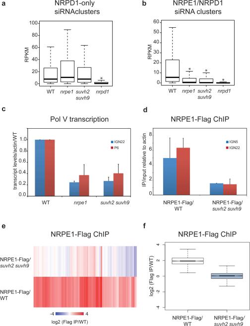
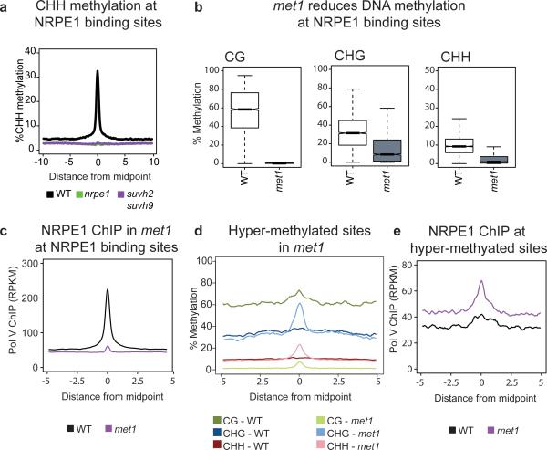
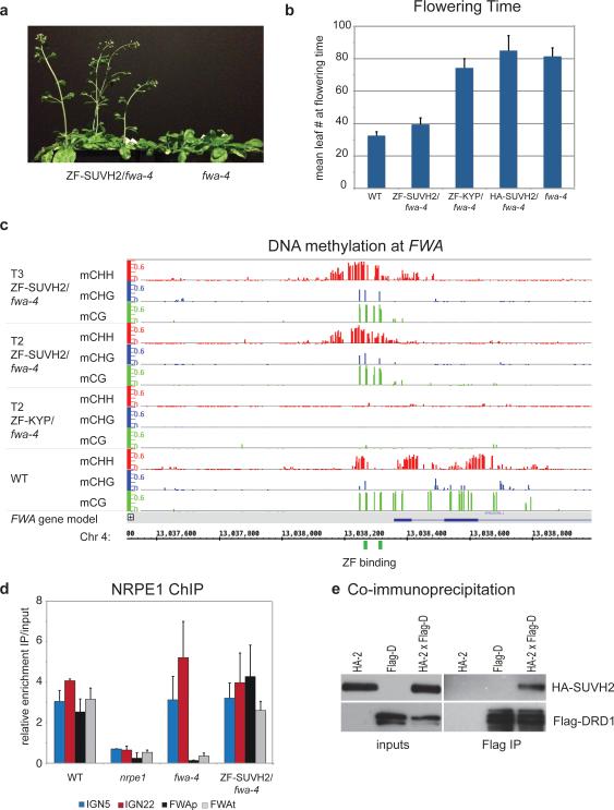
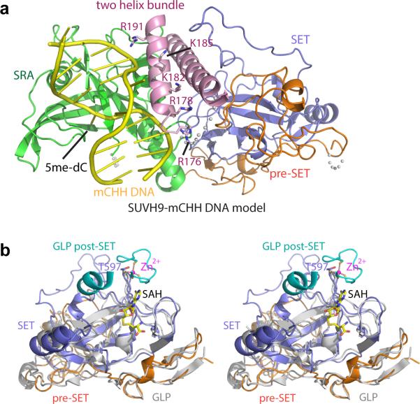
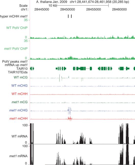
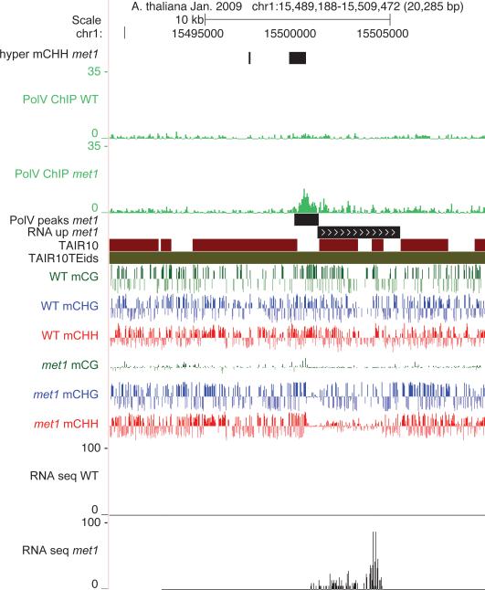
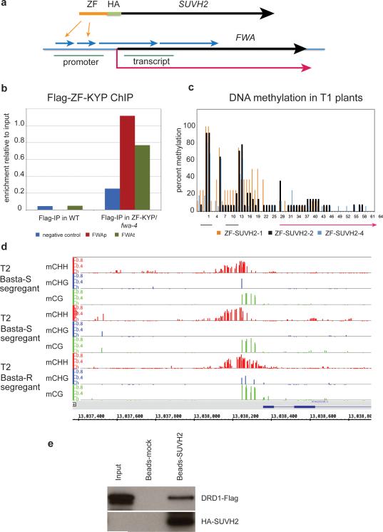
RNA-directed DNA methylation in Arabidopsis thaliana depends on the upstream synthesis of 24-nucleotide small interfering RNAs (siRNAs) by RNA POLYMERASE IV (Pol IV) and downstream synthesis of non-coding transcripts by Pol V. Pol V transcripts are thought to interact with siRNAs which then recruit DOMAINS REARRANGED METHYLTRANSFERASE 2 (DRM2) to methylate DNA. The SU(VAR)3-9 homologues SUVH2 and SUVH9 act in this downstream step but the mechanism of their action is unknown. Here we show that genome-wide Pol V association with chromatin redundantly requires SUVH2 and SUVH9. Although SUVH2 and SUVH9 resemble histone methyltransferases, a crystal structure reveals that SUVH9 lacks a peptide-substrate binding cleft and lacks a properly formed S-adenosyl methionine (SAM)-binding pocket necessary for normal catalysis, consistent with a lack of methyltransferase activity for these proteins. SUVH2 and SUVH9 both contain SRA (SET- and RING-ASSOCIATED) domains capable of binding methylated DNA, suggesting that they function to recruit Pol V through DNA methylation. Consistent with this model, mutation of DNA METHYLTRANSFERASE 1 (MET1) causes loss of DNA methylation, a nearly complete loss of Pol V at its normal locations, and redistribution of Pol V to sites that become hypermethylated. Furthermore, tethering SUVH9 [corrected] with a zinc finger to an unmethylated site is sufficient to recruit Pol V and establish DNA methylation and gene silencing. These results indicate that Pol V is recruited to DNA methylation through the methyl-DNA binding SUVH2 and SUVH9 proteins, and our mechanistic findings suggest a means for selectively targeting regions of plant genomes for epigenetic silencing.


Figures

Figure 1
Figure 1. Crystal structure of SUVH9
a. Ribbon diagram of the SUVH9 crystal structure containing a two-helix bundle, SRA domain, pre-SET domain, and SET domain colored in pink, green, orange, and blue, respectively. The Zn3Cys9 cluster is highlighted in a ball-and-stick representation and disordered regions are shown with dashed lines. b. A superposition of SUVH9 SRA domain (in green) and SUVH5 SRA domain (in silver) shows that both proteins adopt a similar fold. c. Top panel: the crystal structure of human GLP in complex with bound SAH (PDB code: 2IGQ) in a silver ribbon representation. Bottom panel: the SAH binding site in an electrostatic surface representation. The cofactor SAH is shown in a space-filling representation. d. Top panel: the crystal structure of human GLP in complex with SAH and H3K9me2 peptide (PDB code: 2RFI) in silver ribbon representation. Bottom panel: the peptide binding site in an electrostatic surface representation. The post-SET domain and the acidic loop of the SET domain involved in peptide substrate binding are highlighted in cyan and dark blue, respectively. The bound peptide is shown in a space-filling representation in both panels. e. Top panel: the crystal structure of SUVH9 in the free state in a color-coded ribbon representation. Bottom panel: an expanded view of the putative SAH binding site in an electrostatic surface representation. f. Top panel: the crystal structure of SUVH9 in the free state in a color-coded ribbon representation. Bottom panel: an expanded view of the putative peptide-binding site in an electrostatic surface representation. The long insertion loop of the SET domain is highlighted in magenta.
Figure 2
Figure 2. SUVH2 and SUVH9 are required for Pol V dependent siRNA production, chromatin binding, and transcription
a. Boxplot (whiskers extend to ±1.5 interquartile range (IQR)) of RPKM values for 24-nt siRNAs at previously defined siRNA clusters dependent on Pol IV (NRPD1), but not Pol V (NRPE1). 24-nt counts allow for up to 100 identical reads to be counted at any given position. * indicates a significant decrease (P<2.2e-16, Mann-Whitney U test). b. Similar to plot in (a) for clusters defined as dependent on Pol IV and Pol V. c. Quantitative reverse transcription PCR (RT-qPCR) of IGN22 and P6 relative to ACTIN7 and normalized to Columbia-0 (WT). Mean +/− standard deviation (SD) of two biological replicas. d. Quantitative PCR (qPCR) of IGN22 and IGN5 from Flag ChIP shown as enrichment of IP/input relative to ACTIN7 in NRPE1-Flag/WT and NRPE1-Flag/suvh2 suvh9 lines. Mean +/− SD of two biological replicas. e. Heat map of NRPE1 enrichment at defined NRPE1 sites determined by Flag ChIP-Seq in either NRPE1-Flag/WT or NRPE1-Flag/suvh2 suvh9, with Flag ChIP in WT as negative control. f. Box plot of NRPE1 enrichment at sites shown in Fig. 2e for NRPE1-Flag/WT and NRPE1-Flag/suvh2 suvh9.
Figure 3
Figure 3. Pol V binding is dependent on DNA methylation
a. Metaplot of percent CHH methylation at all defined NRPE1 binding sites as determined by BS-seq in wild type (WT), nrpe1, and suvh2 suvh9. b. Box plots showing DNA methylation in each cytosine context at defined NRPE1 binding sites in WT and met1. c. Pol V occupancy in met1 is reduced at NRPE1 sites. d. Metaplot of DNA methylation at sites defined as hyper-methylated in met1. e. Pol V occupancy in met1 is increased at defined hyper-methylated sites.
Figure 4
Figure 4. Tethered SUVH2 recruits Pol V through DRD1, resulting in DNA methylation and a late-flowering phenotype
a. Plants grown side-by-side to illustrate early flowering of ZF-SUVH2 in fwa-4 (T2 plants) compared to fwa-4. b. Flowering time of Columbia-0 (WT), ZF-SUVH2 in fwa-4, ZF-KYP in fwa-4, HA-SUVH2 in fwa-4, and fwa-4. Flowering time was determined by counting all rosette and cauline leaves up until the terminal flower. The average leaf number and standard deviation of between 20-30 plants was determined. Mean +/− SD. c. Percent methylation at each cytosine in the FWA repeat region as determined by BS-seq in T2 and T3 ZF-SUVH2/fwa-4 plants compared to T2 ZF-KYP/fwa-4 (unmethylated) and WT (standard methylation pattern). ZF binding sites are shown in green and the FWA gene in blue. d. NRPE1 ChIP in WT (positive control), nrpe1 mutant (negative control), fwa-4 epiallele, and ZF-SUVH2/fwa-4. qPCR results of two well-characterized NRPE1 binding sites (IGN5 and IGN22) and two regions in FWA (FWAp: promoter; FWAt: transcript) are shown as enrichment of IP/input relative to negative control. Mean +/− SD of two biological replicas. e. Coimmunoprecipitation of HA-SUVH2 in Arabidopsis using Flag-DRD1. Left panels are inputs from the two parental strains (expressing either HA-SUVH2 (HA-2) or Flag-DRD1 (Flag-D)) and an F2 line expressing both HA-SUVH2 and Flag-DRD1 (HA-2xFlag-d). The right panels show elution from Flag-magnetic beads. Top panels are HA western blots, bottom panels are Flag western blots.
Extended Data Figure 1
Extended Data Figure 1. Interdomain interactions of SUVH9
a. Color-coded schematic representation of full length SUVH9 and the N-terminally truncated construct used for crystallization. b. The hydrophobic interactions and charged interactions within the two-helix bundle shown in two alternate views rotated by 180 degree. Residues involved in inter-helix hydrophobic interactions are highlighted in yellow. c. The N-terminal part of the first α-helix forms charged and hydrogen bonding interactions with the SRA domain and the SET domain. The interacting residues are shown in stick representation and the hydrogen-bonding interactions are shown with dashed red lines. d. The C-terminal part of the first α-helix exhibits extensive hydrophobic interactions with the SRA domain and the pre-SET/SET domains. The tip of a long loop from the SET domain covers over the first helix and forms hydrophobic interactions with it. e. The second α-helix forms some interactions with the SRA domain. f. The SRA domain forms a hydrophobic core that interacts with the pre-SET/SET domains. g. A long insertion loop of SUVH9 SET domain (highlighted in magenta) is enriched with hydrophobic residues and forms extensive hydrophobic interactions with the two-helix bundle, the pre-SET and SET domains.
Extended Data Figure 2
Extended Data Figure 2. SUVH9 SRA and pre-SET/SET domains
a. A model positioning the mCHH DNA to the active site of SUVH9 SRA domain following superposition of the structures of the SUVH5 SRA-mCHH complex [PDB code: 3Q0F) and SUVH9 in the free state. SUVH9 domains are depicted with the same color-coding as in Figure 1a and the modeled DNA is colored in yellow. The DNA fits well into the SRA domain without significant steric clashes. Some surrounding residues on the second α-helix of the two-helix bundle, which can potentially be involved in the binding to the DNA, are highlighted in a stick representation. b. A stereo view of the superposition of the structure of SUVH9 in the free state and the structure of human GLP catalytic fragment complexed with SAH (PDB code: 2IGQ). The GLP pre-SET and SET domains are colored in silver and its post-SET domain is colored in cyan. The zinc-binding motif of GLP post-SET domain and SET domain, the bound SAH molecule, and the corresponding Thr597 of SUVH9 are highlighted in a stick representation.
Extended Data Figure 3
Extended Data Figure 3. Structure-based sequence alignment of SUVH family proteins from Arabidopsis
The secondary structural elements of SUVH9 are labeled on the top of the sequence alignment. The domain boundaries are marked on the top and depicted with color-coding as in Figure 1a. Conserved residues involved in the interaction with flipped 5mC base and the DNA backbone available from the published SUVH5-DNA complex structures are highlighted with cyan circles and blue hexagons, respectively. The insertions in the SET domains are highlighted with a purple box. The zinc-coordinating Cys residues are highlighted with black stars in the SET domain and grey stars in the post-SET domain. Two-tyrosine residues that are conserved and normally important for enzymatic activity are highlighted with red dots.
Extended Data Figure 4
Extended Data Figure 4. SUVH2 and SUVH9 act redundantly genome-wide
a. Metaplots of CHH methylation over DMRs identified in the various SUVH mutants. b. Metaplots of CHH methylation over Pol V binding sites. c. Venn diagram detailing the overlaps between CHH hypo-methylated regions in SUVH mutants.
Extended Data Figure 5
Extended Data Figure 5. Pol V occupancy in WT versus met1
Chromosome 1 showing Pol V ChIP in WT versus met1 as mapped over TAIR10 (green genes, red TE).
Extended Data Figure 6
Extended Data Figure 6. Screen shot of Pol V binding in WT versus met1
An example of reduced Pol V binding in met1 at sites that become hypomethylated.
Extended Data Figure 7
Extended Data Figure 7. Screen shot of Pol V binding in WT versus met1.
Reduction in Pol V binding in a met1 hypomethylated site.
Extended Data Figure 8
Extended Data Figure 8. Screen shot of Pol V binding at a hyper-CHH methylated site in WT versus met1
An example of Pol V being redistributed to regions that gain methylation in met1.
Extended Data Figure 9
Extended Data Figure 9. Pol V binding at hyper-CHH methylated site that is also transcribed
Strong Pol V binding was detected at regions in the genome that not only retained high levels of non-CG methylation, but also were transcriptionally activated in met1.
Extended Data Figure 10
Extended Data Figure 10. ZF-SUVH2 construct stably recruits Pol V to FWA through a direct interaction with DRD1
a. Top: Diagram of SUVH2 with Zn Finger (ZF) inserted immediately before the HA tag. Bottom: Schematic of FWA gene showing the two small and two large repeats (blue arrows), the regions amplified by PCR (promoter and transcript: green lines), the start and direction of transcription (red arrow), and the sites bound by the ZF (indicated by two orange arrows). b. Flag-ChIP in WT versus ZF-KYP (flag-tagged) showing enrichment at FWA in both the promoter and transcript region (see above). c. Percent methylation at each C in the FWA repeat region from three individual T1 plants. Percent methylation was determined from 20-25 clones of bisulfite-treated DNA. d. BS-Seq of FWA from a Basta-resistant line containing the ZF-SUVH2 transgene and two Basta-sensitivie siblings which had lost the ZF-SUVH2 transgene. e. Pull-down of DRD1-Flag with ZF-SUVH2. Input: DRD1-Flag extract from Arabidopsis; Beads-mock: elution from DRD1-Flag pull-down using HA-magnetic beads pre-bound with Nicotiana benthamiana extract; Beads-ZF-SUVH2: elution from DRD1-Flag pull-down using HA-magnetic beads pre-bound with Nicotiana benthamiana ZF-SUVH2 extract. Top panel: Flag blot; bottom panel: HA blot.

References

    1. Pelissier T, Wassenegger M. A DNA target of 30 bp is sufficient for RNA-directed DNA methylation. RNA. 2000;6:55–65. - PMC - PubMed
    1. Aufsatz W, Mette MF, van der Winden J, Matzke AJ, Matzke M. RNA-directed DNA methylation in Arabidopsis. Proceedings of the National Academy of Sciences of the United States of America. 2002;99(Suppl 4):16499–16506. - PMC - PubMed
    1. Law JA, Jacobsen SE. Establishing, maintaining and modifying DNA methylation patterns in plants and animals. Nature Reviews. Genetics. 2010;11:204–220. - PMC - PubMed
    1. Pontier D, et al. Reinforcement of silencing at transposons and highly repeated sequences requires the concerted action of two distinct RNA polymerases IV in Arabidopsis. Genes & Development. 2005;19:2030–2040. - PMC - PubMed
    1. Pikaard CS, Haag JR, Ream T, Wierzbicki AT. Roles of RNA polymerase IV in gene silencing. Trends in Plant Science. 2008;13:390–397. - PMC - PubMed

Publication types

MeSH terms

Associated data