. 2014 Feb 27;506(7489):503-6.
doi: 10.1038/nature12902. Epub 2014 Jan 19.

Sessile alveolar macrophages communicate with alveolar epithelium to modulate immunity

Affiliations

Sessile alveolar macrophages communicate with alveolar epithelium to modulate immunity

Kristin Westphalen et al. Nature. .

Abstract

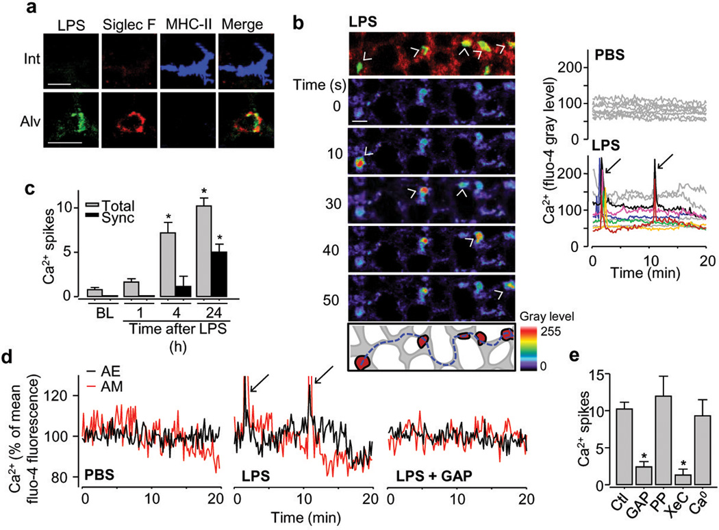
The tissue-resident macrophages of barrier organs constitute the first line of defence against pathogens at the systemic interface with the ambient environment. In the lung, resident alveolar macrophages (AMs) provide a sentinel function against inhaled pathogens. Bacterial constituents ligate Toll-like receptors (TLRs) on AMs, causing AMs to secrete proinflammatory cytokines that activate alveolar epithelial receptors, leading to recruitment of neutrophils that engulf pathogens. Because the AM-induced response could itself cause tissue injury, it is unclear how AMs modulate the response to prevent injury. Here, using real-time alveolar imaging in situ, we show that a subset of AMs attached to the alveolar wall form connexin 43 (Cx43)-containing gap junction channels with the epithelium. During lipopolysaccharide-induced inflammation, the AMs remained sessile and attached to the alveoli, and they established intercommunication through synchronized Ca(2+) waves, using the epithelium as the conducting pathway. The intercommunication was immunosuppressive, involving Ca(2+)-dependent activation of Akt, because AM-specific knockout of Cx43 enhanced alveolar neutrophil recruitment and secretion of proinflammatory cytokines in the bronchoalveolar lavage. A picture emerges of a novel immunomodulatory process in which a subset of alveolus-attached AMs intercommunicates immunosuppressive signals to reduce endotoxin-induced lung inflammation.


Figures

Figure 1
Figure 1. Live confocal microscopy of AMs in situ
(a) Interstitial (arrowhead) and luminal (arrow) YFP+ cells (yellow-green) in autofluorescent alveoli (red). Sketch of imaged field. n = 15. (b) Immunofluorescence and quantification of alveolar (top) and interstitial (bottom) YFP+ cells. n = 4 or 5. (c) Dye (green) injection in alveolus (Alv). Interstitial (Int) MHC-II+ DC (red, arrowhead) and luminal AM (arrow). n = 3. (d) Merge of S. aureus (green, arrow in inset) with AMs (red; arrowhead in inset). n = 4 (e) Ca2+ uncaging in epithelium (dotted circle) spreads Ca2+ (arrow) to AM (arrowhead). Bars show GAP 26/27 effect. n = 4 or 5. (f) Alveoli with Cx43 (red) and calcein (green) show Cx43-low (arrowhead) and Cx43-high AMs (arrow) expressing YFP (yellow-green). High power images show calcein in AMs before (pre) and after (post) photobleaching. Line drawn by linear regression. n = 23 AMs, 4 lungs. (g) Cx43 (red), CD11c (green) and nuclei (blue) in AMs from BAL (B) or lung tissue (T). Bars show protein (left) and mRNA (right) expressions in AMs. n = 4. Scale bars, 15 μm. mean±SEM. *P<0.05.
Figure 2
Figure 2. Communicating Ca2+ spikes in AMs
(a) Fluorescent LPS (green), AM (red) and DC (blue) in interstitium (Int) and alveolar lumen (Alv). n = 3 (b) YFP-expressing AMs (yellow; topmost panel), pseudocolored sequential images show increased fluo-4 fluorescence (arrowheads), 24 h after LPS. Dashes sketch spike path between AMs. Tracings show synchronous AM responses (arrows). n = 4 (c) Ca2+ spikes normalized for 10 AMs per imaging field. Total, all spikes; Sync, Synchronous spikes; BL, baseline (n = 8); 1h, n = 6; 4h, n = 4; 24h, n = 12 (d) Ca2+ oscillations and spikes (arrows) in AMs and adjoining alveolar epithelium (AE) 24 h after LPS or PBS. GAP, GAP26/27. n = 4 (e) Ca2+ spikes are 24h after LPS, normalized for 10 AMs per imaging field.; Ctl, LPS alone (n = 12); PP, PPADS (n = 4); XeC, xestospongin C (n = 4); Ca0, Ca2+ depletion (n = 3). GAP, n = 5. Scale bars, 20 μm. Bars, mean±SEM. *P<0.05 versus Ctl or corresponding BL.
Figure 3
Figure 3. AM-MyD88 in inflammatory signaling
(a-c) Synchronized (Syn) and non-synchronized (Non) Ca2+ spikes (arrows) and oscillations in wild type (WT, n = 12), CD11cMyD88−/− (My−/−, n = 5) and CD11cCx43−/− (Cx−/−, n = 5) mice. (d) Alveoli (green), and Ly6G+ (red) and CD11b+ neutrophils (blue) 24 h after LPS. Scale bar, 30 μm. n = 4 (e) Responses are 24 h after treatments. LPS WT: n = 9, others: n = 4. Bars, mean±SEM. *P<0.05.
Figure 4
Figure 4. AM-epithelial signaling
(a) BAL-cytokine ELISA in CD11cCx43−/− (Cx−/−) and littermate control (LC) mice. nd, not detectable. n = 3 or 4. (b) Kaplan-Meier plots. n = 16 LC, 17 Cx43−/−. (c,d) Data are 24 h after treatment. IP, immunoprecipitation. n = 4 (e) In situ immunofluorescence (red) of alveolar epithelium and YFP+ AMs (yellow-green) 24 h after LPS. n = 4 (f,g) Lung lysate Western blots and BAL leukocyte counts 24 h after LPS in lungs given scrambled (Sc) or CAMKKα-specific (Si) siRNA. n = 4 for PBS+si, n = 5 for LPS+si, others: n = 6. All blots (n = 3) from same sample set. CAMKKα and Actin processed on different gel. Bars, mean±SEM. *P<0.05.

References

    1. Laskin DL, Sunil VR, Gardner CR, Laskin JD. Macrophages and tissue injury: agents of defense or destruction? Annu Rev Pharmacol Toxicol. 2011;51:267–288. - PMC - PubMed
    1. Medzhitov R. Toll-like receptors and innate immunity. Nat Rev Immunol. 2001;1:135–145. - PubMed
    1. Thorley AJ, et al. Differential regulation of cytokine release and leukocyte migration by lipopolysaccharide-stimulated primary human lung alveolar type II epithelial cells and macrophages. J Immunol. 2007;178:463–473. - PubMed
    1. Kuebler WM, Parthasarathi K, Wang PM, Bhattacharya J. A novel signaling mechanism between gas and blood compartments of the lung. J Clin Invest. 2000;105:905–913. - PMC - PubMed
    1. Maus UA, et al. Role of resident alveolar macrophages in leukocyte traffic into the alveolar air space of intact mice. Am J Physiol Lung Cell Mol Physiol. 2002;282:L1245–L1252. - PubMed

Publication types

MeSH terms