. 2014 Apr 1;127(Pt 7):1441-53.
doi: 10.1242/jcs.136390. Epub 2014 Jan 24.

HDAC1 activates FoxO and is both sufficient and required for skeletal muscle atrophy

Affiliations

HDAC1 activates FoxO and is both sufficient and required for skeletal muscle atrophy

Adam W Beharry et al. J Cell Sci. .

Abstract

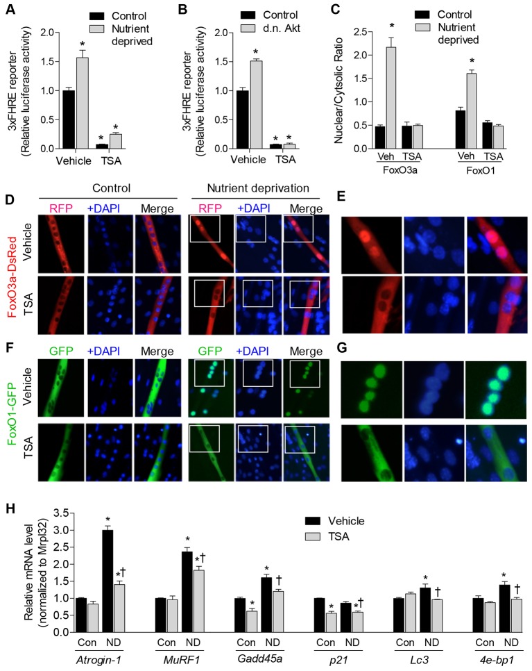
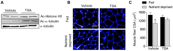
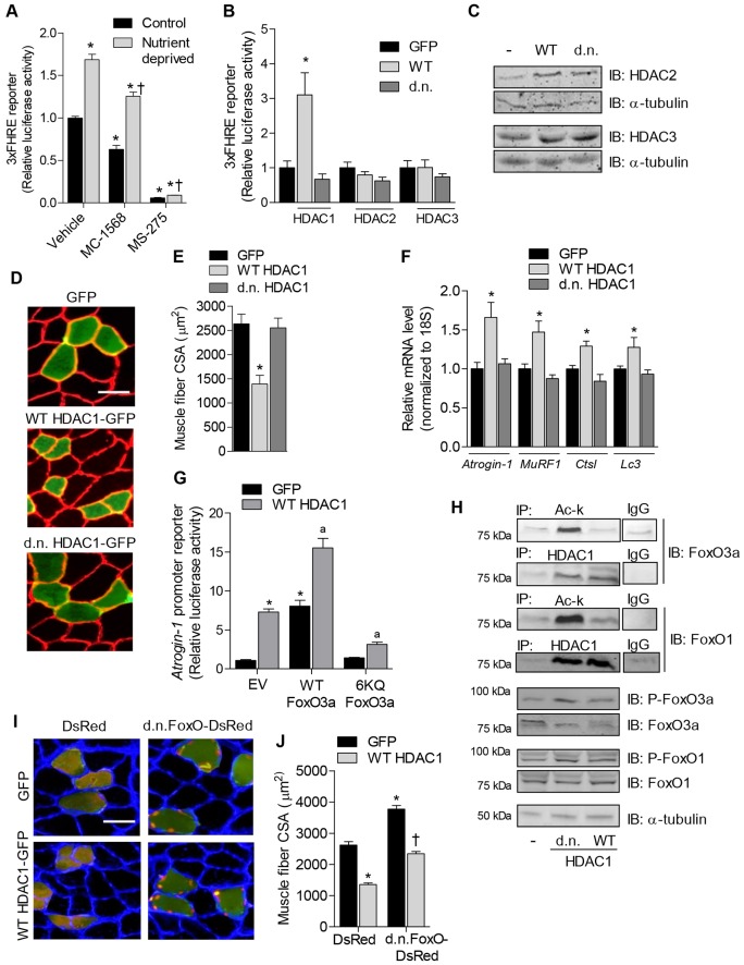
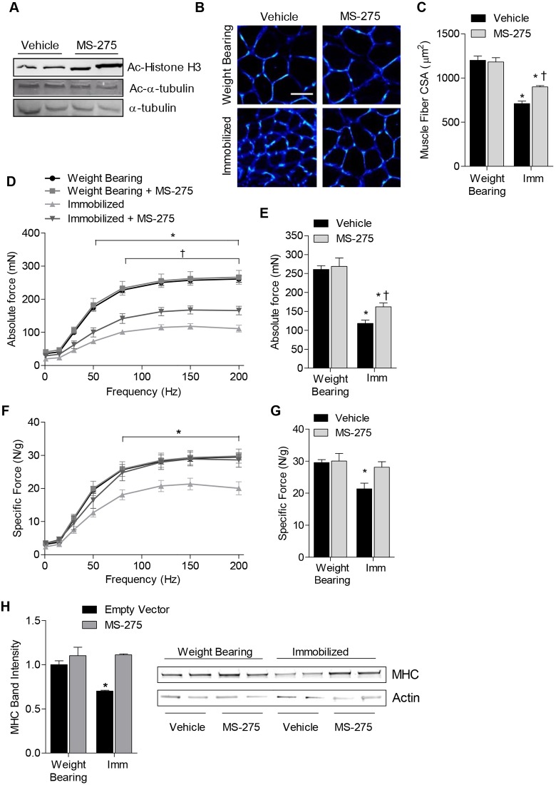
The Forkhead box O (FoxO) transcription factors are activated, and necessary for the muscle atrophy, in several pathophysiological conditions, including muscle disuse and cancer cachexia. However, the mechanisms that lead to FoxO activation are not well defined. Recent data from our laboratory and others indicate that the activity of FoxO is repressed under basal conditions via reversible lysine acetylation, which becomes compromised during catabolic conditions. Therefore, we aimed to determine how histone deacetylase (HDAC) proteins contribute to activation of FoxO and induction of the muscle atrophy program. Through the use of various pharmacological inhibitors to block HDAC activity, we demonstrate that class I HDACs are key regulators of FoxO and the muscle-atrophy program during both nutrient deprivation and skeletal muscle disuse. Furthermore, we demonstrate, through the use of wild-type and dominant-negative HDAC1 expression plasmids, that HDAC1 is sufficient to activate FoxO and induce muscle fiber atrophy in vivo and is necessary for the atrophy of muscle fibers that is associated with muscle disuse. The ability of HDAC1 to cause muscle atrophy required its deacetylase activity and was linked to the induction of several atrophy genes by HDAC1, including atrogin-1, which required deacetylation of FoxO3a. Moreover, pharmacological inhibition of class I HDACs during muscle disuse, using MS-275, significantly attenuated both disuse muscle fiber atrophy and contractile dysfunction. Together, these data solidify the importance of class I HDACs in the muscle atrophy program and indicate that class I HDAC inhibitors are feasible countermeasures to impede muscle atrophy and weakness.

Keywords: Acetylation; Deacetylation; FoxO; Histone deacetylase; Muscle disuse; Nutrient deprivation.


Figures

Fig. 1.
Fig. 1.
Inhibition of class I and II HDACs by TSA blocks induction of the muscle atrophy program. (A) FoxO-dependent luciferase reporter (3×FHRE) activity was normalized to Renilla reniformis luciferase from 3-day-differentiated skeletal myotubes treated with TSA or vehicle during 18 hours of nutrient deprivation or control conditions (Con). (B) FoxO-dependent luciferase reporter activity was normalized to Renilla luciferase from 4-day-differentiated skeletal myotubes transfected as myoblasts with a dominant-negative (d.n.) Akt expression plasmid (or empty vector), and treated with TSA or vehicle 24 hours before harvest. (C–G) 3-day-differentiated myotubes expressing ectopic FoxO3a–DsRed or FoxO1–GFP were deprived of nutrients for 6 hours in the presence of TSA (or vehicle). Cellular localization of the proteins was subsequently determined using fluorescence microscopy following fixation and incubation with DAPI to label cell nuclei. The mean fluorescence of FoxO3a–DsRed and FoxO1–GFP in nuclear and cytoplasmic compartments was calculated for each condition and is expressed as a ratio to indicate the relative localization (C) Representative images from each condition are shown in D–G. The panels in E and G are enlarged images of the areas indicated by white boxes in the corresponding images in D and F, respectively. In F, images taken from control or nutrient-deprived skeletal myotubes are in alignment with those of D. (H) The relative mRNA levels of the FoxO target genes, atrogin-1, MuRF1, Gadd45a, p21, Lc3 and 4e-bp1 in 3-day-differentiated myotubes following 6 hours of nutrient deprivation (or control conditions) in the presence of TSA or vehicle. ND, nutrient deprived. All data represent n = 3 and are reported as means ± s.e.m., normalized to the absolute control group. *P<0.05 (compared with absolute control group). P<0.05 (compared with vehicle within respective treatment group).
Fig. 2.
Fig. 2.
TSA treatment prevents skeletal muscle atrophy in vivo. Mice were treated with TSA or vehicle (sterile PBS) for 3 days during normal (fed) conditions or during nutrient deprivation. (A) Representative western blots for acetylated (Ac) histone H3, acetylated-α-tubulin or α-tubulin as a control, from vehicle- or TSA-treated muscles. (B) Representative muscle cross-sections incubated with wheatgerm agglutinin to visualize muscle fiber membranes (blue). (C) Average plantaris muscle fiber CSA from all groups. Bars represent mean ± s.e.m. for six muscles per group. *P<0.05 (compared with absolute control group).
Fig. 3.
Fig. 3.
HDAC1 deacetylase activity is sufficient to cause skeletal muscle atrophy. (A,B) FoxO-dependent luciferase reporter activity (A) was normalized to Renilla reniformis luciferase from 3-day-differentiated skeletal myotubes treated with vehicle, MC-1568 (class II HDAC inhibitor) or MS-275 (class I HDAC inhibitor) for 6 hours under control conditions or during nutrient deprivation and (B) from soleus muscles of rats transfected with either GFP, WT or dominant-negative HDAC1, HDAC2 or HDAC3 expression plasmids. (C) Overexpression of HDAC2 and HDAC3 was confirmed by western blot. (D–F) Soleus muscles were transfected with GFP, WT HDAC1–GFP or dominant-negative HDAC1–GFP expression plasmids and harvested 7 days after transfection to visualize muscle fiber CSA (D), measure the mean fiber CSA (E), and quantify the relative mRNA levels of atrogin-1, MuRF1, Ctsl and Lc3 normalized to 18S (F). (G) Relative luciferase activity from soleus muscles transfected with a luciferase reporter plasmid driven by the promoter of atrogin-1, plus expression plasmids for GFP or WT HDAC1, plus empty vector (EV), WT FoxO3a or FoxO3a 6KQ. (H) The ability of HDAC1 to interact with, and regulate the acetylation of, endogenous FoxO1 and FoxO3a was determined in soleus muscles injected with GFP, WT HDAC1–GFP or dominant-negative HDAC1–GFP expression plasmids. Equal amounts of protein extract were incubated with either an antibody against acetyl-lysine to immunoprecipitate (IP) total acetylated proteins (Ac-k), an antibody against HDAC1 to IP HDAC1 protein complexes, or IgG as a negative control. Precipitated proteins were subjected to SDS-PAGE and immunoblotted for either FoxO3a or FoxO1. Experiments were independently repeated three times. Western blots were also performed on equal amounts of whole-muscle lysates from the same samples using antibodies for total FoxO1 and FoxO3a or phosphorylated FoxO1 or FoxO3a (P-FoxO1 and P-FoxO3a, respectively). (I) Representative muscle cross-sections and (J) mean muscle fiber CSA of soleus muscles co-transfected with expression plasmids for DsRed plus GFP, DsRed plus WT HDAC1–GFP, dominant-negative FoxO plus GFP or dominant-negative FoxO plus WT HDAC1–GFP. Scale bars: 50 µm. All data represent the mean ± s.e.m. for six muscles per group. *P<0.05 (compared with absolute control group). P<0.05 (compared with control within respective treatment group). aP<0.05 (compared with EV plus WT HDAC1). d.n., dominant negative.
Fig. 4.
Fig. 4.
HDAC1 deacetylase activity is required for skeletal muscle atrophy. (A) Representative muscle cross-sections and (B) mean fiber CSA from 7-day-immobilized soleus muscles injected with GFP or dominant-negative (d.n.) HDAC1–GFP. The dashed line represents the average CSA of weight-bearing GFP-transfected fibers. Scale bar: 50 µm. (C) Relative luciferase activity driven by a FoxO-dependent reporter plasmid and (D) relative mRNA levels of atrogin-1, MuRF1, Ctsl and Lc3 normalized to 18S from the soleus of 3-day-immobilized rats transfected with GFP, WT or dominant-negative HDAC1–GFP expression plasmids. All data from immobilized muscles are normalized to data collected from the absolute control group (weight-bearing, GFP) to reflect the raw data. All data represent mean ± s.e.m. for six muscles per group. *P<0.05 (compared with absolute control group). P<0.05 (compared with control within respective treatment group). (E) Total protein levels of FoxO1 and FoxO3a, or phosphorylated FoxO1 and FoxO3a (P-FoxO1 and P-FoxO3a, respectively) from 7-day-immobilized (Imm) soleus muscles injected with GFP, WT or dominant-negative HDAC1–GFP was measured by western blot. (F) The levels of endogenous HDAC1 in whole-muscle lysate, or cytosolic or nuclear fractions in weight-bearing or immobilized (Imm) muscles were measured using western blot. Western blots for histone H1 and Sod1 on nuclear or cytosolic fractions are included, demonstrating successful separation of the nuclear and cytosolic fractions.
Fig. 5.
Fig. 5.
MS-275 prevents contractile dysfunction and attenuates muscle fiber atrophy associated with muscle disuse. Mice were treated with MS-275 or vehicle for 10 days under normal weight-bearing conditions or 10 days of muscle disuse, induced by cast-immobilization of the hind limbs. (A) Representative western blots are shown for acetylated histone H3 (Ac-histone H3), acetylated-α-tubulin (Ac-α-tubulin) or α-tubulin (as loading control) from vehicle- or MS-275-treated muscles. (B) Representative soleus muscle cross-sections were incubated with wheatgerm agglutinin to visualize muscle fiber membranes (blue). Scale bar: 50 µm. (C) The average muscle fiber CSA from each group was quantified. (D–G) The force-generating capacity of soleus muscles from each group was measured, including (D) absolute force–frequency relationship, (E) maximal absolute force, (F) specific force–frequency relationship (force normalized to muscle weight), and (G) maximal specific force. (H) The relative protein content of myosin heavy chain (MHC) was determined using SDS-PAGE. All data represent the mean ± s.e.m. for six muscles per group. *P<0.05 (compared with the weight-bearing group treated with vehicle). P<0.05 (compared with the vehicle-treated control within respective treatment group). Imm, immobilized.

References

    1. Bertaggia E., Coletto L., Sandri M. (2012). Posttranslational modifications control FoxO3 activity during denervation. Am. J. Physiol. 302, C587–C596 10.1152/ajpcell.00142.2011 - DOI - PubMed
    1. Brunet A., Bonni A., Zigmond M. J., Lin M. Z., Juo P., Hu L. S., Anderson M. J., Arden K. C., Blenis J., Greenberg M. E. (1999). Akt promotes cell survival by phosphorylating and inhibiting a Forkhead transcription factor. Cell 96, 857–868 10.1016/S0092--8674(00)80595--4 - DOI - PubMed
    1. Caiozzo V. J., Haddad F., Baker M. J., Herrick R. E., Prietto N., Baldwin K. M. (1996). Microgravity-induced transformations of myosin isoforms and contractile properties of skeletal muscle. J. Appl. Physiol. 81, 123–132 - PubMed
    1. Caiozzo V. J., Baker M. J., Baldwin K. M. (1998). Novel transitions in MHC isoforms: separate and combined effects of thyroid hormone and mechanical unloading. J. Appl. Physiol. 85, 2237–2248 - PubMed
    1. Campione M., Ausoni S., Guezennec C. Y., Schiaffino S. (1993). Myosin and troponin changes in rat soleus muscle after hindlimb suspension. J. Appl. Physiol. 74, 1156–1160 - PubMed

Publication types