. 2014 Mar 14;289(11):7374-87.
doi: 10.1074/jbc.M113.526111. Epub 2014 Jan 24.

CCN2 suppresses catabolic effects of interleukin-1β through α5β1 and αVβ3 integrins in nucleus pulposus cells: implications in intervertebral disc degeneration

Affiliations

CCN2 suppresses catabolic effects of interleukin-1β through α5β1 and αVβ3 integrins in nucleus pulposus cells: implications in intervertebral disc degeneration

Cassie M Tran et al. J Biol Chem. .

Abstract

The objective of the study was to examine the regulation of CCN2 by inflammatory cytokines, IL-1β, and TNF-α and to determine whether CCN2 modulates IL-1β-dependent catabolic gene expression in nucleus pulposus (NP) cells. IL-1β and TNF-α suppress CCN2 mRNA and protein expression in an NF-κB-dependent but MAPK-independent manner. The conserved κB sites located at -93/-86 and -546/-537 bp in the CCN2 promoter mediated this suppression. On the other hand, treatment of NP cells with IL-1β in combination with CCN2 suppressed the inductive effect of IL-1β on catabolic genes, including MMP-3, ADAMTS-5, syndecan 4, and prolyl hydroxylase 3. Likewise, silencing of CCN2 in human NP cells resulted in elevated basal expression of several catabolic genes and inflammatory cytokines like IL-6, IL-4, and IL-12 as measured by gene expression and cytokine protein array, respectively. Interestingly, the suppressive effect of CCN2 on IL-1β was independent of modulation of NF-κB signaling. Using disintegrins, echistatin, and VLO4, peptide inhibitors to αvβ3 and α5β1 integrins, we showed that CCN2 binding to both integrins was required for the inhibition of IL-1β-induced catabolic gene expression. It is noteworthy that analysis of human tissues showed a trend of altered expression of these integrins during degeneration. Taken together, these results suggest that CCN2 and inflammatory cytokines form a functional negative feedback loop in NP cells that may be important in the pathogenesis of disc disease.

Keywords: Cartilage Biology; Chondrocytes; Cytokine; Extracellular Matrix Proteins; Integrins.


Figures
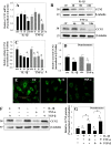
FIGURE 1.
FIGURE 1.
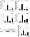
IL-1β and TNF-α decrease CCN2 expression in NP cells. A, real time RT-PCR analysis of NP cells treated with IL-1β or TNF-α for 4, 8, and 24 h shows that CCN2 is significantly decreased at 24 h. B, Western blot analysis of cells treated with IL-1β or TNF-α for 4, 8, and 24 h shows a decrease in CCN2 protein levels. C, densitometric analysis of several independent experiments as shown in B confirms that CCN2 is significantly decreased by IL-1β or TNF-α treatment. D, densitometric analysis of several Western blots of conditioned media from IL-1β- or TNF-α-treated cells shows significantly decreased levels of secreted CCN2 with IL-1β or TNF-α treatment. E, immunofluorescence microscopy of NP cells treated with IL-1β or TNF-α for 24 h shows that CCN2 protein is decreased with treatment compared with untreated control. TGF-β counteracts suppression of CCN2 by IL-1β and TNF-α F, Western blot of NP cells treated with IL-1β and TNF-α with and without TGF-β. IL-1β and TNF-α suppress CCN2; however, the addition of TGF-β together with either IL-1β and TNF-α negates the suppressive effects of the inflammatory cytokines on CCN2 expression. G, densitometry analysis of at least three Western blot experiments as shown in F reveals that although IL-1β and TNF-α suppress CCN2, the addition of TGF-β abrogates this suppressive effect. The values shown are means ± S.E. from at least three independent experiments. *, p < 0.05. Ctr or ctr, control.
FIGURE 2.
FIGURE 2.
IL-1β and TNF-α suppress CCN2 expression through NF-κB signaling. A and B, Western blot analysis of NP cells treated with IL-1β (Α) or TNF-α (B) with or without pretreatment with NF-κB (SM7368), p38 (SB203580), JNK (SP60025), or ERK (PD98059) inhibitors used at 10 μm each. Note that suppression of CCN2 by IL-1β or TNF-α treatment is ablated only by pretreatment with the NF-κB inhibitor. C, densitometric analysis of several independent experiments as shown in A above confirms that suppression of CCN2 by IL-1β is abrogated only by treatment with NF-κB inhibitor. D, immunofluorescence image of NP cells transduced with a lentivirus co-expressing shRNA against p65 and YFP (ShP65-LV) demonstrates high transduction efficiency. E, Western blot analysis of NP cells transduced with lentivirally mediated shRNA against a scrambled control sequence (Ctr-LV) or p65 (Shp65-LV) that were treated with IL-1β or TNF-α. CCN2 is suppressed by IL-1β or TNF-α, but with p65 knockdown this effect is abrogated. F, densitometric analysis of several independent experiments shown in D shows that p65 was significantly knocked down when cells were transduced with Shp65-LV. F and G, densitometric analysis of several independent experiments shows that the suppression of CCN2 protein levels by IL-1β (G) or TNF-α (H) is rescued by knockdown of p65. The values shown are means ± S.E. from at least three independent experiments. *, p < 0.05. Ctr, control.
FIGURE 3.
FIGURE 3.
Suppression of CCN2 promoter activity by IL-1β and TNF-α requires p65 binding to NF-κB binding sites. A, Multiz alignment of two NF-κB binding sites at −93/86 and −546/−537 bp on human CCN2 promoter shows relatively high conservation between vertebrate species. B, schematic of WT and mutant (MT1, MT2, or MT1,2) human CCN2 promoter constructs with NF-κB binding sites located at −93/−86 bp and −546/−537 bp. C, NP cells were transfected with WT and mutant (MT1, MT2, or MT1,2) human CCN2 promoter constructs and treated with IL-1β or TNF-α. Only the WT promoter shows a decrease in promoter activity in response to IL-1β and TNF-α treatment, whereas the mutant promoter constructs show no significant change (ns). D, ChIP analysis shows binding of p65 to both the κB sites within CCN2 promoter in presence or absence of IL-1β. Negative primers show little or no amplification, indicating the high specificity of the ChIP assay. The asterisk indicates significant increase in binding/enrichment over negative control (Neg. Ctr). The values shown are means ± S.E. from three independent experiments. *, p < 0.05.
FIGURE 4.
FIGURE 4.
CCN2 treatment decreases the IL-1β-mediated induction of several catabolic genes. A, real time RT-PCR analysis of NP cells treated with IL-1β (10 ng/ml), CCN2 (10 ng/ml), or a both IL-1β and CCN2 together. A–D, IL-1β causes the induction in catabolic genes MMP-3 (A), ADAMTS-5 (B), syndecan 4 (SDC4) (C), and PHD3 (D), whereas CCN2 alone has no significant effect. When the cells are treated with both CCN2 and IL-1β, CCN2 suppressed IL-1β-dependent induction of catabolic genes compared with IL-1β alone. E, Western blot analysis of conditioned media from NP cells treated with IL-1β, CCN2, or both IL-1β and CCN2 together shows that the level of secreted MMP-3 is induced by IL-1β treatment and unchanged by CCN2 treatment. Compared with MMP3 levels induced by IL-1β alone, MMP-3 levels are decreased when cells are treated with CCN2 and IL-1β together. F, densitometric analysis of at least three independent experiments as in E shows a statistically significant increase MMP-3 protein levels with IL-1β treatment that is significantly decreased when cells are treated with both IL-1β and CCN2 together. The values shown are means ± S.E. from at least three independent experiments. *, p < 0.05. ctr, control.
FIGURE 5.
FIGURE 5.
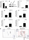
Silencing of CCN2 results in the increase in basal inflammatory gene expression by NP cells. A, real time RT-PCR analysis of human NP cells transduced with a lentiviral construct expressing shRNA against a scrambled control sequence (Ctr-LV) or CCN2 (shCCN2-LV) shows that CCN2 expression was significantly suppressed in cells receiving shCCN2-LV. B, Western blot analysis confirms silencing of CCN2 by shCCN2. C–G, knockdown of CCN2 expression resulted in the basal increase in MMP3 (C), MMP13 (D), ADAMTS-4 (E), ADAMTS-5 (F), and PHD3 (G). H, conditioned medium from human NP cells transduced with Ctr-LV or shCCN2-LV was probed for inflammatory cytokine production using an inflammatory cytokine array. Control cells show basal levels of inflammatory cytokine production, including IL-6 and IL-10, whereas knockdown of CCN2 causes an increase in the production of several inflammatory cytokines, which are marked with red ovals and whose fold change is indicated in parentheses. Positive controls (+ctr) and negative controls (−ctr) indicated that the array was properly performed. MCP-3, monocyte chemotactic protein 3; GCSF, granulocyte colony-stimulating factor; CXCL9, chemokine (CXC motif) ligand 9; RANTES, chemokine (CC motif) ligand 5; VEGF, vascular endothelial growth factor. The values shown are means ± S.E. from at least three independent experiments. *, p < 0.05. Ctr or ctr, control.
FIGURE 6.
FIGURE 6.
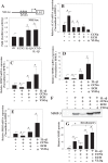
Anti-catabolic effect of CCN2 requires binding to integrin receptors. A, NP cells were transfected with a prototypic reporter responsive to NF-κB activity (NRE-luc) and treated with CCN2, IL-1β, or both together. IL-1β treatment alone induces NF-κB activity, whereas CCN2 has no significant effect. When CCN2 is added in combination with IL-1β, there is no significant difference in reporter activity compared with IL-1β alone. B–E, real time RT-PCR analysis of aggrecan (ACAN) gene expression with treatment of CCN2 with and without inhibitors of integrin binding, ECH, which inhibits αvβ3, or VLO4, which inhibits α5β1. CCN2 treatment alone increases aggrecan expression, whereas pretreatment with either inhibitor ablates this effect. Note that inhibitors alone have no significant effect on ACAN expression. Real time RT-PCR analysis following treatment of NP cells with IL-1β or IL-1β and CCN2 together, with or without ECH or VLO4. Treating cells with CCN2 in combination with IL-1β causes a significant decrease in the expression of MMP3 (C), MMP13 (D), and PHD3 (E) compared with IL-1β alone. Interestingly, pretreatment with ECH or VLO4 before IL-1β and CCN2 treatment not only abrogates the suppressive effect of CCN2 on IL-1β but also results in further induction in catabolic gene expression. F, Western blot analysis of conditioned media of NP cells treated with IL-1β, CCN2, or both together, with and without ECH or VLO4 shows that IL-1β induction of secreted MMP3 levels are decreased when cells are treated with CCN2 together with IL-1β. Integrin αvβ3 or α5β1 inhibition by ECH or VLO4 abolishes the decrease in MMP3 by the addition of CCN2. G, densitometric analysis of at least three independent experiments as shown in F reveals that the increase in MMP3 by IL-1β is significantly decreased by CCN2. When cells are pretreated with ECH or VLO4, not only is this effect abolished, but also MMP3 levels are significantly elevated over IL-1β treatment alone. The values shown are means ± S.E. from at least three independent experiments. *, p < 0.05. ctr, control.
FIGURE 7.
FIGURE 7.
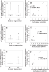
Expression analysis of β1, α5, and αV integrin subunits in human degenerative disc samples. A–C, real time RT-PCR analysis of human degenerate disc samples (n = 34, grades ≤3 = 11, grades 4 and 5 = 23) shows a trend of increasing expression of integrin subunits β1 (A), α5 (B), and αV (C). The data are represented as box and whisker plots. Each box represents the 75th–25th percentile of values, the line inside each box represents the median value separating the upper and lower quartiles, whiskers show the maximum and minimum values excluding outliers in each set, and dots denote outliers that fall outside of 1.5 times the upper or lower quartile range, respectively. D–F, a positive correlation in mRNA levels between β1 and α5 (p = 0.0038) (D), β1 and αV (p = 0.0038) (E), and αV and α5 (p = 2E-07) (F) was found in human degenerate disc samples.
FIGURE 8.
FIGURE 8.
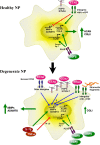
The role of CCN2 in healthy and degenerate NP. In the healthy NP, CCN2 interacts with α5β1 and αvβ3 integrins to maintain basal transcription of extracellular matrix genes, aggrecan (ACAN) and collagen II (COLII), while keeping the transcription of catabolic genes, MMPs, and ADAMTS, in check. TGF-β in the healthy state also promotes CCN2 and the matrix gene expression, resulting in an anabolic contribution toward matrix homeostasis. In the degenerate NP, an increase in inflammatory cytokines, IL-1β and TNF-α, drives the increase in catabolic, matrix degrading enzymes, MMPs and ADAMTS, and suppresses CCN2 through NF-κB signaling. Increased TGF-β in degeneration can override CCN2 suppression by cytokines and lead to excess CCN2, which will interact with a differential set of receptors induced in degeneration, including the heparan sulfate proteoglycan (HSPG), syndecan 4 (SYND4), other integrins, and increased α5β1. At the same time, the production of fibronectin fragments is induced in degeneration and induces catabolic gene expression by the interaction with α5β1, reducing the availability of this integrin for CCN2 interaction as well. It is possible that downstream signaling from these differential interactions, involving CCN2, could contribute toward the overall shift to catabolism in degeneration, unlike in the healthy state, where CCN2 interaction with α5β1 and αvβ3 integrins promotes anti-catabolic/anabolic effects. Thus, the differential effects of CCN2 could be due to the difference in the receptors it engages.

References

    1. Setton L. A., Chen J. (2006) Mechanobiology of the intervertebral disc and relevance to disc degeneration. J. Bone Joint Surg. Am. 88, 52–57 - PubMed
    1. Feng H., Danfelter M., Strömqvist B., Heinegård D. (2006) Extracellular matrix in disc degeneration. J. Bone Joint Surg. Am. 88, 25–29 - PubMed
    1. Wang J., Markova D., Anderson D. G., Zheng Z., Shapiro I. M., Risbud M. V. (2011) TNF-α and IL-1β promote a disintegrin-like and metalloprotease with thrombospondin type I motif-5-mediated aggrecan degradation through syndecan-4 in intervertebral disc. J. Biol. Chem. 286, 39738–39749 - PMC - PubMed
    1. Tian Y., Yuan W., Fujita N., Wang J., Wang H., Shapiro I. M., Risbud M. V. (2013) Inflammatory cytokines associated with degenerative disc disease control aggrecanase-1 (ADAMTS-4) expression in nucleus pulposus cells through MAPK and NF-κB. Am. J. Pathol. 182, 2310–2321 - PMC - PubMed
    1. Le Maitre C. L., Hoyland J. A., Freemont A. J. (2007) Catabolic cytokine expression in degenerate and herniated human intervertebral discs. IL-1β and TNFα expression profile. Arthritis Res. Ther. 9, R77. - PMC - PubMed

Publication types

MeSH terms

LinkOut - more resources