. 2014 Mar 14;289(11):7799-811.
doi: 10.1074/jbc.M113.525352. Epub 2014 Jan 24.

Crystal structure of a Schistosoma mansoni septin reveals the phenomenon of strand slippage in septins dependent on the nature of the bound nucleotide

Affiliations

Crystal structure of a Schistosoma mansoni septin reveals the phenomenon of strand slippage in septins dependent on the nature of the bound nucleotide

Ana E Zeraik et al. J Biol Chem. .

Abstract

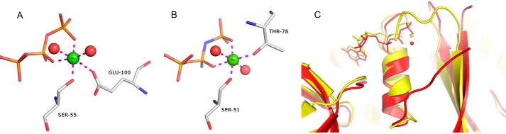
Septins are filament-forming GTP-binding proteins involved in important cellular events, such as cytokinesis, barrier formation, and membrane remodeling. Here, we present two crystal structures of the GTPase domain of a Schistosoma mansoni septin (SmSEPT10), one bound to GDP and the other to GTP. The structures have been solved at an unprecedented resolution for septins (1.93 and 2.1 Å, respectively), which has allowed for unambiguous structural assignment of regions previously poorly defined. Consequently, we provide a reliable model for functional interpretation and a solid foundation for future structural studies. Upon comparing the two complexes, we observe for the first time the phenomenon of a strand slippage in septins. Such slippage generates a front-back communication mechanism between the G and NC interfaces. These data provide a novel mechanistic framework for the influence of nucleotide binding to the GTPase domain, opening new possibilities for the study of the dynamics of septin filaments.

Keywords: Conformational Change; Crystal Structure; Filament; GTPase; GTPase Domain; Protein Conformation; Protein Structure; Schistosoma; Septin; X-ray Crystallography.


Figures

FIGURE 1.
FIGURE 1.
SmSEPT10 displays no detectable GTPase activity. Shown is the profile of the elution of guanine nucleotides from the DEAE-5PW anion exchange column as measured at 253 nm after incubating 15 μm SmSEPT10 and 45 μm GTP for different time intervals. AU, absorbance units.
FIGURE 2.
FIGURE 2.
SmSEPT10G is able to bind GTP and GDP. Shown are raw ITC data (top) and binding isotherms (bottom) from the guanine nucleotide binding assay utilizing the following: SmSEPT10G (24 μm) in standard buffer in the presence of 5 mm MgCl2 titrated with 2 mm GTP (A) or SmSEPT10G (15 μm) in standard buffer titrated with 3 mm GDP (B). Each titration was carried out with 45 injections of 5 μl.
FIGURE 3.
FIGURE 3.
SmSEPT10-GDP complex is predominantly magnesium-free at physiological concentrations. Shown are 31P{1H} NMR spectra recorded at 290 K and operating at 31P frequency of 243.94 MHz (11.4 T) of SmSEPT10G-GDP complex in the absence of Mg2+ (A) and in the presence of 1 mm (B) and 20 mm (C) of Mg2+. The chemical shift scale was referenced in 0.00 ppm from signal of 85% external phosphoric acid contained in a capillary tube, and the resonances were assigned to the α- and β-phosphate (free and bound), according to John et al. (54).
FIGURE 4.
FIGURE 4.
Ribbon diagram of the asymmetric unit of the SmSEPT10G-GDP complex. The nomenclature for the secondary structure elements follows that adopted by Sirajuddin et al. (12) and is shown on the right.
FIGURE 5.
FIGURE 5.
LIGPLOT+ representation of contacts within the GTP binding site. Hydrogen bonds made between the GTP molecule and the B subunit are shown explicitly, and the majority are conserved in both complexes (GTP and GDP). Water molecules (which are observed interacting with both the phosphate groups and the base) are shown as light blue spheres, and the Mg2+ ion is shown as a green sphere.
FIGURE 6.
FIGURE 6.
SmSEPT10 lacks catalytic activity. Both p21 H-ras (A) and SEPT2 (B) display the classical “loaded spring” mechanism by which main chain amide groups from both switch regions (SW1 and SW2) form direct hydrogen bonds to the γ-phosphate. In the case of switch 1, this is provided by a threonine residue that is not conserved in SmSEPT10. This threonine also secures a water molecule (red sphere in A and B), which is poised for in-line attack during catalysis. In SmSEPT10 (C), no direct hydrogen bonds are formed with the γ-phosphate by residues from the switch regions. SW2 interacts via a water molecule (small red sphere in C), and SW1 is completely absent due to the lack of the threonine. As a consequence, there is no water adequately poised for catalysis. In SmSEP10, a standard 2FoFc electron density map for this region is also shown.
FIGURE 7.
FIGURE 7.
The Mg2+ binding site. A, Mg2+ ion coordination as observed in the SmSEPT10 complex with GTP. B, Mg2+ binding site in the GppNHp complex with SEPT2. C, superposition of the SmSEPT10 (red) and SEPT2 (yellow) complexes, showing that the switch I region in the former does not participate in Mg2+ coordination. This leads to a significant difference from the metal coordination sphere, as can be seen in A and B.
FIGURE 8.
FIGURE 8.
Electron density of the β3 strand. FoFc electron density omit maps (contoured at 2.5σ) for the β3 strand in the GDP complex (A) and the GTP complex (B). In the latter, all residues are shifted by two to the right bring Glu100 into a position for coordinating the Mg2+ ion. Slippage appears to be facilitated by the existence of a Lys-Leu-Lys-Leu repeat and the presence of many small β-branched amino acids.
FIGURE 9.
FIGURE 9.
β-strand slippage. Shown are HERA hydrogen bonding diagrams for part of the β-sheet composed of strands 1–3. The situation in the GDP complex (A) is different to that of the GTP complex (B), but the total number of main chain hydrogen bonds is almost identical in both cases. In C, the result of slippage is seen to be the communication between the G interface to the left and the NC interface to the right. Part of a filament of the GTP complex is shown (dark blue) with a single subunit from the GDP complex superimposed at the center (light blue). The sliding (β3) strand is shown in yellow (GTP complex) and orange (GDP complex).
FIGURE 10.
FIGURE 10.
The consequences of strand slippage. Part of the heterocomplex of SEPT2-SEPT6-SEPT7 is shown (red, dark blue, and yellow, respectively) with the SmSEPT10G structure, as observed when complexed to GTP (light blue), superimposed on SEPT6. The consequence of strand slippage is the extension of the β-hairpin connecting β2 to β3 and thereby the coverage of the N-terminal helix α0. This region corresponds to the polybasic region known to be important for membrane association in mammalian septins. The implication is that strand slippage may affect membrane binding in a nucleotide-dependent fashion.
FIGURE 11.
FIGURE 11.
Comparison between SmSEPT10 and Arf6. Septins may operate via a mechanism analogous to that seen in Arf proteins, which use β-strand slippage as a means to dislodge the N-terminal helix and so influence membrane association. A, superposition of Arf6 in its GDP complex (blue) and GTPγS complex (yellow), showing the slippage of both strands β2 and β3 with respect to β1, in the direction of the short N-terminal helix. B, in SmSEPT10, a similar color scheme is adopted, but in this case, β3 slips with respect to both β1 and β2.

Similar articles

Cited by

References

    1. Mostowy S., Cossart P. (2012) Septins. The fourth component of the cytoskeleton. Nat. Rev. Mol. Cell Biol. 13, 183–194 - PubMed
    1. Nishihama R., Onishi M., Pringle J. R. (2011) New insights into the phylogenetic distribution and evolutionary origins of the septins. Biol. Chem. 392, 681–687 - PMC - PubMed
    1. Versele M., Thorner J. (2005) Some assembly required. Yeast septins provide the instruction manual. Trends Cell Biol. 15, 414–424 - PMC - PubMed
    1. Russell S. E., Hall P. A. (2011) Septin genomics. A road less travelled. Biol. Chem. 392, 763–767 - PubMed
    1. Cao L., Yu W., Wu Y., Yu L. (2009) The evolution, complex structures and function of septin proteins. Cell Mol. Life Sci. 66, 3309–3323 - PMC - PubMed

Publication types

MeSH terms

LinkOut - more resources