. 2014 Apr 1;306(7):H1032-40.
doi: 10.1152/ajpheart.00911.2013. Epub 2014 Jan 24.

Novel role of aminopeptidase-A in angiotensin-(1-7) metabolism post myocardial infarction

Affiliations

Novel role of aminopeptidase-A in angiotensin-(1-7) metabolism post myocardial infarction

Mahmoud S Alghamri et al. Am J Physiol Heart Circ Physiol. .

Abstract

Aminopeptidase-A (APA) is a less well-studied enzyme of the renin-angiotensin system. We propose that it is involved in cardiac angiotensin (ANG) metabolism and its pathologies. ANG-(1-7) can ameliorate remodeling after myocardial injury. The aims of this study are to (1) develop mass spectrometric (MS) approaches for the assessment of ANG processing by APA within the myocardium; and (2) investigate the role of APA in cardiac ANG-(1-7) metabolism after myocardial infarction (MI) using sensitive MS techniques. MI was induced in C57Bl/6 male mice by ligating the left anterior descending (LAD) artery. Frozen mouse heart sections (in situ assay) or myocardial homogenates (in vitro assay) were incubated with the endogenous APA substrate, ANG II. Results showed concentration- and time-dependent cardiac formation of ANG III from ANG II, which was inhibited by the specific APA inhibitor, 4-amino-4-phosphonobutyric acid. Myocardial APA activity was significantly increased 24 h after LAD ligation (0.82 ± 0.02 vs. 0.32 ± 0.02 ρmol·min(-1)·μg(-1), MI vs. sham, P < 0.01). Both MS enzyme assays identified the presence of a new peptide, ANG-(2-7), m/z 784, which accumulated in the MI (146.45 ± 6.4 vs. 72.96 ± 7.0%, MI vs. sham, P < 0.05). Use of recombinant APA enzyme revealed that APA is responsible for ANG-(2-7) formation from ANG-(1-7). APA exhibited similar substrate affinity for ANG-(1-7) compared with ANG II {Km (ANG II) = 14.67 ± 1.6 vs. Km [ANG-(1-7)] = 6.07 ± 1.12 μmol/l, P < 0.05}. Results demonstrate a novel role of APA in ANG-(1-7) metabolism and suggest that the upregulation of APA, which occurs after MI, may deprive the heart of cardioprotective ANG-(1-7). Thus APA may serve as a potentially novel therapeutic target for management of tissue remodeling after MI.

Keywords: MALDI-imaging; aminopeptidase-A; angiotensin peptides; myocardial infarction; renin-angiotensin system.


Figures

Fig. 1.
Fig. 1.
Matrix-assisted laser desorption ionization (MALDI) imaging of ANG II metabolism in the myocardium. A: transverse myocardial section stained with hematoxylin and eosin (H and E) showing different anatomical areas within the myocardium. B: photographic image of a myocardium section before incubation with ANG II for spatial visualization of the generated peptides. C: MALDI imaging of ANG III [mass-to-charge (m/z) 931] generated in myocardium. D: MALDI imaging of ANG-(1–7) m/z 899 generated in the myocardium. E: overlay of ANG III and ANG-(1–7) signals. F: ANG-(1–7) formation after incubation with ANG II (10–1,000 μmol/l) at 5, 10, and 15 min. G: ANG III formation after incubation with ANG II (10–1,000 μmol/l) at 5, 10, and 15 min. Data are means ± SE. LV, left ventricle; RV, right ventricle; LVW, left ventricular wall; IVW, interventricular septal wall.
Fig. 2.
Fig. 2.
MALDI TOF/TOF of m/z 931 and m/z 899 formed in situ in the myocardium after incubation with ANG II. A: MS/MS of ANG-(1–7) standard. B: MS/MS of the product m/z 899 generated in the myocardium. C: MS/MS of ANG III standard. D: MS/MS of the generated product m/z 931. E: reproducibility of ANG III and ANG-(1–7) formation within the myocardium. There was little variation for ANG III and ANG-(1–7) generation in the myocardial sections within the same animal (intra-animal) or between animals (interanimal).
Fig. 3.
Fig. 3.
Inhibition of ANG-(1–7) and ANG III formation by specific enzyme inhibitors. A: ANG-(1–7) formation was inhibited by angiotensin-converting enzyme 2 (ACE2) inhibitor MLN-4760 in a concentration-dependent manner. B: quantitation of generated ANG-(1–7) after treatment with MLN-4760. C: ANG III formation was inhibited by the selective aminopeptidase-A (APA) inhibitor, 4-amino-phosphonobutyric acid (4-APBA). D: quantitation of ANG III formation after treatment with 4-APBA. Data were analyzed using one-way ANOVA. Modified Tukey post hoc test was used to compare means. Values are means ± SE (*P < 0.05 vs. 0 μmol/l; n = 3).
Fig. 4.
Fig. 4.
Increase of ANG-(1–7) formation after APA inhibition. A: ANG-(1–7) formation after incubation of myocardial sections with 100 μmol/l ANG II and 0, 1, 10, 100 μmol/l APA inhibitor 4-APBA. B: quantitation of ANG-(1–7) formation after APA inhibition using the MALDI imaging approach. Data were analyzed using one-way ANOVA. Modified Tukey post hoc test was used to compare means (*P < 0.05 vs. 0 μmol/l; n = 3 per group). C: quantitation of ANG-(1–7) formation before and after treatment with 50 μmol/l 4-APBA using the in vitro MALDI approach. Myocardial homogenates were incubated with 50 μmol/l ANG II for 30 min in MES buffer (pH = 6.5) at 37°C. The formed peptide was quantitated based on ANG-(1–7) stable-isotope-labeled standard. Data were analyzed using Student's t-test. Values are means ± SE (*P < 0.05; n = 3/group).
Fig. 5.
Fig. 5.
Role of ACE2 in APA-mediated increase of ANG-(1–7). A: intensity of the substrate ANG II after incubation with 100 μmol/l ANG II (baseline) or with 100 μmol/l ANG II and 100 μmol/l 4-APBA (4-APBA). B: ANG III generation in wild-type (WT) and ACE2 knockout (KO) after incubation with 100 μmol/l ANG II (baseline) or with 100 μmol/l ANG II and 100 μmol/l 4-APBA (4-APBA) using the in situ MALDI assay. C: ANG-(1–7) generation in WT and ACE2 KO after incubation with 100 μmol/l ANG II (baseline) or with 100 μmol/l ANG II and 100 μmol/l 4-APBA (4-APBA) using the in situ MALDI assay. Data were analyzed using two-way ANOVA. Modified Tukey post hoc test was used to compare means. Values are means± SE (*P < 0.05 vs. WT, †P < 0.05 vs. baseline; n = 4–5 per group).
Fig. 6.
Fig. 6.
In vitro metabolism of ANG-(1–7) and ANG II by APA. Recombinant APA (rAPA) enzyme was incubated with ANG-(1–7) or ANG II for 30 min at 37°C in assay buffer. A: ANG-(1–7) in assay buffer. B: ANG-(1–7)+ 1 ng rAPA. C: ANG-(1–7)+ 5 ng rAPA. D: ANG-(1–7) + 1 ng APA + 0.3 nmol/l 4-APBA. E: ANG-(1–7) + 1 ng APA + 3 nmol/l 4-APBA. F: ANG II in assay buffer. G: ANG II + 1 ng rAPA. H: ANG II + 5 ng rAPA. I: ANG II + 1 ng APA + 0.3 nmol/l 4-APBA. J: ANG II + 1 ng APA + 3 nmol/l 4-APBA.
Fig. 7.
Fig. 7.
Michaelis-Menten enzyme kinetics for APA and the substrates ANG II and ANG-(1–7) using the in vitro MALDI enzyme assay. A: rAPA (1 ng) was incubated with 1–90 μmol/l ANG II for 30 min at 37°C. B: rAPA (1 ng) was incubated with 1–90 μmol/l ANG-(1–7) for 30 min at 37°C. Results show similar binding affinities for APA to ANG-(1–7) (Km = 6.07 ± 1.12) and ANG II (Km = 14.67 ± 1.6). Values are means ± SE; n = 3 per group.
Fig. 8.
Fig. 8.
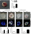
MALDI imaging after myocardial infarction (MI) and sham operation. A: representative 2,3,5-triphenyltetrazolium chloride (TTC) staining performed 24 h post-MI. Scale bar, 1 cm. (*infarct region). B: Western blot of APA of myocardial segments (a 3-mm myocardial segment was excised from the ventricular area of the heart at the plane of the papillary muscles) after MI or sham operation. APA expression was increased in MI compared with sham. (*P < 0.05; n = 3/group). C: in situ MALDI imaging assay of ANG III formed on a myocardial section after sham surgery using ANG II as a substrate. D: in situ MALDI imaging assay of ANG III on a myocardial section after MI surgery using ANG II as a substrate. The infarct area (*) is traced in red. E: in situ MALDI imaging assay of ANG-(2–7) formed on a myocardial section after sham surgery using ANG-(1–7) as a substrate. F: in situ MALDI APA imaging assay of ANG-(2–7) formed on a myocardial section after MI using ANG-(1–7) as a substrate The infarct area (*) is traced in red. G: quantitation of formed ANG III and ANG-(2–7). Data were analyzed using two-way ANOVA. Modified Tukey post hoc test was used to compare means. Values are means ± SE (*P < 0.05 vs. sham; n = 6/group). Signals were quantitated as integrated intensity and normalized to the area of the whole heart section. H: quantitation of the in vitro MALDI enzyme assay using homogenates from sham and MI hearts with ANG II as substrate. I: quantitation of the in vitro MALDI enzyme assay using homogenates from sham and MI hearts with ANG-(1–7) as substrate. Data were analyzed using Student's t-test. Values are means ± SE (*P < 0.05; n = 3–4/group).

References

    1. Alghamri MS, Weir NM, Anstadt MP, Elased KM, Gurley SB, Morris M. Enhanced angiotensin II-induced cardiac and aortic remodeling in ACE2 knockout mice. J Cardiovasc Pharmacol Ther 18: 138–151, 2013 - PubMed
    1. Bodineau L, Frugiere A, Marc Y, Claperon C, Llorens-Cortes C. Aminopeptidase A inhibitors as centrally acting antihypertensive agents. Heart Fail Rev 13: 311–319, 2008 - PubMed
    1. Bodineau L, Frugiere A, Marc Y, Inguimbert N, Fassot C, Balavoine F, Roques B, Llorens-Cortes C. Orally active aminopeptidase A inhibitors reduce blood pressure: a new strategy for treating hypertension. Hypertension 51: 1318–1325, 2008 - PubMed
    1. Camelliti P, Devlin GP, Matthews KG, Kohl P, Green CR. Spatially and temporally distinct expression of fibroblast connexins after sheep ventricular infarction. Cardiovasc Res 62: 415–425, 2004 - PubMed
    1. Clark MA, Nguyen C, Tran H. Angiotensin III induces c-Jun N-terminal kinase leading to proliferation of rat astrocytes. Neurochem Res 37: 1475–1481, 2012 - PubMed

Publication types

MeSH terms

LinkOut - more resources